08 Jul IVF: Single Cell Analysis Allows Extremely Early Prediction Of Embryo Abnormalities
 MedicalResearch.com Interview with:
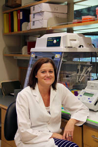
Shawn L. Chavez, Ph.D
Assistant Scientist/Professor
Oregon National Primate Research Center
OHSU | Oregon Health & Science University
Medical Research: What is the background for this study?
Dr. Chavez: This study builds upon a previous study also published in Nature Communications in 2012, which demonstrated that chromosomally normal and abnormal 4-cell human embryos can be largely distinguished by combining the timing intervals of the first three cell divisions with the presence or absence of a dynamic process called cellular fragmentation. The current study further combines time-lapse imaging of embryo development and full chromosome analysis with high throughout single-cell gene expression profiling to assess the chromosomal status of human embryos up to the 8-cell stage.
Medical Research: What are the main findings?
Dr. Chavez: The key findings of this research were that by measuring the duration of the first cell division, one can identify which embryos are chromosomally normal versus abnormal even earlier in development. By examining gene expression at a single-cell level, we were able to correlate the chromosomal make-up of an embryo to a subset of 12 genes that are activated prior to the first cell division. These genes likely came from either the egg or sperm and can be used to predict whether an embryo will be chromosomally normal or abnormal within the first 30 hours of development.
Medical Research: What should clinicians and patients take away from your report?
Dr. Chavez: A big debate in the in vitro fertilization (IVF) community right now is whether time-lapse imaging is actually beneficial and can replace pre-implantation screening (PGS). Personally, I think that by combining these two techniques and now potentially extending this to gene expression profiling, will provide the most comprehensive information for both clinicians and patients in the decision of which embryo(s) to transfer. The main goal of the prediction model was not to predict chromosomal abnormalities by itself, but to identify cellular pathways and related molecules indicative of embryo viability in culture medium or via other methods.
Medical Research: What recommendations do you have for future research as a result of this study?
Dr. Chavez: Although the predictive model has been tested on embryos from several different patients and IVF clinics, it will likely be necessary to test the model on other embryo cohorts in order to further substantiate our findings. Additional studies should also focus on the 1-cell embryo as a potential source of biomarkers that can prospectively predict embryo chromosomal status and obviously, this needs to be non-invasive at this stage of development.
Citation:
Maria Vera-Rodriguez, Shawn L. Chavez, Carmen Rubio, Renee A. Reijo Pera, Carlos Simon. Prediction model for aneuploidy in early human embryo development revealed by single-cell analysis. Nature Communications, 2015; 6: 7601 DOI: 10.1038/ncomms8601
[wysija_form id=”3″]
Shawn L. Chavez, Ph.D, & Assistant Scientist/Professor (2015). IVF: Single Cell Analysis Allows Extremely Early Prediction Of Embryo Abnormalities
Last Updated on July 8, 2015 by Marie Benz MD FAAD
