
06 Apr Scientists Generate Functional Skin From Stem Cells
MedicalResearch.com Interview with:
Takashi Tsuji, PhD
Team Leader of Laboratory for Organ Regeneration
RIKEN Center fo
r Developmental Biology
Chuo-ku, Kobe, Hyogo Japan
MedicalResearch.com: What was the impetus for this research? What made you think about creating a skin model?
Answer. Previously, we successfully demonstrated the functional organ regeneration including tooth (PNAS 2009), hair follicles (Nature Communications 2012), salivary gland (Nature Communications 2013a) and lachrymal gland (Nature Communications 2013b). We focused onto a complex organogenesis through the epithelial and mesenchymal cell interaction. In the current study as a continuous work, we would like to regenerate organ system by using multipotent stem cells such as ES and iPS cells. In this study, we first demonstrated the generation of a functional bioengineered 3D integumentary organ system from murine iPS cells.
MedicalResearch.com: Can you describe what you created in layperson terms? How big is it, what does it look like and what is it capable of doing?
Answer. We succeeded to demonstrate the proof-of-concept to generate 3D integumentary organ system, complete skin, which has skin appendages such as hair follicle and sebaceous gland, by mimicking the organogenesis during embryogenesis. In this work, we performed in murine system, so, the transplantable skin size is small as 1 mm2 /1 site. We think that further studies for humanization and the development of in vitro culture system would lead to realize of clinical applications for severe burned patients and severe hair loss. Furthermore, this method will contribute to understand the onset of dermoid tumor, which has ectodermal organs such as tooth and hair follicle, in human.
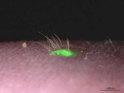
Figure demonstrates successful transplantation of
the bioengineered 3D integumentary system derived from GFP-expressed iPS
cells. Green fluorescence indicates the bioengineered skin including
iPS-derived hair shafts.
MedicalResearch.com: Why were mouse gum cells used and could any other type of mouse cell be used successfully in this regard?
Answer. We confirmed this bioengineered whole skin using other iPS cell clones, including a gingiva-derived iPS cell clone (ectodermal origin) and the stomach-derived clone (endodermal origin). These iPS cell clones generated the bioengineered 3D integumentary organ system, including hair follicles and sebaceous gland. These results indicate that the generation of a bioengineered 3D integumentary organ system is not dependent on the origin of iPS cell clones from different germ layers because heterogeneity exists among iPS cell clones.
MedicalResearch.com: What is Wnt10b and what role did it play in this skin development process?
Answer. Wnt10b is one of the ligand for the Wnt receptor and activates the canonical Wnt signaling pathway. Wnt signaling pathway plays important roles in hair folliculogenesis and various organ development
MedicalResearch.com: What sort of chemicals were used to transform cells from mouse gums into stem-like iPS cells?
Answer. The mouse iPS cell line ‘gingiva (mouse gum cells)-derived iPS’ was established by Dr. Egusa at Tohoku University. His group demonstrated that iPS cell generation from adult wild-type mouse gingival fibroblasts via introduction of four factors (Oct3/4, Sox2, Klf4 and c-Myc; GF-iPS-4F cells) or three factors (the same as GF-iPS-4F cells, but without the c-Myc oncogene; GF-iPS-3F cells) without drug selection.
MedicalResearch.com: What is the significance of the cultured skin having hair follicles and sebaceous glands and were these functional?
Answer. Natural skin structure has appendage organs such as hair follicle, dermis, sebaceous gland, and sweat gland and subcutaneous adipose tissue. These components play important roles to maintain homeostasis including moisturization, fat secretion and protect for noxious stimulations. This bioengineered 3D integumentary organ system was fully functional following transplantation into immunodeficient mice and could be properly connected to surrounding host tissues, such as the epidermis, arrector pili muscles and nerve fibres, without tumorigenesis.
MedicalResearch.com: Might a similar technique be successful in humans and, if so, how long might it be before clinical trials might begin?
Answer. In current our study, we used murine iPS cells but not human cells. Thus, we further develop for the future clinical application in human. We also developed a CDB method by using in vivo transplantation system and isolated the 3D integumentary system from the explants. In future study, we would like to develop the in vitro culture system to generate the system but not in vivo transplantation model. Thus, we cannot answer the correct period to develop human clinical application. We would like to realize the application within 10 years.
MedicalResearch.com: How did you show that the transplanted skin formed functional connections with the host nerve and muscle fibers?
Answer. We isolated cystic tissues with hair follicles in the explants, cut them into small specimens containing 10-20 follicular units, and transplanted them onto the backs of female nude mice using a follicular unit transplantation method. Then, we examined the functional connection between the transplanted skin and host skin by histological analysis.
MedicalResearch.com: How can this model be useful? Did you essentially create transplantable skin in mice?
Answer. We successfully regenerate a transplantable whole skin including skin appendages in mice and confirmed their functions. Our method would be useful for several clinical applications and basic oncology as described in Question 3.
MedicalResearch.com: How long did this transplanted skin last?
Answer. The transplanted whole skin could engraft and function more than 70 days in recipient nude mice. In addition, we have confirmed to repeat the hair cycle like a natural hair.
MedicalResearch.com: Why wouldn’t this approach prompt a potentially damaging immune response?
Answer. In our work, we used immunodeficient nude mice as a model. Thus, our whole skin did not prompt immune rejection in host mice. In general, the transplants will acceptable for the transplantation if we will use the perfect match of major histocompatibility antigens between donor and host animals.
MedicalResearch.com: How does your study advance researchers’ efforts to generate human skin organs that could actually be used in humans?
Answer. Up to now, the skin substitute had already been developed and applied clinical cure to save a lot of serious burn patients from death. However, past developed skin substitute, which has been composed of one or two layers, each independent-cultured keratinized epithelium and dermis, did not have any types of skin appendage organs such as hair follicle, sebaceous gland, and sweat gland, by tissue-engineering technology. These skin appendage organs, which are induced by reciprocal epithelial and mesenchymal interactions, play important roles for skin homeostasis such as eruption of hair shaft, fat secretion and moisture of skin as skin organ system, 3D integumentary organ system. Organ generation, which is a complex process of organogenesis during embryogenesis, is a next-generation of regenerative medicine. In our study, we succeed to reproduce of skin development during embryo genesis and regenerate skin field, 3D integumentary system including skin appendages from iPS cells. Our present outcomes indicate a proof of concept of regenerative therapy of fully functional and integrated skin organ system that will have a potential for the application of the future clinical treatment.
MedicalResearch.com: What else needs to be done/improved to generate such skin organs?
Answer. Importantly, to regenerate 3D integumentary organ system, it is essential to mimic to the developmental process of skin field. These skin appendage organs arise from their organ germs, which are induced by reciprocal epithelial and mesenchymal interactions, in the skin field. Thus, we thought that the induction of immature epithelial and mesenchymal tissues of skin field in iPS cells is essential. Furthermore, to induce skin appendage organs, we induced organogenesis by using Wnt10b-signalling, which plays important roles for hair follicle development during embryogenesis.
MedicalResearch.com: Was there anything that was particularly challenging for you during the experiments done for this new study? How did you overcome that challenge?
Answer. In the present study, we used murine iPS cells but not human cells. Thus, we further develop for the future clinical application in human. We also developed a CDB method by using in vivo transplantation system and isolated the 3D integumentary system from the explants. We succeeded to demonstrated the proof-of-concept to generate 3D integumentary organ system by mimicking the organogenesis during embryogenesis. In future study, we would like to develop the in vitro culture system to generate the system but not in vivo transplantation model.
MedicalResearch.com: What is the take-home message here for the public?
Answer. In the current study, we successfully demonstrated the proof-of-concept to regenerate organ system from iPS cells. Although we have several obstacles to realize for human clinical application, this work will contribute to understanding of principals for the development for human organ system and further clinical application techniques.
MedicalResearch.com: What is the potential here for humans? And what are some of the obstacles in terms of making this work in people?
Answer. In this work, we reported the successful induction of bioengineered skin including skin appendages from murine iPS cells. This work demonstrated the proof-of-concept to induce a bioengineered organ system by the mimicking to organogenesis during embryogenesis. In the future study, we would like to develop of the human bioengineered skin from iPS cells. Furthermore, in the current study, we performed to generate the whole skin by using in vivo transplantation model. To realize for the application in human, we have to establish a method for the generation of human 3D integumentary system in an in vitro organoid culture.
MedicalResearch.com: What is next for research? Do you have funding for the next phase? How long may it take to move to human testing and beyond?
Answer. We are now trying to induce other ectodermal organs such as tooth and secretory organs such as salivary gland and lachrymal gland in in vitro organoid culture. These works will contribute the understanding of the molecular mechanisms of organogenesis and the development of next-generation organ replacement regenerative therapy. We would like to realize the clinical testing for human within 10 years.
MedicalResearch.com: Is there anything else you would like to add?
. Our present study successfully demonstrate the proof-of-concept to induce skin field from iPS cells. We are now trying to induce other ectodermal organs such as tooth and secretory organs such as salivary gland and lachrymal gland in in vitro organoid culture. These work will contribute the understanding of the molecular mechanisms of organogenesis and the development of next-generation organ replacement regenerative therapy.
CITATION:
Egusa, K. Okita, H. Kayashima, G. Yu, S. Fukuyasu, M. Saeki, T. Matsumoto, S. Yamanaka, H. Yatani, Gingival fibroblasts as a promising source of induced pluripotent stem cells. PLoS One. 5(9), e12743 (2010)
[wysija_form id=”5″]
Takashi Tsuji, PhD (2016). Scientists Generate Functional Skin From Stem Cells MedicalResearch.com
Last Updated on April 6, 2016 by Marie Benz MD FAAD