
28 Nov neuroinflammation
Posted at 21:27h
in
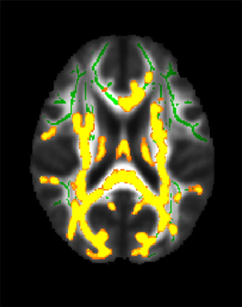
This figure shows increased neuroinflammation (yellow colors) associated with higher hidden fat (visceral fat) in the cohort of 54 participants with an average age of 50 years in the brain’s white matter. The green colors are the normal white matter