
12 May How Micro X Ray Fluorescence Helps Researchers Study Trace Metals in Tissue Samples
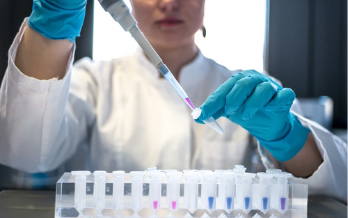
Medical research often focuses on cells, proteins, genes, and biomarkers. Yet there is another layer of information that can quietly shape how scientists understand disease: the tiny distribution of elements inside tissue. Metals such as iron, zinc, copper, calcium, and manganese are present in very small amounts, but they can still influence inflammation, oxidative stress, brain function, tumor behavior, and tissue repair. For researchers, the challenge is simple to describe and hard to solve: how do you study these elements without destroying the sample or losing its spatial context?
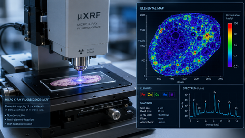
This is where methods like xrf become useful in biomedical laboratories. Micro X-ray fluorescence allows scientists to map elements across very small areas of a sample, often with minimal preparation. Companies such as Merkel Technologies work in this field because modern labs increasingly need tools that can show where elements are located, not only how much of each element is present.
Why Trace Metals Matter in Medical Research
Trace metals are easy to overlook because they are measured in tiny quantities. Still, they are involved in many biological processes. Iron helps carry oxygen and supports cell metabolism. Zinc is connected with immune function and enzyme activity. Copper plays a role in blood vessel formation and oxidative balance. Calcium is central to signaling between cells.
In healthy tissue, these elements usually follow a controlled pattern. In diseased tissue, the pattern may shift. A tumor may show unusual metal accumulation. Brain tissue affected by neurodegenerative disease may show changes in iron or copper distribution. Inflamed tissue may display different mineral behavior compared with nearby healthy tissue.
For researchers, these patterns can raise important questions:
- Do certain elements collect around damaged cells?
- Are metal changes linked to disease progression?
- Can elemental maps help explain why one tissue area behaves differently from another?
- Could trace metals become supportive biomarkers in future studies?
These questions do not always lead to immediate clinical answers. Research is a slow process. Still, mapping elements can add another layer of evidence to histology, microscopy, molecular testing, and imaging.
The Value of Seeing Where Elements Are Located
Traditional chemical analysis can tell scientists that a tissue sample contains a certain amount of iron or zinc. That information is useful, but it may hide important details. A whole sample measurement averages everything together. If one tiny region has a high concentration and another region has almost none, the average may look normal.
Micro X-ray fluorescence helps solve this problem by creating elemental maps. Instead of giving only one number, it can show how an element is spread across the sample surface. This matters because biology is spatial. Cells are organized in layers, clusters, vessels, membranes, and damaged zones. Disease also has geography inside tissue.
For example, in cancer research, scientists may want to compare elemental signals inside a tumor core, at the tumor edge, and in nearby normal tissue. In neuroscience, researchers may study whether metals collect in specific brain regions. In bone or dental research, calcium and phosphorus mapping can help describe mineral structure. In toxicology, elemental imaging may help show where a harmful metal has accumulated after exposure.
The key advantage is context. A researcher can look at an elemental map beside a microscope image and ask better questions. The image may show a lesion, while the elemental map shows whether that lesion has a different chemical profile.
Why Sample Preservation Is Important
Many biomedical samples are rare, expensive, or difficult to collect. Some come from animal models. Some come from archived clinical material. Some are tiny biopsies. In these cases, destructive testing is not always ideal.
Micro XRF is valuable because it can often analyze samples with little damage. This can allow the same sample to be used for other types of study, depending on the preparation method and research workflow. A tissue section may be examined visually, mapped for elements, and then compared with other laboratory findings.
This is especially useful when researchers work with:
- Thin tissue sections
- Biopsy material
- Bone and mineralized tissue
- Implanted medical device surfaces
- Small experimental samples
- Archived research specimens
Careful sample handling still matters. The substrate, fixation method, section thickness, and contamination risk can all affect results. For example, if a sample is placed on a material that contains the same element being studied, the background signal may confuse the analysis. Good laboratory planning is just as important as the instrument itself.
Where This Method Fits With Other Tools
Micro XRF does not replace histology, genomics, proteomics, or clinical imaging. It works best when used as part of a wider research question. A pathologist may identify tissue structure. A molecular test may show gene expression. A microscope may show cell morphology. Micro XRF can then add elemental information to the same story.
This combined approach is useful because disease rarely has one simple cause. A change in metal distribution might reflect inflammation, blood flow, cell death, oxidative stress, or exposure to an outside substance. Researchers need other methods to interpret what the elemental signal means.
In practical terms, micro XRF can support early-stage research in several areas. In cancer biology, it may help compare tumor and non-tumor tissue. In neurobiology, it can support studies of metal imbalance in brain regions. In pharmacology, it may help examine how metal-containing compounds or nanoparticles are distributed in tissue. In pathology research, it can help describe unusual deposits or mineral changes.
The strength of the method is its ability to create a bridge between chemistry and structure. It gives scientists a way to ask, “Where exactly is this element, and what is happening in that place?”
A Careful Tool for Complex Questions
The most interesting part of micro XRF in medical research is that it does not force the sample into a single number. It respects the complexity of tissue. It shows patterns, borders, clusters, and gradients. For researchers who work with small samples, this can be a powerful way to find details that would otherwise stay hidden.
At the same time, the results must be interpreted carefully. An elemental map is evidence, not a diagnosis. It can suggest a direction, support a hypothesis, or reveal an unexpected pattern. The real value comes when scientists combine it with good experimental design, proper controls, and other biological data.
As medical research becomes more precise, tools that can study tiny areas with high detail will become more important. Micro XRF is one of those tools. It helps researchers look at tissue in a different way, through the quiet language of elements. For a field that often depends on small clues, that extra layer of information can make a real difference.
Disclaimer: The information on MedicalResearch.com is provided for educational purposes only, and is in no way intended to diagnose, cure, or treat any medical or other condition. Some links are sponsored. Products, services and providers are not warranted or endorsed by MedicalResearch.com or Eminent Domains Inc. Always seek the advice of your physician or other qualified health and ask your doctor any questions you may have regarding a medical condition. In addition to all other limitations and disclaimers in this agreement, service provider and its third party providers disclaim any liability or loss in connection with the content provided on this website.
Last Updated on May 12, 2026 by Marie Benz MD FAAD