
21 Jul cell-structures
Posted at 14:00h
in
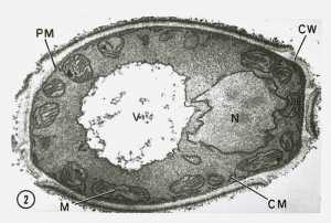
This transmission electron microscopic (TEM) image revealed some of the ultrastructural morphology displayed by a Candida sp. fungal organism. CW = cell wall, PM = plasma membrane, M = mitochondria, V = vacuole, and N = nucleus.