

16 Jul An Important Guide to Mole Monitoring and Early Skin Cancer Detection
Most people have moles. They’re a normal part of having skin. Yet many people feel uncertain about whether their moles are concerning or just another benign spot. This uncertainty is exactly why systematic mole monitoring has become essential to skin cancer prevention.
Understanding what makes a mole risky, how to track changes over time and when to seek professional evaluation can literally save your life. Early detection of melanoma increases five-year survival rates to over 90 percent, compared to much lower rates when diagnosis happens at advanced stages.
The relationship between moles and melanoma is straightforward but often misunderstood. Not all moles become melanoma. Most moles remain benign throughout a person’s lifetime. However, melanoma can arise from existing moles or appear as completely new lesions. This means your moles deserve regular attention and professional monitoring if you have risk factors. The good news is that systematic monitoring catches changes early when treatment is most effective. This article breaks down everything you need to know about monitoring moles, understanding your risk and getting the right screening.
Key Takeaways
- Regular mole monitoring is one of the most effective strategies for early melanoma detection, with early-stage melanomas showing survival rates exceeding 90 percent.
- Understanding the ABCDE rule helps you recognize potentially concerning changes that warrant professional evaluation.
- Digital mole mapping technology allows dermatologists to track subtle changes over time that might be invisible to the naked eye.
- People with more than 50 moles, a family history of melanoma or fair skin should have professional screening at least annually.
- Monthly self-examinations combined with annual dermatology appointments create the ideal monitoring routine for most people.
Why Mole Monitoring Matters More Than You Think
Melanoma is the most dangerous form of skin cancer because it can spread to other parts of the body more readily than other skin cancers. The statistics are stark. While melanoma accounts for only about five percent of all skin cancers, it’s responsible for about 75 percent of skin cancer deaths.
This disparity exists precisely because early detection changes everything. When melanoma is caught before it spreads, treatment is straightforward and survival rates are excellent. When it’s discovered after spreading, treatment becomes much more complex and outcomes worsen significantly.
The challenge is that melanoma can be difficult to spot. Unlike basal cell or squamous cell carcinoma, which typically announce themselves through persistent scaling or bleeding, melanoma can hide among a person’s normal moles. Someone with dozens of moles might easily miss the one that’s changing in subtle ways. Without systematic monitoring, new melanomas might go unnoticed until they’ve progressed to a more dangerous stage. Rarely melanoma can develop in an atypical fashion, or even from an internal organ, but most melanomas develop from new or changing skin lesions.
Studies show that people who have regular skin screenings by dermatologists catch melanomas at thinner, earlier stages compared to those who don’t have routine screening. Those thinner melanomas have higher survival rates and require less aggressive treatment. Early detection prevents not just deaths but also unnecessary surgery, chemotherapy and the anxiety that comes with advanced-stage diagnosis. Investing time in mole monitoring is genuinely life-saving prevention.
Understanding Mole Types and What Changes Matter
Not all moles carry equal risk. Dermatologists recognize several categories of moles, each with different characteristics and implications. Common moles are the most frequent type — typically uniform in color, well-defined, smaller than a pencil eraser and flat or only slightly raised. Common moles rarely become melanoma, though they are still worth monitoring for changes. Atypical moles, also called dysplastic nevi, are larger, have irregular shapes and may show color variation. People with atypical moles have higher melanoma risk and need more frequent screening.
Congenital nevi are moles you’re born with. While they’re usually benign, large congenital nevi carry slightly elevated melanoma risk, so they warrant monitoring. Acquired moles develop throughout life from sun exposure and genetics. The number of acquired moles matters. People with more than 50 moles have higher melanoma risk than those with fewer moles.
The key to productive mole monitoring is understanding what changes matter. The ABCDE rule provides a framework. A stands for asymmetry — normal moles are roughly symmetrical. B is for border irregularity — healthy moles have clean, even borders. C stands for color — moles should be a uniform tan, brown or black, and multiple colors within one mole are concerning. D is for diameter — moles larger than a pencil eraser (about six millimeters) deserve monitoring. E stands for evolution — any mole that changes in size, shape, color or symptoms is concerning and needs professional evaluation.
Beyond the ABCDE rule, dermatologists watch for the “ugly duckling sign.” If you have multiple similar moles but one looks notably different from the others, that different one deserves attention even if it doesn’t clearly meet the ABCDE criteria. Trust that instinct and have it evaluated by a professional.
The Role of Professional Screening and Digital Technology
While self-monitoring is important, professional screening catches things that home examination can’t. Dermatologists have trained eyes and use diagnostic tools that reveal details invisible to the naked eye. The handheld dermatoscope magnifies skin lesions and reveals microscopic patterns — sometimes called dermoscopy — that help distinguish benign moles from melanoma. Research published in JAMA Dermatology has shown that autonomous total body photography combined with digital dermoscopy reliably delivers images comparable to specialist-captured photography for skin cancer screening — a finding that underpins the rapid adoption of digital mole mapping.
Total body photography combined with sequential digital dermoscopy imaging creates a comprehensive baseline of all your moles. When you return for follow-up appointments, the computer software overlays new images onto previous images, highlighting subtle changes in size, shape, color or surface characteristics. This technology detects changes so small that the human eye would miss them. Studies show that structured surveillance using mole mapping identifies melanomas at earlier stages and thinner thicknesses compared to routine clinical examination alone.
For people at higher risk of melanoma, systematic mole mapping represents a major advancement in early detection. This systematic approach to monitoring is why services like mole mapping Sydney have become standard of care in dermatology. The technology represents a real leap forward in melanoma prevention.
When to Seek Professional Evaluation
If you notice any mole that meets ABCDE criteria or seems different from your other moles, see a dermatologist. You don’t need to wait for a perfect diagnosis. Dermatologists are trained to evaluate borderline lesions and determine which ones need biopsy and which ones can be safely monitored. Many concerning-looking moles turn out to be benign, but the process is the correct way to manage uncertainty in cancer screening. A biopsy answers questions definitively.
If you have no risk factors for melanoma and no concerning moles, annual screening is reasonable for adults. If you have atypical moles, multiple moles, fair skin, family history of melanoma or a personal history of severe sunburns, more frequent screening makes sense. Many dermatologists recommend every six months for higher-risk patients.
It’s also worth noting that melanoma can appear in locations you might not expect. Most melanomas appear on areas exposed to sun, but they can also develop in places with minimal sun exposure, including the soles of feet, between toes, under fingernails and even on the scalp or in the eye. This is another reason systematic professional screening matters — dermatologists can examine your entire body, not just the obvious spots.
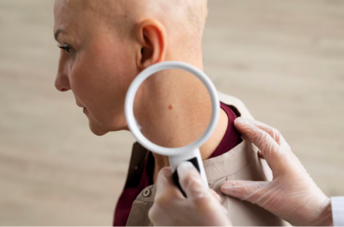
Monthly Self-Examinations: Upgrade Your Safety Net
Between professional appointments, monthly self-examinations provide an important safety net. Choose a consistent time each month to examine your skin systematically. You’ll need good lighting and a mirror, or have a partner help you examine hard-to-see areas like your back. Start with your face and move downward. Examine your arms, legs, hands, feet and spaces between your toes. Look at your scalp, ears and neck.
As you examine, look for new moles or changes in existing ones. Use your phone to take photos of moles you want to track. Comparing photos month to month sometimes reveals subtle changes that might not be obvious otherwise. If you see something new or something changing, don’t panic — but do schedule a dermatology appointment.
The key is perspective. You’re not looking for perfection. You’re watching for change.
Treatment Options When Concerns Arise
If a biopsy confirms melanoma, treatment options depend on the stage and thickness of the cancer. Early-stage, thin melanomas are typically treated with surgical excision alone. The dermatologist removes the mole with a margin of normal skin, sending it to pathology to confirm that all cancer cells have been removed. Most patients with early-stage melanoma are cured with this single procedure.
For thicker melanomas or those showing signs of spread, additional treatment might include sentinel lymph node biopsy, immunotherapy or targeted therapy. The contrast between early-stage and advanced melanoma treatment is dramatic. Someone with a thin melanoma caught early might need just one outpatient procedure and recover completely. Someone with advanced melanoma might face months or years of systemic therapy with significant side effects.
Research on primary care screening shows that regular monitoring catches melanomas at earlier, more treatable stages, significantly improving outcomes and reducing the need for aggressive interventions.
Frequently Asked Questions
How often should I have a dermatology screening for moles?
For people without risk factors, annual screening is generally recommended. For people with atypical moles, numerous moles (more than 50), fair skin, family history of melanoma, or personal history of sun damage, screening every six months is more appropriate. Your dermatologist can recommend the ideal frequency based on your individual risk profile.
Can a mole be cancerous without showing any ABCDE warning signs?
Yes, though this is less common. The ABCDE rule catches the majority of concerning moles, but some melanomas — particularly the rare amelanotic melanomas that lack pigment — may not fit the classic pattern. This is another reason professional screening matters. If something about a mole seems off to you even without clear ABCDE changes, professional evaluation is appropriate.
Do all atypical moles need to be removed?
No. Atypical moles don’t automatically become melanoma. Most remain benign throughout life. The significance of atypical moles is that they indicate higher melanoma risk, so people with atypical moles warrant more frequent screening. Specific atypical moles are biopsied only if they show changes or if the dermatologist has specific concerns about a particular lesion.
Is it safe to have moles biopsied?
Biopsies are safe and standard medical procedures. The procedure creates a small wound that requires a few stitches and leaves a small scar, but serious complications are extremely rare. The benefit of a definitive answer about whether a lesion is cancerous far outweighs the minor risk. If a dermatologist recommends a biopsy, the potential information gained justifies the minor procedural risk.
Disclaimer: The information on MedicalResearch.com is provided for educational purposes only, and is in no way intended to diagnose, cure, or treat any medical or other condition. Some links are sponsored. Products, services and providers are not warranted or endorsed by MedicalResearch.com or Eminent Domains Inc. Always seek the advice of your physician or other qualified health provider and ask your doctor any questions you may have regarding a medical condition. In addition to all other limitations and disclaimers in this agreement, service provider and its third party providers disclaim any liability or loss in connection with the content provided on this website.
Last Updated on August 14, 2026 by Marie Benz MD FAAD