Author Interviews, CDC, Infections / 20.05.2024

MedicalResearch.com Interview with: Cathryn Haigh, Ph.D. Chief Prion Cell Biology Unit Laboratory of Neurological Infections and Immunity National Institute of Allergy and Infectious Diseases Division of Intramural Research, Rocky Mountain Laboratories National Institutes of Health Hamilton, MT 59840 MedicalResearch.com: What is the background for this study,  ie what are prions/prion-related diseases?  Where are prions found? Response: Prion diseases are infectious neurodegenerative diseases of humans and animals.  In humans these diseases often manifest as rapidly progressing dementias but are rarely caused by a known exposure to the infectious agents (prions).  More commonly they are sporadic (no known cause) or hereditary. One form of human disease is believed to have arisen from eating beef contaminated with bovine spongiform encephalopathy (as known as mad cow disease).  This has resulted in concerns that chronic wasting disease (CWD), a prion disease affecting deer, elk and moose, might also have the potential to cross the species barrier and cause disease in humans.  To date, transmissions of CWD prions to cynomolgus macaques have been negative, a good sign that crossing the species barrier would not be easy, but macaques are not human so we wanted to test whether CWD could infect human brain tissue. To do this we used a human cerebral organoid model (mini human brain tissues grown from skin cells in a laboratory) and directly exposed the organoids to prions from the brains of animals that had died of CWD. (more…)
Author Interviews / 23.01.2019

MedicalResearch.com Interview with: Byron Caughey, Ph.D.  Senior Investigator Chief, TSE/prion Biochemistry Section Laboratory of Persistent Viral Diseases NIH/NIAID Rocky Mountain Laboratories . Hamilton, MT 59840 USA MedicalResearch.com: What is the background for this study? Would you briefly explain the significance of prion-induced diseases and why they have been difficult to diagnosis?  Response: Although prion diseases such as Creutzfeldt-Jakob disease (CJD) are rarer than many other neurodegenerative diseases in humans, they are rapidly fatal, untreatable and transmissible. Therefore it is important to be able to diagnose prion diseases as early as possible, not only to accurately inform patients, families and caregivers, but also to reduce risks of transmission and  improve prospects for developing therapeutics. Toward these goals, we have shown that our RT-QuIC prion seed amplification assays are highly accurate for diagnosing sporadic CJD using patients’ spinal fluid and/or nasal brushings in the clinical phase of disease. However, these particular specimens may not always be available, and it remains unclear how early they become RT-QuIC-positive in infected individuals in the months or years prior to the onset of overt clinical signs. We also showed recently that skin samples obtained post-mortem from sCJD patients are RT-QuIC positive. In the current study, we determined how early prion seeds appear in the rodents infected with prions in order to gain clues as to whether analyses of skin biopsies might provide a means of early preclinical detection of prion infections in humans. (more…)
Author Interviews, Infections, NIH, Ophthalmology / 05.12.2018

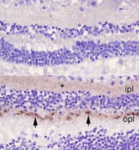
MedicalResearch.com Interview with: Top, retina of a control patient. Bottom, retina from a patient with CJD. Arrowheads point to abnormal prions in the outer plexiform layer (opl), and the asterisk (*) marks more diffuse prions in the inner plexiform layer (ipl).Orrù et al., mBioByron Caughey, Ph.D. Senior Investigator Chief, TSE/prion Biochemistry Section Laboratory of Persistent Viral Diseases NIH/NIAID Rocky Mountain Laboratories Hamilton, MT 59840 USA  MedicalResearch.com: What is the background for this study? What are the main findings?  Response: Corneal transplants have caused the transmission of Creutzfeldt-Jakob disease (CJD) in at least two cases, and pathological prion protein has been detected in the retinas of the eyes of sporadic CJD cases. To build on these previous indications of prions in eye tissue, we tested the distribution of prions in various components of eyes from 11 sCJD decedents. We applied a highly sensitive surrogate test for prions (RT-QuIC) that indicated that all of the sCJD cases had prions in multiple parts of their eye, including the cornea and sclera, which is the white outer surface of the eye. Retinas were usually contained the highest levels, in some cases approaching levels in the brain. Some other parts such as the cornea, lens and vitreous had much lower, but detectable, levels.  (more…)