14 Apr Clinical Findings and Brain Calcifications of Zika Babies Described
MedicalResearch.com Interview with:

Team of Doctors: Ana van Der Linden, Alessandra Brainer, Maria de Fatima Aragao, Vanessa va Der Linden e Arthur Cesário
Maria de Fatima Vasco Aragao MD, PhD
Radiologist and Neuroradiologist
Professor of Radiology, Mauricio de Nassau University, Recife, Brazil
Scientific Director of Multimagem Radiology Clinic, Recife – PE, Brazil
President of Pernambuco Radiology Society
MedicalResearch.com: What is the background for this study?
Response: The new Zika virus epidemic in Brazil was recognized as starting in the first half of 2015 and the microcephaly epidemic was detected in the second half of that same year.

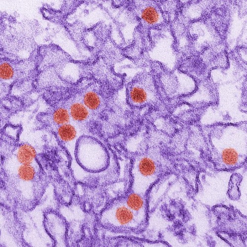
This is a transmission electron micrograph (TEM) of Zika virus, which is a member of the family Flaviviridae. Virus particles are 40 nm in diameter, with an outer envelope, and an inner dense core.
MedicalResearch.com: What are the main findings?
- Response: In our study of the 23 mothers, only one did not report rash during pregnancy (rash is a sign that can happen in Zika virus infection). However, Zika virus infection can be asymptomatic in three of every four infected patients. All of the 23 babies had the same clinical and epidemiological characteristics and other congenital infection diseases had been excluded. Of these 23 babies, six were tested for IgM antibodies, specific to Zika virus and all six proved positive. So, by deduction, the other 17 babies on whom it was not possible to make the IgM test, were considered as also having presumed congenital infection related to the Zika virus, after other congenital infections being excluded.
- All the babies showed malformations of cortical development and sulcation. The most frequent cortical malformation were: Microcephaly with a simplified cortical gyral pattern and areas of thick cortex of polymicrogyria or pachygyria which were located predominantly in the frontal lobes.
- Abnormalities of the corpus callósum (hypogenesis and hypoplasia) were common.
- Decreased brain volume was a common finding. Ventriculomegaly was present in all the babies, with a predominant enlargement of the posterior portions of the lateral ventricles,
- Delayed myelination were also common. The cisterna magna was enlarged in most of the cases, with or without cerebellar hypoplasia.
- Some of the babies showed a symmetrical enlargement of the anterior subarachnoid space of the supratentorial compartment, associated with severe ventriculomegaly.
MedicalResearch.com: What should patients and clinicians take away from this report?
- Although calcification can be found in many congenital infections and other diseases, the knowledge of the preferred calcification location of each disease can help in suggesting one etiology rather than another. So, the radiological findings, taken together in the context of a clinical scenario, allows a diagnosis to be made in many cases.
- The brain calcifications in most babies with presumed Congenital Zika Virus Infection were predominant in the junction between the cortex and the subcortical white matter. These calcifications associated with malformations of cortical development. may be used as a radiological indication of a suspect Zika vírus congenital infection.
- It is not known the exact mechanism of how or why this presumed causal factor (Congenital Zika Virus Infection) is now causing such serious brain damage to most oh these babies. The hypothesis is that the presumed Zika virus infection destroys brain cells, forming many lesions similar to “scars” on which calcium is deposited over the course of time.
- No mother of our study had rash in late pregnancy. So, we can suppose that, probably, in this disease also, the earlier the infection during pregnancy, the more severe the brain lesions and microcephaly. However, our study has only a small sample (23 children). So we cannot confirm statistically this hypothesis at the moment.
MedicalResearch.com: Is there anything else you would like to add?
Response: A limitation for the study was that the supposed Zika virus infection of the mothers was based exclusively on the mothers’ recall of rash during pregnancy. Therefore, the mothers cannot remember the exact month that they had rash, symptom of Zika infection, during pregnancy and this undermines the analysis for this hypothesis that was not the objective of this study.
Another limitation, for the moment, is that there is no routinely available test for definitive confirmation of the ZIKV infection; for this reason, in the majority of cases, the diagnosis of the Zika virus related congenital infection was established by clinical, epidemiological and radiological criteria, after the exclusion of other congenital infections.Citation:
Clinical features and neuroimaging (CT and MRI) findings in presumed Zika virus related congenital infection and microcephaly: retrospective case series study
MedicalResearch.com: Is there anything else you would like to add?
Response: This is was a retrospective study of patients from the Associação de Assistência a Criança Deficiente” (AACD) that is one of four rehabilitation center reference for congenital infection presumed by ZIKV in Pernambuco, Brazil. Dr. Vanessa van Der Linden, Director Clinic of AACD is the second author of this work. The CT and MR images were first evaluated by neurologists of this work. Later, the CT and MRI images were reevaluated for two also experienced neuroradiologists and also authors of this work.
Last Updated on April 14, 2016 by Marie Benz MD FAAD
