
19 Nov Recognising Acute Peripheral Vision Warnings Protects Central Sight
Be sure to have a regular eye examination for glaucoma, cataracts, retinal disease and other eye issues.
Brief flashes of light at the edge of your vision, or new dark specks and cobweb-like shapes drifting across it, are easy to dismiss as tiredness or screen strain. Usually these changes are harmless. Sometimes, however, they are the first warning of a retinal tear, which can lead to a retinal detachment if it is not treated.
A retinal detachment is a medical emergency that can cause permanent vision loss. Knowing which symptoms need urgent attention, and acting on them quickly, gives you the best chance of simpler treatment and good vision afterwards.
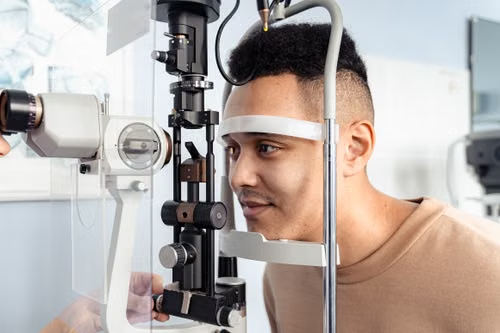
Photo by Antoni Shkraba via Pexels
If you notice persistent flashes, new floaters or a shadow in your vision, an urgent dilated eye examination can show whether you have a retinal tear or Retinal Detachment, and whether treatment is needed. The NEI (National Eye Institute) describes the warning signs and treatment of retinal detachment in more detail.
Why Flashes and Floaters Happen
The inside of the eye is filled with a clear gel called the vitreous. With age, this gel gradually becomes more liquid and shrinks, and eventually it separates from the retina, the light-sensitive layer at the back of the eye. This is called a posterior vitreous detachment (PVD). It is very common, especially after age 50, and it often happens earlier in people who are very short-sighted.
For most people, a PVD causes new floaters and occasional flashes but no lasting harm. In a minority of cases, though, the gel pulls hard enough on the retina to tear it. Fluid can then pass through the tear and lift the retina away from the wall of the eye. That is a retinal detachment.
Because a harmless PVD and a retinal tear can cause the same symptoms, the only way to tell them apart is a dilated eye examination by an eye doctor.
Symptoms That Need Urgent Attention
- A shadow, curtain or veil moving across any part of your vision: seek emergency eye care the same day.
- Sudden new flashes of light, especially at the edge of your vision: arrange an urgent dilated examination,
- A sudden shower of new floaters, or a large increase in floaters: arrange an urgent dilated examination.
- Sudden blurring or loss of vision in one eye: seek emergency care.
A single floater that has been present and unchanged for a long time is usually not urgent, but any new floaters or flashes should be checked. If you have already been examined and told you have a PVD, contact your eye doctor promptly if your symptoms suddenly change, or if a shadow appears.
Why You Should Not Wait for Blurry Vision
A common mistake is waiting until central vision becomes blurry before seeking help. The retina has no pain receptors, so tears and early detachments are painless. A detachment that starts at the edge of the retina may not affect central vision until it spreads to the macula, the small central area responsible for reading and seeing detail.
Outcomes are generally better when a detachment is repaired before the macula detaches. Once the macula is involved, some loss of sharpness or distortion may remain even after successful surgery. Early action matters for that reason.
How Retinal Tears and Detachments Are Treated
A retinal tear found before the retina detaches can often be treated in the clinic with laser treatment or freezing treatment (cryotherapy). These create a seal around the tear to help prevent fluid from getting underneath.
A retinal detachment usually needs surgery. Depending on the type and location of the detachment, options include:
- Pneumatic retinopexy: a gas bubble is injected into the eye, combined with laser or freezing treatment to seal the tear.
- Scleral buckle: a band is placed on the outside wall of the eye to support the retina.
- Vitrectomy: the vitreous gel is removed, the retina is reattached, and the eye is filled with a gas bubble or, in some complex cases, silicone oil.
Your surgeon will recommend the approach that best suits your eye. After a gas bubble, you may be asked to hold a particular head position for a period of time. You must not fly, travel to high altitudes or have nitrous oxide anaesthesia until the gas has fully absorbed, because the bubble can expand and dangerously raise the pressure in the eye. Silicone oil does not carry the same travel restriction, but it is usually removed in a second operation and is generally reserved for more complex detachments. Follow your surgeon’s specific instructions closely.
Who Is at Higher Risk?
Anyone can develop a retinal tear, but the risk is higher for people who:
- Are very short-sighted (highly myopic)
- Have had a retinal tear or detachment in either eye before
- Have a family history of retinal detachment
- Have had cataract surgery or other eye surgery
- Have had a significant eye injury
- Have known thinning in the outer retina, such as lattice degeneration
If any of these apply to you, ask your eye doctor how often you should have a dilated examination. Learn the warning signs so you can act quickly. Wearing protective eyewear during sports and activities with a risk of eye injury is also sensible.
A Simple Habit: Check One Eye at a Time
Because the other eye can compensate, a problem in one eye is easy to miss. If you have had symptoms or are at higher risk, occasionally cover one eye and then the other, and look for any new shadow, blank area or distortion. This does not replace an examination, but it can help you notice a change early.
Most flashes and floaters turn out to be harmless. The few that signal a retinal tear are far easier to treat when they are caught early, so a prompt examination is always worthwhile.
For a broader overview of eye health, see this MedicalResearch.com overview of retina and eye disease research.
The information on MedicalResearch.com is provided for educational purposes only, and is in no way intended to diagnose, cure, or treat any medical or other condition. Some links are sponsored. MedicalResearch.com and Eminent Domains Inc. do not warrant or endorse products or claims made by third party links. Always seek the advice of your physician or other qualified health provider and ask your doctor any questions you may have regarding a medical condition. In addition to all other limitations and disclaimers in this agreement, service provider and its third party providers disclaim any liability or loss in connection with the content provided on this website.
Last Updated on November 19, 2026 by Marie Benz MD FAAD