Author Interviews, Cancer Research, JAMA, MRI, Prostate Cancer, UCLA / 12.09.2019
Prostate Cancer: MRI-Ultrasound Fusion Technology Improves Biopsy Assessment During Active Surveillance
MedicalResearch.com Interview with:
Rajiv Jayadevan, MD
and Leonard S. Marks, MD
Department of Urology
UCLA
MedicalResearch.com: What is the background for this study?
Response: Men with low risk prostate cancer often enter “active surveillance” programs. These programs allow patients to defer definitive treatment (and avoid their associated side effects) until more aggressive disease is detected, if at all. Patients typically undergo a “confirmatory biopsy” 6 to 12 months after diagnosis to verify that their disease is low risk, and then undergo repeat biopsies every 1 to 2 years. These biopsies have traditionally been performed under the guidance of transrectal ultrasonography.
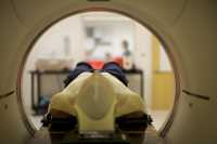
Transrectal ultrasonography is unable to accurately visualize tumors within the prostate, necessitating that biopsy cores be obtained systematically from all parts of the prostate. MRI-ultrasonography fusion biopsy is a newer technology that has been shown to characterize biopsy findings more accurately than transrectal ultrasonography, leading to improved disease detection. This technology also allows us to visualize tumors within the prostate, and directly target these tumors during a biopsy session. (more…)


