CT Scanning, Medical Imaging, MRI, Radiology / 02.06.2025
MRI Compared to a CT Scan: What Are the Differences You Should Know?

[caption id="attachment_68888" align="aligncenter" width="500"] Photo by MART PRODUCTION[/caption]
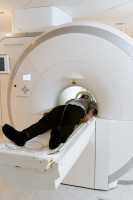
Imagine two powerful, non-invasive imaging tools, each with its own unique set of capabilities. Both MRI (Magnetic Resonance Imaging) and CT (Computed Tomography) scans are essential in the medical field for diagnosing and treating a wide range of conditions. Yet, they operate on entirely different principles and are used in distinct scenarios. Understanding the differences between MRI and CT scans can empower patients to make informed decisions about their healthcare. This article will delve into the workings of MRI and CT scans, their benefits and drawbacks, and why a healthcare professional might choose one over the other.
Photo by MART PRODUCTION[/caption]
Imagine two powerful, non-invasive imaging tools, each with its own unique set of capabilities. Both MRI (Magnetic Resonance Imaging) and CT (Computed Tomography) scans are essential in the medical field for diagnosing and treating a wide range of conditions. Yet, they operate on entirely different principles and are used in distinct scenarios. Understanding the differences between MRI and CT scans can empower patients to make informed decisions about their healthcare. This article will delve into the workings of MRI and CT scans, their benefits and drawbacks, and why a healthcare professional might choose one over the other.


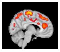
 Dr. Callaghan[/caption]