
MRI
09 Dec Cerebral Perfusion Is Perturbed by Preterm Birth and Brain Injury
MedicalResearch.com Interview with: Eman S. Mahdi, MD, MBChB Pediatric Radiology Fellow [caption id="attachment_30368" align="alignleft" width="133"] Dr. Catherine Limperopoulos[/caption]
Catherine Limperopoulos, PhD
Director, Developing Brain Research Laboratory
Co-Director of Research, Division of Neonatology
Diagnostic Imaging and Radiology
Children’s National Health System
Washington, DC
MedicalResearch.com: What is the background for this study? What are the main findings?
Response: Premature birth is a major public health concern in the United States affecting 1 in 10 infants each year. Prematurity-related brain injury is very common and associated with a high prevalence of brain injury and accompanying lifelong neurodevelopmental morbidities.
Early disturbances in systemic and cerebral hemodynamics are thought to mediate prematurity-related brain injury. The extent to which cerebral blood flow (CBF) is disturbed in preterm birth is poorly understood, in large part because of the lack of monitoring techniques that can directly and non-invasively measure cerebral blood flow.
We report for the first time early disturbances in global and regional cerebral blood flow in preterm infants following brain injury on conventional magnetic resonance imaging (MRI) over the third trimester of ex-uterine life using arterial spin labelling images. In terms of regional differences, we saw a marked decrease in blood flow to the thalamus and the pons, regions known to be metabolically active during this time.
Dr. Catherine Limperopoulos[/caption]
Catherine Limperopoulos, PhD
Director, Developing Brain Research Laboratory
Co-Director of Research, Division of Neonatology
Diagnostic Imaging and Radiology
Children’s National Health System
Washington, DC
MedicalResearch.com: What is the background for this study? What are the main findings?
Response: Premature birth is a major public health concern in the United States affecting 1 in 10 infants each year. Prematurity-related brain injury is very common and associated with a high prevalence of brain injury and accompanying lifelong neurodevelopmental morbidities.
Early disturbances in systemic and cerebral hemodynamics are thought to mediate prematurity-related brain injury. The extent to which cerebral blood flow (CBF) is disturbed in preterm birth is poorly understood, in large part because of the lack of monitoring techniques that can directly and non-invasively measure cerebral blood flow.
We report for the first time early disturbances in global and regional cerebral blood flow in preterm infants following brain injury on conventional magnetic resonance imaging (MRI) over the third trimester of ex-uterine life using arterial spin labelling images. In terms of regional differences, we saw a marked decrease in blood flow to the thalamus and the pons, regions known to be metabolically active during this time.
02 Dec God Activates Reward Centers In Brain
MedicalResearch.com Interview with: [caption id="attachment_30097" align="alignleft" width="133"] Dr. Jeffrey S. Anderson[/caption]
Jeffrey S. Anderson, MD, PhD
Director the fMRI Neurosurgical Mapping Service
Principal Investigator for the Utah Functional Neuroimaging Laboratory
University of Utah
MedicalResearch.com: What is your study about?
Response: Billions of people find meaning in life and make choices based on religious and spiritual experiences. These experiences range from epiphanies that change the lives of celebrated mystics to subtle feelings of peace and joy in the lives of neighbors, friends, or family members that are interpreted as spiritual, divine, or transcendent.
Astonishingly, with all we understand about the brain, we still know very little about how the brain participates in these experiences. We set out to answer what brain networks are involved in representing spiritual feelings in one group of people, devout Mormons.
Dr. Jeffrey S. Anderson[/caption]
Jeffrey S. Anderson, MD, PhD
Director the fMRI Neurosurgical Mapping Service
Principal Investigator for the Utah Functional Neuroimaging Laboratory
University of Utah
MedicalResearch.com: What is your study about?
Response: Billions of people find meaning in life and make choices based on religious and spiritual experiences. These experiences range from epiphanies that change the lives of celebrated mystics to subtle feelings of peace and joy in the lives of neighbors, friends, or family members that are interpreted as spiritual, divine, or transcendent.
Astonishingly, with all we understand about the brain, we still know very little about how the brain participates in these experiences. We set out to answer what brain networks are involved in representing spiritual feelings in one group of people, devout Mormons.
28 Nov Virtual Reality Systems Can Generate Immersive 3D Images of Fetuses
[caption id="attachment_30002" align="alignleft" width="200"]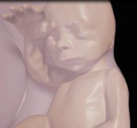 Close-up of fetus at 26 weeks[/caption]
MedicalResearch.com Interview with:
Dr. Heron Werner Junior
Clínica de Diagnóstico por Imagem – CDPI
Rio de Janeiro - Brazil
MedicalResearch.com: What is the background for this study?
Response: A growing number of technological advancements in obtaining and viewing images through noninvasive techniques have brought major breakthroughs in fetal medicine.
In general, two main technologies are used to obtain images within the uterus during pregnancy i.e. ultrasound (US) and magnetic resonance imaging (MRI).
Close-up of fetus at 26 weeks[/caption]
MedicalResearch.com Interview with:
Dr. Heron Werner Junior
Clínica de Diagnóstico por Imagem – CDPI
Rio de Janeiro - Brazil
MedicalResearch.com: What is the background for this study?
Response: A growing number of technological advancements in obtaining and viewing images through noninvasive techniques have brought major breakthroughs in fetal medicine.
In general, two main technologies are used to obtain images within the uterus during pregnancy i.e. ultrasound (US) and magnetic resonance imaging (MRI).
08 Nov Brain Gray Matter Volume Predictive of Weight Loss Success
MedicalResearch.com Interview with: Fatemeh Mokhtari Medical Imaging PhD Student VT-WFU SBES MedicalResearch.com: What is the background for this study? What are the main findings? Response: The objective of this study was to use baseline anatomical brain MRI scans to prospectively predict weight loss success following an intensive lifestyle intervention. In the study, 52 participants, age 60 to 79, were recruited from the Cooperative Lifestyle Interventions Programs II (CLIP-II) project. The participants were overweight or obese (BMI greater than 28 and less than 42) and had a history of either cardiovascular disease or metabolic syndrome. All participants had a baseline MRI scan, and then were randomized to one of three groups – diet only, diet plus aerobic exercise training or diet plus resistance exercise training. The goal of the 18-month diet and exercise program was a weight loss of 7 to 10 percent of body mass. Basic brain structure information garnered from the MRIs was classified using a support vector machine, a type of computerized predictive algorithm. Specifically, we trained a computational predictive model which mapped each subject’s brain scan to weight loss performance. Predictions were based on baseline brain gray and white matter volume from the participants’ MRIs and compared to the study participants’ actual weight loss after the 18 months. The accuracy of the model was then tested, and our prediction algorithms were 78% accurate in predicting successful weight loss. Brain gray matter volume provided higher prediction accuracy compared with white matter and the combination of the two outperformed either one alone.19 Sep Computer Bests Neuroradiologists in Distinguishing Tumor Recurrence From Radiation Necrosis
Posted at 21:39h
in Author Interviews, Brain Cancer - Brain Tumors, Case Western, MRI, Radiology, Technology
MedicalResearch.com Interview with:
[caption id="attachment_28107" align="alignleft" width="200"] Dr. Pallavi Tiwari[/caption]
Dr. Pallavi Tiwari PhD
Assistant Professor biomedical engineering
Case Western Reserve University
MedicalResearch.com: What is the background for this study? What are the main findings?
Response: One of the biggest challenges in neuro-oncology currently is distinguishing radionecrosis, a side-effect of aggressive radiation, from tumor recurrence on imaging. Surgical intervention is the only means of definitive diagnosis, but suffers from considerable morbidity and mortality. The treatments for radionecrosis and cancer recurrence are very different. Early identification of the two conditions can help speed prognosis, therapy, and improve patient outcomes.
The purpose of this feasibility study was to evaluate the role of machine learning algorithms along with computer extracted texture features, also known as radiomic features, in distinguishing radionecrosis and tumor recurrence on routine MRI scans (T1w, T2w, FLAIR). The radiomic algorithms were trained on 43 studies from our local collaborating institution - University Hospitals Case Medical Center, and tested on 15 studies at a collaborating institution, University of Texas Southwest Medical Center. We further compared the performance of the radiomic techniques with two expert readers.
Our results demonstrated that radiomic features can identify subtle differences in quantitative measurements of tumor heterogeneity on routine MRIs, that are not visually appreciable to human readers. Of the 15 test studies, the radiomics algorithm could identify 12 of 15 correctly, while expert 1 could identify 7 of 15, and expert 2, 8 of 15.
Dr. Pallavi Tiwari[/caption]
Dr. Pallavi Tiwari PhD
Assistant Professor biomedical engineering
Case Western Reserve University
MedicalResearch.com: What is the background for this study? What are the main findings?
Response: One of the biggest challenges in neuro-oncology currently is distinguishing radionecrosis, a side-effect of aggressive radiation, from tumor recurrence on imaging. Surgical intervention is the only means of definitive diagnosis, but suffers from considerable morbidity and mortality. The treatments for radionecrosis and cancer recurrence are very different. Early identification of the two conditions can help speed prognosis, therapy, and improve patient outcomes.
The purpose of this feasibility study was to evaluate the role of machine learning algorithms along with computer extracted texture features, also known as radiomic features, in distinguishing radionecrosis and tumor recurrence on routine MRI scans (T1w, T2w, FLAIR). The radiomic algorithms were trained on 43 studies from our local collaborating institution - University Hospitals Case Medical Center, and tested on 15 studies at a collaborating institution, University of Texas Southwest Medical Center. We further compared the performance of the radiomic techniques with two expert readers.
Our results demonstrated that radiomic features can identify subtle differences in quantitative measurements of tumor heterogeneity on routine MRIs, that are not visually appreciable to human readers. Of the 15 test studies, the radiomics algorithm could identify 12 of 15 correctly, while expert 1 could identify 7 of 15, and expert 2, 8 of 15.
15 Sep Brain Scans Can Predict Specific Spontaneous Emotions
Posted at 01:27h
in Author Interviews, Duke, Medical Imaging, MRI, PLoS, Psychological Science, Social Issues
MedicalResearch.com Interview with:
[caption id="attachment_27929" align="alignleft" width="133"] Dr. Kevin LaBar[/caption]
Kevin S. LaBar, Ph.D.
Professor and Head, Cognition & Cognitive Neuroscience Program
Co-Director of Undergraduate Studies in Neuroscience
Center for Cognitive Neuroscience
Duke University
Durham, NC
MedicalResearch.com: What is the background for this study? What are the main findings?
Response: Emotion research is limited by a lack of objective markers of emotional states. Most human research relies on self-report, but individuals may not have good insight into their own emotions. We have developed a new way to identify emotional states using brain imaging and machine learning tools. First, we induced emotional states using film and music clips while individuals were in an MRI scanner. We trained a computer algorithm to identify the brain areas that distinguished 7 emotions from each other (fear, anger, surprise, sadness, amusement, contentment, and a neutral state). This procedure created a brain map for each of the 7 emotions. Then, a new group of participants self-reported their emotional state every 30 seconds in an MRI scanner while no stimuli were presented. We could predict which emotion was spontaneously reported by the subjects by comparing their brain scans to each of the 7 emotion maps. Finally, in a large group of 499 subjects, we found that the presence of the fear map during rest predicted state and trait anxiety while the presence of the sadness map predicted state and trait depression.
Dr. Kevin LaBar[/caption]
Kevin S. LaBar, Ph.D.
Professor and Head, Cognition & Cognitive Neuroscience Program
Co-Director of Undergraduate Studies in Neuroscience
Center for Cognitive Neuroscience
Duke University
Durham, NC
MedicalResearch.com: What is the background for this study? What are the main findings?
Response: Emotion research is limited by a lack of objective markers of emotional states. Most human research relies on self-report, but individuals may not have good insight into their own emotions. We have developed a new way to identify emotional states using brain imaging and machine learning tools. First, we induced emotional states using film and music clips while individuals were in an MRI scanner. We trained a computer algorithm to identify the brain areas that distinguished 7 emotions from each other (fear, anger, surprise, sadness, amusement, contentment, and a neutral state). This procedure created a brain map for each of the 7 emotions. Then, a new group of participants self-reported their emotional state every 30 seconds in an MRI scanner while no stimuli were presented. We could predict which emotion was spontaneously reported by the subjects by comparing their brain scans to each of the 7 emotion maps. Finally, in a large group of 499 subjects, we found that the presence of the fear map during rest predicted state and trait anxiety while the presence of the sadness map predicted state and trait depression.
15 Sep MRI Not Always Better Than Regular X Rays For Knee Pain Evaluation
MedicalResearch.com Interview with: [caption id="attachment_27918" align="alignleft" width="155"] Dr. Muyibat Adelani[/caption]
Muyibat Adelani, MD
Assistant Professor of Orthopaedic Surgery
Washington University
St. Louis
MedicalResearch.com: What is the background for this study? What are the main findings?
Response: In our practice, we noticed that more patients are coming in already having had MRIs. We wanted to know how many people actually had weight-bearing knee x-rays before the MRI. We found that only a quarter of patients had weight-bearing x-rays before the MRI. We found that half of the MRIs obtained prior to referral to an orthopaedic surgeon did not contribute to the patient's treatment.
Dr. Muyibat Adelani[/caption]
Muyibat Adelani, MD
Assistant Professor of Orthopaedic Surgery
Washington University
St. Louis
MedicalResearch.com: What is the background for this study? What are the main findings?
Response: In our practice, we noticed that more patients are coming in already having had MRIs. We wanted to know how many people actually had weight-bearing knee x-rays before the MRI. We found that only a quarter of patients had weight-bearing x-rays before the MRI. We found that half of the MRIs obtained prior to referral to an orthopaedic surgeon did not contribute to the patient's treatment.
08 Sep MRI Generally Safe For Fetus in First Trimester, But Gadolinium Should Be Avoided Unless Strongly Indicated
MedicalResearch.com Interview with: Dr. Joel G. Ray MD, MS, FRCPC Professor, Department of Medicine, University of Toronto Professor Department of Obstetrics and Gynecology St. Michael’s Hospital MedicalResearch.com: What is the background for this study? What are the main findings? Response: We have little information about the fetal safety to of MRI in the first trimester of pregnancy, or that of MRI with gadolinium contrast performed at any point in pregnancy.02 Aug Risk of Background Changes on Breast MRI Reexamined
MedicalResearch.com Interview with: [caption id="attachment_26671" align="alignleft" width="75"] Dr. Bennani-Baiti[/caption]
Barbara Bennani-Baiti, MD, MS and
[caption id="attachment_26672" align="alignleft" width="69"]
Dr. Bennani-Baiti[/caption]
Barbara Bennani-Baiti, MD, MS and
[caption id="attachment_26672" align="alignleft" width="69"] Dr. P. Baltzer[/caption]
Pascal Andreas Baltzer MD
Departement of Biomedical Imaging and Nuclear Medicine
Medical University of Vienna
Vienna, Austria
MedicalResearch.com: What is the background for this study? What are the main findings?
Response: Breast MRI ist the most sensitive method for detecting breast cancer. It is currently routinely used in the screening of high-risk patients and as an additional imaging technique in case of inconclusive conventional imaging (mammography and ultrasound).
Besides its high sensitivity for detection of breast cancer, breast MRI further provides functional information about normal breast tissue perfusion. Background parenchymal enhancement (BPE) reflects the perfusion or vascularization of the breast and is generally higher in active breast tissue. High-risk patients harbor breast tissue that is at an elevated risk for breast cancer due to several factors (i.e. mutations such as BRCA1, high familial risk, previous radiation of the chest wall, etc.). After a connection between increased breast cancer odds and elevated BPE has been shown in high-risk patients, the community has since assumed that an elevated background enhancement at breast MRI equates an elevated risk for breast cancer for all women. We have shown that this not true for women that are not considered high-risk. In fact, the only risk factor for women undergoing breast MRI without additional risk factors is age.
Dr. P. Baltzer[/caption]
Pascal Andreas Baltzer MD
Departement of Biomedical Imaging and Nuclear Medicine
Medical University of Vienna
Vienna, Austria
MedicalResearch.com: What is the background for this study? What are the main findings?
Response: Breast MRI ist the most sensitive method for detecting breast cancer. It is currently routinely used in the screening of high-risk patients and as an additional imaging technique in case of inconclusive conventional imaging (mammography and ultrasound).
Besides its high sensitivity for detection of breast cancer, breast MRI further provides functional information about normal breast tissue perfusion. Background parenchymal enhancement (BPE) reflects the perfusion or vascularization of the breast and is generally higher in active breast tissue. High-risk patients harbor breast tissue that is at an elevated risk for breast cancer due to several factors (i.e. mutations such as BRCA1, high familial risk, previous radiation of the chest wall, etc.). After a connection between increased breast cancer odds and elevated BPE has been shown in high-risk patients, the community has since assumed that an elevated background enhancement at breast MRI equates an elevated risk for breast cancer for all women. We have shown that this not true for women that are not considered high-risk. In fact, the only risk factor for women undergoing breast MRI without additional risk factors is age.
13 Jul Analysis of Multiple MRI and PET Images Detects Earliest Signs of Alzheimer’s Disease
MedicalResearch.com Interview with: [caption id="attachment_26102" align="alignleft" width="200"] Dr. Y. M. Medina[/caption]
Dr. Yasser Iturria Medina PhD
Post-doctoral fellow
Montreal Neurological Institute
MedicalResearch.com: What is the background for this study? What are the main findings?
Response: We used over 200 peripheral molecular biomarkers, five different neuroimaging modalities and cognitive/clinical measurements to detect spatiotemporal abnormalities in subjects with dementia or with mild signs of cognitive deterioration. By means of a mathematical framework, we reordered all the biomarkers/descriptors considered, according to how much they change during the disease process. The results suggested that, contrary as suggested by more traditional clinical analyses, there are multiple early signs of neurodegeneration, at the molecular level and at the brain’s macroscopic and cognitive state. In particular, we observed notable early signs of generalized vascular dysregulation, which may be supporting the vascular hypothesis of Alzheimer’s disease. However, we still need to perform deeper analyzes, in order to clarify the complex causal mechanisms that trigger the disease.
Dr. Y. M. Medina[/caption]
Dr. Yasser Iturria Medina PhD
Post-doctoral fellow
Montreal Neurological Institute
MedicalResearch.com: What is the background for this study? What are the main findings?
Response: We used over 200 peripheral molecular biomarkers, five different neuroimaging modalities and cognitive/clinical measurements to detect spatiotemporal abnormalities in subjects with dementia or with mild signs of cognitive deterioration. By means of a mathematical framework, we reordered all the biomarkers/descriptors considered, according to how much they change during the disease process. The results suggested that, contrary as suggested by more traditional clinical analyses, there are multiple early signs of neurodegeneration, at the molecular level and at the brain’s macroscopic and cognitive state. In particular, we observed notable early signs of generalized vascular dysregulation, which may be supporting the vascular hypothesis of Alzheimer’s disease. However, we still need to perform deeper analyzes, in order to clarify the complex causal mechanisms that trigger the disease.
12 Jul MRI Images Demonstrate Why Drinking Water Sometimes Curbs Appetite
MedicalResearch.com Interview with: [caption id="attachment_26058" align="alignleft" width="160"] Guido Camps[/caption]
Guido Camps, MSc PhD candidate
Wageningen University and Research Centre
The Netherlands
Editor's note: The researcher would like readers to be aware that this work is preliminary and has not yet been published in a peer-reviewed journal.
MedicalResearch.com: What is the background for this study? What are the main findings?
Response: The background was that we wanted to study gastric distension with actual food. Because using different foods would also change the caloric content, we added water. We wanted to see if we could measure both the stomach and the brain, and what the added distension would feel like to the subjects and what brain effects we could see.
Guido Camps[/caption]
Guido Camps, MSc PhD candidate
Wageningen University and Research Centre
The Netherlands
Editor's note: The researcher would like readers to be aware that this work is preliminary and has not yet been published in a peer-reviewed journal.
MedicalResearch.com: What is the background for this study? What are the main findings?
Response: The background was that we wanted to study gastric distension with actual food. Because using different foods would also change the caloric content, we added water. We wanted to see if we could measure both the stomach and the brain, and what the added distension would feel like to the subjects and what brain effects we could see.
06 Jul No Increased Risk of Parkinson’s From MRI Gadolinium Exposure
MedicalResearch.com Interview with: [caption id="attachment_25906" align="alignleft" width="115"] Dr. Blayne Welk[/caption]
Blayne Welk, MD, MSc,FRCSC
Assistant Professor of Surgery
Western University
London, Canada
MedicalResearch.com: What is the background for this study? What are the main findings?
Response: Prior research has demonstrated that gadolinium, which may be used during MRI scans to help visualise the body organs, can be deposited in the body, and remain there for years. The US FDA released a notice last year stating that further research was needed to evaluate the clinical implications of these brain deposits. One of the areas that gadolinium is deposited is the brain, specifically in two regions which control voluntary movement (the globus pallidus and dentate nucleus). Damage to these areas could cause symptoms of Parkinsonism. We used administrative data from Ontario, Canada to evaluate whether people who underwent MRI scans with gadolinium had a higher risk of developing Parkinsonism in the future. In this study, we did not demonstrate an increased risk of Parkinsonism in patients exposed to gadolinium.
Dr. Blayne Welk[/caption]
Blayne Welk, MD, MSc,FRCSC
Assistant Professor of Surgery
Western University
London, Canada
MedicalResearch.com: What is the background for this study? What are the main findings?
Response: Prior research has demonstrated that gadolinium, which may be used during MRI scans to help visualise the body organs, can be deposited in the body, and remain there for years. The US FDA released a notice last year stating that further research was needed to evaluate the clinical implications of these brain deposits. One of the areas that gadolinium is deposited is the brain, specifically in two regions which control voluntary movement (the globus pallidus and dentate nucleus). Damage to these areas could cause symptoms of Parkinsonism. We used administrative data from Ontario, Canada to evaluate whether people who underwent MRI scans with gadolinium had a higher risk of developing Parkinsonism in the future. In this study, we did not demonstrate an increased risk of Parkinsonism in patients exposed to gadolinium.
24 Jun Breast Cancer Surgery Can Be Improved By Turning Patient Over For MRI
Posted at 15:29h
in Author Interviews, Breast Cancer, Brigham & Women's - Harvard, MRI, Surgical Research
MedicalResearch.com Interview with:
[caption id="attachment_25507" align="alignleft" width="120"] Dr. Eva Gombos[/caption]
Eva C. Gombos, MD
Assistant Professor, Radiology
Harvard Medical School
Brigham and Women’s Hospital
MedicalResearch.com: What is the background for this study?
Response: Treatment of early stage breast cancer, breast-conserving therapy (BCT), which consists of lumpectomy followed by whole-breast irradiation, requires re-excision 20 %–40% of patients due to positive margins.
Breast MR is the imaging modality with the highest sensitivity to detect breast cancer. However, patients who undergo breast MR imaging have not experienced reduced re-excision or improved survival rates.
Our hypothesis is that supine (performed with patient lying on her back) MR imaging within the operating room can be used to plan the extent of resection, to detect residual tumor immediately after the first attempt at definitive surgery, and to provide feedback to the surgeon within the surgical suite. The aim of this study was to use intraoperative supine MR imaging to quantify breast tumor deformation and displacement secondary to the change in patient positioning from imaging (prone performed the patient lying on her stomach) to surgery (supine) and to evaluate the residual tumor immediately after BCT.
Dr. Eva Gombos[/caption]
Eva C. Gombos, MD
Assistant Professor, Radiology
Harvard Medical School
Brigham and Women’s Hospital
MedicalResearch.com: What is the background for this study?
Response: Treatment of early stage breast cancer, breast-conserving therapy (BCT), which consists of lumpectomy followed by whole-breast irradiation, requires re-excision 20 %–40% of patients due to positive margins.
Breast MR is the imaging modality with the highest sensitivity to detect breast cancer. However, patients who undergo breast MR imaging have not experienced reduced re-excision or improved survival rates.
Our hypothesis is that supine (performed with patient lying on her back) MR imaging within the operating room can be used to plan the extent of resection, to detect residual tumor immediately after the first attempt at definitive surgery, and to provide feedback to the surgeon within the surgical suite. The aim of this study was to use intraoperative supine MR imaging to quantify breast tumor deformation and displacement secondary to the change in patient positioning from imaging (prone performed the patient lying on her stomach) to surgery (supine) and to evaluate the residual tumor immediately after BCT.
22 Jun MRI Brain Scans Can Predict Disruption of Blood-Brain Barrier in Stroke Patients
MedicalResearch.com Interview with: [caption id="attachment_25426" align="alignleft" width="200"] Dr. Richard Leigh[/caption]
Dr. Richard Leigh MD
Neuro Vascular Brain Imaging Unit
National Institute of Neurological Disorders and Stroke
National Institutes of Health, Bethesda, MD
MedicalResearch.com: What is the background for this study? What are the main findings?
Response: Patients who suffer an ischemic stroke have limited treatment options. One of the reasons for this is that our treatments can sometimes make the stroke worse by transforming the ischemic stroke into a hemorrhagic stroke. In our study we identified a new piece of information that we can extract from the patient’s MRI scan that informs us on the risk of having a hemorrhage.
Dr. Richard Leigh[/caption]
Dr. Richard Leigh MD
Neuro Vascular Brain Imaging Unit
National Institute of Neurological Disorders and Stroke
National Institutes of Health, Bethesda, MD
MedicalResearch.com: What is the background for this study? What are the main findings?
Response: Patients who suffer an ischemic stroke have limited treatment options. One of the reasons for this is that our treatments can sometimes make the stroke worse by transforming the ischemic stroke into a hemorrhagic stroke. In our study we identified a new piece of information that we can extract from the patient’s MRI scan that informs us on the risk of having a hemorrhage.
09 Jun MRI Imaging Links Saturated Fat In Breasts with Aggressive Breast Cancer
MedicalResearch.com Interview with: Sungheon G. Kim, PhD Associate Professor Department of Radiology NYU Langone and Researcher at the Center for Advanced Imaging, Innovation, and Research MedicalResearch.com: What is the background for this study? Dr. Kim: The role of fat in breast cancer development and growth has been studied extensively using body mass index (BMI), a measure of whole body fatness, and dietary fat intake in a number of epidemiological studies. However, there is a paucity of studies to assess the role of breast fat itself in breast cancer due to lack of a non-invasive and fast measurement method. Since breast fibroglandular cells are surrounded by breast fat cells, the characteristics of breast fat may have a stronger relationship with breast cancer development and growth than BMI and/or dietary fat. However, it is not trivial to study the role of breast fat, mainly due to the lack of a non-invasive and fast measurement method sensitive enough to important features of breast fat, such as types of fat.30 May Alterations in Brain Matter Loss and Gain Identified in Schizophrenia
MedicalResearch.com Interview with: [caption id="attachment_24781" align="alignleft" width="113"] Dr. Lena Palaniyappan[/caption]
Lena Palaniyappan
Medical Director
Prevention & Early Intervention Program for Psychoses (PEPP)
London, Ontario
MedicalResearch.com: What is the background for this study? What are the main findings?
Response: It is now well established that patients with schizophrenia show reduced thickness of brain's grey matter in Magnetic Resonance Imaging studies, indicating either a developmental or an acquired deficit in the amount of brain tissue. Such reductions are seen both in treated and untreated patients, suggesting that current treatments do not reverse the process of tissue loss, if at all this is occurring in patients. We wanted to study if subtle increase in brain tissue also accompanied this reduction. We observed that across the group of 98 medicated patients, reduced thickness was consistently accompanied by subtle, but nevertheless noticeable increases in thickness. Such increases were more pronounced in those with a longer duration of illness.
Dr. Lena Palaniyappan[/caption]
Lena Palaniyappan
Medical Director
Prevention & Early Intervention Program for Psychoses (PEPP)
London, Ontario
MedicalResearch.com: What is the background for this study? What are the main findings?
Response: It is now well established that patients with schizophrenia show reduced thickness of brain's grey matter in Magnetic Resonance Imaging studies, indicating either a developmental or an acquired deficit in the amount of brain tissue. Such reductions are seen both in treated and untreated patients, suggesting that current treatments do not reverse the process of tissue loss, if at all this is occurring in patients. We wanted to study if subtle increase in brain tissue also accompanied this reduction. We observed that across the group of 98 medicated patients, reduced thickness was consistently accompanied by subtle, but nevertheless noticeable increases in thickness. Such increases were more pronounced in those with a longer duration of illness.
11 May MRI Improves Detection of Prostate Cancer
MedicalResearch.com Interview with: [caption id="attachment_24053" align="alignleft" width="133"] Dr. Vikas Gulani[/caption]
Dr. Vikas Gulani MD, PhD
Director, MRI, University Hospitals Case Medical Center
Associate Professor, Radiology
CWRU School of Medicine
Cleveland, OH
MedicalResearch.com: What is the background for this study?
Dr. Gulani: For men that have a suspicion for prostate cancer either via the prostate specific antigen (PSA) test or a digital rectal exam, the current standard of care is to perform a transrectal ultrasound (TRUS) guided biopsy to detect cancer. The problem with TRUS biopsy is that most tumors are not visible on ultrasound and hence many significant cancers are missed. At the same time this strategy detects a high number of low risk, indolent cancers, and leads to overtreatment of disease that would be better left untreated.
Diagnostic MRI and MRI-guided biopsy (cognitive, ultrasound-MR fusion, or in-gantry) have been shown to be effective in detecting clinically significant prostate cancer. However, despite these advantages there is reluctance to incorporate MRI into standard practice because it is perceived to be expensive. Our goal was to determine if this presumption is true, and evaluate the cost-effectiveness of the MRI-guided techniques most commonly used.
MedicalResearch.com: What are the main findings?
Dr. Gulani: We found that every MRI strategy we evaluated was cost-effective compared to standard biopsy. Cognitive MRI guided biopsy – where the operator performs an ultrasound biopsy based on knowledge of lesion location from the MRI – was the most cost-effective strategy compared to standard biopsy. In-gantry MRI yielded the highest net health benefits as measured in quality adjusted life years.
Dr. Vikas Gulani[/caption]
Dr. Vikas Gulani MD, PhD
Director, MRI, University Hospitals Case Medical Center
Associate Professor, Radiology
CWRU School of Medicine
Cleveland, OH
MedicalResearch.com: What is the background for this study?
Dr. Gulani: For men that have a suspicion for prostate cancer either via the prostate specific antigen (PSA) test or a digital rectal exam, the current standard of care is to perform a transrectal ultrasound (TRUS) guided biopsy to detect cancer. The problem with TRUS biopsy is that most tumors are not visible on ultrasound and hence many significant cancers are missed. At the same time this strategy detects a high number of low risk, indolent cancers, and leads to overtreatment of disease that would be better left untreated.
Diagnostic MRI and MRI-guided biopsy (cognitive, ultrasound-MR fusion, or in-gantry) have been shown to be effective in detecting clinically significant prostate cancer. However, despite these advantages there is reluctance to incorporate MRI into standard practice because it is perceived to be expensive. Our goal was to determine if this presumption is true, and evaluate the cost-effectiveness of the MRI-guided techniques most commonly used.
MedicalResearch.com: What are the main findings?
Dr. Gulani: We found that every MRI strategy we evaluated was cost-effective compared to standard biopsy. Cognitive MRI guided biopsy – where the operator performs an ultrasound biopsy based on knowledge of lesion location from the MRI – was the most cost-effective strategy compared to standard biopsy. In-gantry MRI yielded the highest net health benefits as measured in quality adjusted life years.
26 Apr MRI-Guided Prostate Biopsies Have Potentially Higher Yield With Fewer Samples
MedicalResearch.com Interview with: [caption id="attachment_23792" align="alignleft" width="200"] Dr. Nelly Tan[/caption]
Dr. Nelly Tan MD
David Geffen School of Medicine
Department of Radiology
UCLA
MedicalResearch.com: What is the background for this study? What are the main findings?
Dr. Tan: Standard of care for prostate cancer diagnosis has been to perform ultrasound guided random (non-targeted) prostate biopsy (TRUS) which is neither sensitive or specific. The main limitation had been our inability to detect and localize prostate cancer through imaging.
Over the past 10 years, MRI has taken center stage for detection and localization of prostate cancer and has shown to improve prostate cancer diagnosis, risk stratification, and staging of the disease. Over the past few years, MRI guided biopsy techniques (in the form of Ultrasound-MRI (US-MRI) fusion and in-bore direct MRI guided biopsy) have been reported. We reported our performance of direct in-bore MRI guided biopsy at UCLA. Our study showed a prostate cancer diagnosis of 59% in all patients and 80% of patients with prostate cancer had clinically significant cancer.
Dr. Nelly Tan[/caption]
Dr. Nelly Tan MD
David Geffen School of Medicine
Department of Radiology
UCLA
MedicalResearch.com: What is the background for this study? What are the main findings?
Dr. Tan: Standard of care for prostate cancer diagnosis has been to perform ultrasound guided random (non-targeted) prostate biopsy (TRUS) which is neither sensitive or specific. The main limitation had been our inability to detect and localize prostate cancer through imaging.
Over the past 10 years, MRI has taken center stage for detection and localization of prostate cancer and has shown to improve prostate cancer diagnosis, risk stratification, and staging of the disease. Over the past few years, MRI guided biopsy techniques (in the form of Ultrasound-MRI (US-MRI) fusion and in-bore direct MRI guided biopsy) have been reported. We reported our performance of direct in-bore MRI guided biopsy at UCLA. Our study showed a prostate cancer diagnosis of 59% in all patients and 80% of patients with prostate cancer had clinically significant cancer.
22 Dec Hemodynamic Imaging Helps Predict Stroke Risk in Posterior Circulation Stroke
[caption id="attachment_20247" align="alignleft" width="135"] Dr. Amin Hanjani[/caption]
MedicalResearch.com Interview with:
Sepideh Amin-Hanjani, MD FAANS FACS FAHA
Professor & Program Director
Co-Director, Neurovascular Surgery
Department of Neurosurgery
University of Illinois at Chicago
Past Chair, AANS/CNS Cerebrovascular Section
Medical Research: What is the background for this study? What are the main findings?
Dr. Amin-Hanjani: Posterior circulation strokes account for up to 30% of all ischemic strokes, and atherosclerotic occlusive disease of the vertebrobasilar (VB) is responsible for approximately one third of these cases. Symptomatic atherosclerotic VB occlusive disease is associated with a high risk of recurrent stroke despite medical therapy, in the range of 10-15% within 2 years. There have been advances in treatment options, particularly endovascular angioplasty and stenting, aimed at reverting the blockage; however these procedures themselves carry risks, and are likely to benefit only selected patients who are at highest risk without intervention. Our study, VERiTAS, aimed to determine if measurement of blood flow in the posterior circulation vessels could identify the high risk patients. Flow measurements were performed using the technique of quantitative magnetic resonance angiography (QMRA) relying on standard MR sequences and the commercial software NOVA. These flow measurements were used to designate patients presenting with symptomatic vertebrobasilar disease as flow compromised or not, and patients were then followed for a median of 23 months in a blinded fashion to determine the risk of subsequent strokes. We found that among 72 such patients, only one quarter (18 patients) demonstrated flow compromise on QMRA, but that this group had a significantly higher risk of subsequent stroke at one year, 22% vs only 4% in the other group. The hazard ratio for subsequent stroke was markedly elevated at 11.5 even after adjusting for age and other stroke risk factors.
Dr. Amin Hanjani[/caption]
MedicalResearch.com Interview with:
Sepideh Amin-Hanjani, MD FAANS FACS FAHA
Professor & Program Director
Co-Director, Neurovascular Surgery
Department of Neurosurgery
University of Illinois at Chicago
Past Chair, AANS/CNS Cerebrovascular Section
Medical Research: What is the background for this study? What are the main findings?
Dr. Amin-Hanjani: Posterior circulation strokes account for up to 30% of all ischemic strokes, and atherosclerotic occlusive disease of the vertebrobasilar (VB) is responsible for approximately one third of these cases. Symptomatic atherosclerotic VB occlusive disease is associated with a high risk of recurrent stroke despite medical therapy, in the range of 10-15% within 2 years. There have been advances in treatment options, particularly endovascular angioplasty and stenting, aimed at reverting the blockage; however these procedures themselves carry risks, and are likely to benefit only selected patients who are at highest risk without intervention. Our study, VERiTAS, aimed to determine if measurement of blood flow in the posterior circulation vessels could identify the high risk patients. Flow measurements were performed using the technique of quantitative magnetic resonance angiography (QMRA) relying on standard MR sequences and the commercial software NOVA. These flow measurements were used to designate patients presenting with symptomatic vertebrobasilar disease as flow compromised or not, and patients were then followed for a median of 23 months in a blinded fashion to determine the risk of subsequent strokes. We found that among 72 such patients, only one quarter (18 patients) demonstrated flow compromise on QMRA, but that this group had a significantly higher risk of subsequent stroke at one year, 22% vs only 4% in the other group. The hazard ratio for subsequent stroke was markedly elevated at 11.5 even after adjusting for age and other stroke risk factors.
12 Dec Brains of Troubled Youth Demonstrate Key Grey Matter Changes
[caption id="attachment_20038" align="alignleft" width="110"] Dr. De Brito[/caption]
MedicalResearch.com Interview with:
Stephane De Brito, PhD
Birmingham Fellow
School of Psychology
Robert Aitken Building, Room 337a
University of Birmingham UK
Medical Research: What is the background for this study? What are the main findings?
Dr. De Brito: In the last decade, an increasing number of neuroimaging studies have used structural magnetic resonance imaging (sMRI) to examine the brains of youths who show behavioural problems that include antisocial and aggressive behaviour. Those studies have mostly relied on a method called voxel-based morphometry (or VBM), which is a whole-brain and automated technique that allows researchers to objectively assess the local composition of brain tissue, such as grey matter volume. The main problem is that the findings from those sMRI studies have been quite disparate and few have been replicated, partly due to differences in sample sizes and characteristics across studies. Therefore, we set out to carry out a meta-analysis of the available data to provide a clearer account of the literature on this topic. A particular strength of our meta-analysis is that we used the original brain imaging maps (also referred to as statistical parametric maps) from 11 of the 13 studies, which makes our analysis more accurate and reliable. The final sample comprised of 394 youths with behavioural problems and 350 typically developing youths, making it the largest study on this topic to date.
Our results showed that, compared to typically developing youths, those with behavioural problems show reduced grey matter volume in the amygdala, the insula, and the prefrontal cortex. These brain areas have been shown to be important for decision-making, empathic responses, processing facial expressions and emotion regulation; key cognitive and affective processes that are shown to be deficient in youths with behavioural problems.
Dr. De Brito[/caption]
MedicalResearch.com Interview with:
Stephane De Brito, PhD
Birmingham Fellow
School of Psychology
Robert Aitken Building, Room 337a
University of Birmingham UK
Medical Research: What is the background for this study? What are the main findings?
Dr. De Brito: In the last decade, an increasing number of neuroimaging studies have used structural magnetic resonance imaging (sMRI) to examine the brains of youths who show behavioural problems that include antisocial and aggressive behaviour. Those studies have mostly relied on a method called voxel-based morphometry (or VBM), which is a whole-brain and automated technique that allows researchers to objectively assess the local composition of brain tissue, such as grey matter volume. The main problem is that the findings from those sMRI studies have been quite disparate and few have been replicated, partly due to differences in sample sizes and characteristics across studies. Therefore, we set out to carry out a meta-analysis of the available data to provide a clearer account of the literature on this topic. A particular strength of our meta-analysis is that we used the original brain imaging maps (also referred to as statistical parametric maps) from 11 of the 13 studies, which makes our analysis more accurate and reliable. The final sample comprised of 394 youths with behavioural problems and 350 typically developing youths, making it the largest study on this topic to date.
Our results showed that, compared to typically developing youths, those with behavioural problems show reduced grey matter volume in the amygdala, the insula, and the prefrontal cortex. These brain areas have been shown to be important for decision-making, empathic responses, processing facial expressions and emotion regulation; key cognitive and affective processes that are shown to be deficient in youths with behavioural problems.
02 Dec MRI May Detect More Early Contralateral Breast Cancer But Not Prevent Advanced Disease
[caption id="attachment_19717" align="alignleft" width="125"] Dr. Wang[/caption]
MedicalResearch.com Interview with:
Shiyi Wang, MD, PhD
Assistant Professor of Epidemiology (Chronic Diseases)
Yale School of Public Health
Medical Research: What is the background for this study?
Dr. Wang: As magnetic resonance imaging (MRI) of the breast has become part of medical care, there is increasing concern that this highly sensitive test might identify health problems that otherwise would not have had an impact on the patient – so called “overdiagnosis”. However, even if MRI use leads to overdiagnosis, the main “theoretical” benefit of early detection by MRI is to prevent future advanced diseases, the prognosis of which is deleterious. A systematic literature review found that, compared to mammography and/or ultrasound, MRI had a 4.1% incremental contralateral breast cancer (breast cancer in the opposite breast) detection rate. At this point, the impact of MRI on long-term contralateral breast cancer outcomes remains unclear.
Medical Research: What are the main findings?
Dr. Wang: Analyzing the Surveillance, Epidemiology, and End Results-Medicare dataset, we compared two groups of women who had breast cancer (one group receiving an MRI, and the other not) in terms of stage-specific contralateral breast cancer occurrences. We found that after five years, the MRI group had a higher detection rate of cancer in the opposite breast than the non-MRI group (7.2 % vs. 4.0%). Specifically, MRI use approximately doubles the detection rate of early stage contralateral breast cancer, but does not decrease the incidence of advanced stage contralateral breast cancer occurrences after a 5-year follow-up. Our results indicate that nearly half of additional breast cancers detected by the preoperative MRI were overdiagnosed, which means that many of these occult cancers not detected by MRI would not have become clinically evident over the subsequent 5 years. There was no evidence that MRI use was benefiting women because the rate of advanced cancer was similar in the MRI and the non-MRI groups.
Dr. Wang[/caption]
MedicalResearch.com Interview with:
Shiyi Wang, MD, PhD
Assistant Professor of Epidemiology (Chronic Diseases)
Yale School of Public Health
Medical Research: What is the background for this study?
Dr. Wang: As magnetic resonance imaging (MRI) of the breast has become part of medical care, there is increasing concern that this highly sensitive test might identify health problems that otherwise would not have had an impact on the patient – so called “overdiagnosis”. However, even if MRI use leads to overdiagnosis, the main “theoretical” benefit of early detection by MRI is to prevent future advanced diseases, the prognosis of which is deleterious. A systematic literature review found that, compared to mammography and/or ultrasound, MRI had a 4.1% incremental contralateral breast cancer (breast cancer in the opposite breast) detection rate. At this point, the impact of MRI on long-term contralateral breast cancer outcomes remains unclear.
Medical Research: What are the main findings?
Dr. Wang: Analyzing the Surveillance, Epidemiology, and End Results-Medicare dataset, we compared two groups of women who had breast cancer (one group receiving an MRI, and the other not) in terms of stage-specific contralateral breast cancer occurrences. We found that after five years, the MRI group had a higher detection rate of cancer in the opposite breast than the non-MRI group (7.2 % vs. 4.0%). Specifically, MRI use approximately doubles the detection rate of early stage contralateral breast cancer, but does not decrease the incidence of advanced stage contralateral breast cancer occurrences after a 5-year follow-up. Our results indicate that nearly half of additional breast cancers detected by the preoperative MRI were overdiagnosed, which means that many of these occult cancers not detected by MRI would not have become clinically evident over the subsequent 5 years. There was no evidence that MRI use was benefiting women because the rate of advanced cancer was similar in the MRI and the non-MRI groups.
14 Nov MRI Screening and Treatment Options Improve Survival for Triple Negative Breast Cancer
[caption id="attachment_19206" align="alignleft" width="197"] Dr. Podo[/caption]
MedicalResearch.com Interview with:
Dr. Franca Podo, Dr Sci
Former Director of the Molecular and Cellular Imaging Unit
Department of Cell Biology and Neurosciences
Istituto Superiore di Sanità
Rome, Italy
Medical Research: What is the background for this study? What are the main findings?
Dr. Podo: Population-based studies showed that triple negative breast cancers (TNBCs), i.e. those which are negative for estrogen and progesterone receptors without HER-2/neu overexpression, have a more aggressive clinical course and a 2-to-3 fold higher likelihood of distant recurrence and death from breast cancer within 5 years from diagnosis, compared with non-TNBCs.
In a study published in Clinical Cancer Research (Online First 26 October 2015) Dr. F. Podo and Dr. F. Santoro (Istituto Superiore di Sanità, Rome) and Prof. F. Sardanelli (Università degli Studi di Milano, IRCCS Policlinico San Donato) in collaboration with other Italian co-authors, compared phenotype features and survival rates of invasive TNBCs versus non-TNBCs detected during the HIBCRIT-1 screening study of 501 asymptomatic women at high genetic-familial risk for breast cancer. The screening included BRCA1 and BRCA2 mutation carriers, as well as women with a strong family history of breast and/or ovarian cancer, enrolled between 2000 and 2008 in 18 centers. Data analysis from a median 9.7-year follow-up until June 2015 showed that, combining an annual screening including magnetic resonance imaging (MRI) with adequate treatment options, the mean 5-year overall survival of triple negative breast cancers was not significantly different from that of non-TNBCs (86% vs 93%), in spite of a 3-fold higher rate of cases of grade 3 invasive ductal carcinoma in the former subgroup (71% in TNBCs vs 23% in non-TNBCs). The mean disease-free survival rates were also very similar (77% vs 76%, respectively).
Dr. Podo[/caption]
MedicalResearch.com Interview with:
Dr. Franca Podo, Dr Sci
Former Director of the Molecular and Cellular Imaging Unit
Department of Cell Biology and Neurosciences
Istituto Superiore di Sanità
Rome, Italy
Medical Research: What is the background for this study? What are the main findings?
Dr. Podo: Population-based studies showed that triple negative breast cancers (TNBCs), i.e. those which are negative for estrogen and progesterone receptors without HER-2/neu overexpression, have a more aggressive clinical course and a 2-to-3 fold higher likelihood of distant recurrence and death from breast cancer within 5 years from diagnosis, compared with non-TNBCs.
In a study published in Clinical Cancer Research (Online First 26 October 2015) Dr. F. Podo and Dr. F. Santoro (Istituto Superiore di Sanità, Rome) and Prof. F. Sardanelli (Università degli Studi di Milano, IRCCS Policlinico San Donato) in collaboration with other Italian co-authors, compared phenotype features and survival rates of invasive TNBCs versus non-TNBCs detected during the HIBCRIT-1 screening study of 501 asymptomatic women at high genetic-familial risk for breast cancer. The screening included BRCA1 and BRCA2 mutation carriers, as well as women with a strong family history of breast and/or ovarian cancer, enrolled between 2000 and 2008 in 18 centers. Data analysis from a median 9.7-year follow-up until June 2015 showed that, combining an annual screening including magnetic resonance imaging (MRI) with adequate treatment options, the mean 5-year overall survival of triple negative breast cancers was not significantly different from that of non-TNBCs (86% vs 93%), in spite of a 3-fold higher rate of cases of grade 3 invasive ductal carcinoma in the former subgroup (71% in TNBCs vs 23% in non-TNBCs). The mean disease-free survival rates were also very similar (77% vs 76%, respectively).
10 Sep Advanced MRI Methods Can Predict Academic Difficulties in Preterm Children
 MedicalResearch.com Interview with:
Henrik Ullman, MD, PhD Candidate
Department of Neuroscience
Karolinska Institutet
Stockholm, Sweden
MedicalResearch.com Interview with:
Henrik Ullman, MD, PhD Candidate
Department of Neuroscience
Karolinska Institutet
Stockholm, Sweden
 Megan Spencer-Smith, PhD
School of Psychological Sciences
Monash University
Melbourne, Australia
Medical Research: What is the background for this study? What are the main findings?
Response: Infants born preterm are at risk for school-age cognitive and academic impairments. While some will suffer severe impairments, many more will experience mild impairments, and it is these children who might not raise sufficient concern for referral and intervention. Identifying early markers and methods for classifying preterm infants at risk for school-age impairments, many years before difficulties emerge, would provide important information for clinicians in advising families regarding intervention and ongoing monitoring.
Brain alterations are common in preterm populations. Any brain alterations associated with school-age impairments are likely already present in the neonatal period but are not detected with the current standard clinical and radiological evaluations.
In this study we wanted to see how well we could use advanced analysis of volumetric and diffusion MRI collected in the neonatal period from 224 very preterm children to predict cognitive functions at five and seven years of age. We used statistical models to look for localised regions as well as machine learning methods to correlate patterns in the neonatal MRI data that could predict school-age outcomes.
We found that localised volumes in the insula and basal ganglia as well as a distributed patterns of diffusion MRI could predict working memory and early mathematical skills even after co-varying for important perinatal clinical factors.
It has previously been shown that quantitative and pattern analysis can catch subtle patterns in MRI data not easily detected by eye and may predict cognitive development. The current study builds further on these results showing clinically relevant predictions in preterm children.
Megan Spencer-Smith, PhD
School of Psychological Sciences
Monash University
Melbourne, Australia
Medical Research: What is the background for this study? What are the main findings?
Response: Infants born preterm are at risk for school-age cognitive and academic impairments. While some will suffer severe impairments, many more will experience mild impairments, and it is these children who might not raise sufficient concern for referral and intervention. Identifying early markers and methods for classifying preterm infants at risk for school-age impairments, many years before difficulties emerge, would provide important information for clinicians in advising families regarding intervention and ongoing monitoring.
Brain alterations are common in preterm populations. Any brain alterations associated with school-age impairments are likely already present in the neonatal period but are not detected with the current standard clinical and radiological evaluations.
In this study we wanted to see how well we could use advanced analysis of volumetric and diffusion MRI collected in the neonatal period from 224 very preterm children to predict cognitive functions at five and seven years of age. We used statistical models to look for localised regions as well as machine learning methods to correlate patterns in the neonatal MRI data that could predict school-age outcomes.
We found that localised volumes in the insula and basal ganglia as well as a distributed patterns of diffusion MRI could predict working memory and early mathematical skills even after co-varying for important perinatal clinical factors.
It has previously been shown that quantitative and pattern analysis can catch subtle patterns in MRI data not easily detected by eye and may predict cognitive development. The current study builds further on these results showing clinically relevant predictions in preterm children.
18 May Patient Motion During MRI Scanning Can Lead To Significant Added Costs
 MedicalResearch.com Interview with:
Jalal B. Andre, MD
Director of Neurological MRI
Harborview Medical Center
Assistant Professor of Radiology
University of Washington
Seattle, WA 98195-7115
Medical Research: What is the background for this study? What are the main findings?
Dr. Andre: Patient motion during clinical magnetic resonance (MR) examinations occurs frequently, can result in artifacts that degrade image quality, and has the potential to mask underlying pathology and affect patient care. Surprisingly, the frequency of motion artifacts in clinical MR examinations has been poorly documented in the literature, as has been the cost associated with obtaining such exams, specifically those that do not meet diagnostic criteria. To better quantify these observations, we performed a retrospective study evaluating the prevalence of motion artifacts during a randomly selected week of clinical MR examinations.
We devised a graded 5-tier scale to quantify patient motion, which incorporated the potential for clinical impact Using this scale, two neuroradiologists performed a consensus evaluation at a picture archiving and communication system station of 192 MR examinations performed during a single calendar week. This evaluation revealed that significant motion artifact (defined as artifact that could impact image interpretation and potentially change diagnosis) was present in 7.5% of outpatient and nearly 30% of inpatient and/or emergency department MR examinations, and that repeated sequences (subcomponents of an MR examination) were present in nearly 20% of completed MR examinations. In addition, we found that the specific imaged body part was less predictive of subsequent patient motion than was patient disposition (if they were imaged as a hospital inpatient and/or emergency department patient). Using a base-case cost estimate derived from fiscal year 2012 outpatient Medicare reimbursement rates and institutional cost estimates, our analysis suggested that a potential cost of $592 per hour could be lost in hospital revenue secondary to patient motion. Extrapolated over a calendar year, the cost of patient motion (as potential forgone institutional revenue) approached $115,000 per scanner per year.
MedicalResearch.com Interview with:
Jalal B. Andre, MD
Director of Neurological MRI
Harborview Medical Center
Assistant Professor of Radiology
University of Washington
Seattle, WA 98195-7115
Medical Research: What is the background for this study? What are the main findings?
Dr. Andre: Patient motion during clinical magnetic resonance (MR) examinations occurs frequently, can result in artifacts that degrade image quality, and has the potential to mask underlying pathology and affect patient care. Surprisingly, the frequency of motion artifacts in clinical MR examinations has been poorly documented in the literature, as has been the cost associated with obtaining such exams, specifically those that do not meet diagnostic criteria. To better quantify these observations, we performed a retrospective study evaluating the prevalence of motion artifacts during a randomly selected week of clinical MR examinations.
We devised a graded 5-tier scale to quantify patient motion, which incorporated the potential for clinical impact Using this scale, two neuroradiologists performed a consensus evaluation at a picture archiving and communication system station of 192 MR examinations performed during a single calendar week. This evaluation revealed that significant motion artifact (defined as artifact that could impact image interpretation and potentially change diagnosis) was present in 7.5% of outpatient and nearly 30% of inpatient and/or emergency department MR examinations, and that repeated sequences (subcomponents of an MR examination) were present in nearly 20% of completed MR examinations. In addition, we found that the specific imaged body part was less predictive of subsequent patient motion than was patient disposition (if they were imaged as a hospital inpatient and/or emergency department patient). Using a base-case cost estimate derived from fiscal year 2012 outpatient Medicare reimbursement rates and institutional cost estimates, our analysis suggested that a potential cost of $592 per hour could be lost in hospital revenue secondary to patient motion. Extrapolated over a calendar year, the cost of patient motion (as potential forgone institutional revenue) approached $115,000 per scanner per year.
14 May Hospital System Efficiently Uses MRI To Screen For Stroke and Shorten Treatment Window
 MedicalResearch.com Interview with:
Amie W. Hsia, MD
Medical Director, Comprehensive Stroke Center
MedStar Washington Hospital Center
NIH Stroke Program at MWHC
Associate Professor, Neurology
Georgetown University Washington, DC 20010
Medical Research: What is the background for this study? What are the main findings?
Dr. Hsia: Acute stroke is a common presenting problem in the emergency department. We know that “time is brain” and that for patients experiencing an ischemic or “blockage” type of stroke, the most common type, the sooner we can administer tPA, a clot-busting medication and the only FDA-approved medication to treat acute stroke, the better chance for a good outcome. Therefore, there is a goal national benchmark time of administering the drug to appropriate acute stroke patients within 60 minutes of their arrival to the emergency department. There are many steps that are necessary in the evaluation of an acute stroke patient in the emergency department before tPA can be given. This includes a brain scan to make sure a patient is not having the less common bleeding type of stroke. A CT or “CAT” scan is the typical type of brain scan that is performed in emergency departments across the country and the world to screen a patient before giving tPA. The primary purpose of the CT scan is to exclude bleeding; it is difficult to visualize an early stroke on CT. Though an MRI can give more complete information including showing the stroke as it is happening in these first few hours and though most hospitals have an MRI scanner, an MRI takes longer to perform and has not traditionally been used in an emergency setting.
At the two hospitals included in this study, MedStar Washington Hospital Center in D.C. and Suburban Hospital in Maryland, we are fortunate to serve as the sites for the NINDS intramural stroke clinical research program and use MRI routinely to screen acute stroke patients to learn more about stroke and develop new treatments for stroke. It is upon this foundation that we performed independent hospital-wide quality improvement initiatives engaging multidisciplinary committees with leadership from all the departments involved in the care of the acute stroke patient in that critical first 60 minutes. Inspired by our colleagues at Washington University in St. Louis led by Dr. Andria Ford who used similar methods in reducing treatment times with CT screening, we used lean manufacturing principles to streamline our processes that include MRI screening and dramatically reduced our treatment times from a baseline of 93 minutes down to 55 minutes while still maintaining safety. Through these efficiency improvements, we were able to achieve a 4-fold increase in the percentage of stroke patients treated with tPA within 60 minutes.
MedicalResearch.com Interview with:
Amie W. Hsia, MD
Medical Director, Comprehensive Stroke Center
MedStar Washington Hospital Center
NIH Stroke Program at MWHC
Associate Professor, Neurology
Georgetown University Washington, DC 20010
Medical Research: What is the background for this study? What are the main findings?
Dr. Hsia: Acute stroke is a common presenting problem in the emergency department. We know that “time is brain” and that for patients experiencing an ischemic or “blockage” type of stroke, the most common type, the sooner we can administer tPA, a clot-busting medication and the only FDA-approved medication to treat acute stroke, the better chance for a good outcome. Therefore, there is a goal national benchmark time of administering the drug to appropriate acute stroke patients within 60 minutes of their arrival to the emergency department. There are many steps that are necessary in the evaluation of an acute stroke patient in the emergency department before tPA can be given. This includes a brain scan to make sure a patient is not having the less common bleeding type of stroke. A CT or “CAT” scan is the typical type of brain scan that is performed in emergency departments across the country and the world to screen a patient before giving tPA. The primary purpose of the CT scan is to exclude bleeding; it is difficult to visualize an early stroke on CT. Though an MRI can give more complete information including showing the stroke as it is happening in these first few hours and though most hospitals have an MRI scanner, an MRI takes longer to perform and has not traditionally been used in an emergency setting.
At the two hospitals included in this study, MedStar Washington Hospital Center in D.C. and Suburban Hospital in Maryland, we are fortunate to serve as the sites for the NINDS intramural stroke clinical research program and use MRI routinely to screen acute stroke patients to learn more about stroke and develop new treatments for stroke. It is upon this foundation that we performed independent hospital-wide quality improvement initiatives engaging multidisciplinary committees with leadership from all the departments involved in the care of the acute stroke patient in that critical first 60 minutes. Inspired by our colleagues at Washington University in St. Louis led by Dr. Andria Ford who used similar methods in reducing treatment times with CT screening, we used lean manufacturing principles to streamline our processes that include MRI screening and dramatically reduced our treatment times from a baseline of 93 minutes down to 55 minutes while still maintaining safety. Through these efficiency improvements, we were able to achieve a 4-fold increase in the percentage of stroke patients treated with tPA within 60 minutes.
20 Feb Pediatric Oncology: Radiation Free Imaging Test as Alternative to PET/CT Scans
Posted at 05:32h
in Author Interviews, CT Scanning, Lancet, Medical Imaging, MRI, Pediatrics, Stanford
 MedicalResearch.com: Interview with:
Dr Heike Daldrup-Link
Associate Professor of Radiology
Stanford University School of Medicine, Palo Alto
MedicalResearch.com: What are the main findings of the study?
Answer: We use magnetic resonance imaging, a technology based on magnetic fields rather than radiotracers or x-rays. The underlying technology is not new – it has been used for tumor staging for many years. This is an advantage as MR scanners are available in nearly every major Children’s Hospital where children with cancer are treated. What is new about our approach is that we combined anatomical and functional images, similar to current approaches that use radiotracers and CT (PET/CT): We first acquired scans that showed the anatomy of the patient very well and we then acquired scans that depict tumors as bright spots with little or no background information. We did that by using an iron supplement as a contrast agent: The iron supplement can be detected by the MRI magnet and improved tumor detection and vessel delineation MR scans. We then fused the anatomical scans with the tumor scans.
MedicalResearch.com: Interview with:
Dr Heike Daldrup-Link
Associate Professor of Radiology
Stanford University School of Medicine, Palo Alto
MedicalResearch.com: What are the main findings of the study?
Answer: We use magnetic resonance imaging, a technology based on magnetic fields rather than radiotracers or x-rays. The underlying technology is not new – it has been used for tumor staging for many years. This is an advantage as MR scanners are available in nearly every major Children’s Hospital where children with cancer are treated. What is new about our approach is that we combined anatomical and functional images, similar to current approaches that use radiotracers and CT (PET/CT): We first acquired scans that showed the anatomy of the patient very well and we then acquired scans that depict tumors as bright spots with little or no background information. We did that by using an iron supplement as a contrast agent: The iron supplement can be detected by the MRI magnet and improved tumor detection and vessel delineation MR scans. We then fused the anatomical scans with the tumor scans.
18 Sep MRIs of the Knee: How big a role is doctor’s financial stake?
 MedicalResearch.com Interview with:
Matthew P. Lungren, MD
Duke University Medical Center
MedicalResearch.com: What are the main findings of the study?
Dr. Lungren: In the single center study, knee MRIs are more likely to be normal when the referring doctor has a financial stake in the imaging center or the equipment used; these data suggest that some of these examinations may be unnecessary.
MedicalResearch.com Interview with:
Matthew P. Lungren, MD
Duke University Medical Center
MedicalResearch.com: What are the main findings of the study?
Dr. Lungren: In the single center study, knee MRIs are more likely to be normal when the referring doctor has a financial stake in the imaging center or the equipment used; these data suggest that some of these examinations may be unnecessary.
17 Sep Stroke Risk: Increased Risk with Intraplaque Carotid Artery Hemorrhage
 MedicalResearch.com Interview with:
Tobias Saam, MD
Institute of Clinical Radiology
Ludwig-Maximilians-Univ Hosp
Munich, Germany
MedicalResearch.com: What are the main findings of the study?
Dr. Saam: The results of our meta-analysis suggest that despite a large degree of detected heterogeneity of the published studies, the presence of intraplaque hemorrhage by MRI in patients with carotid artery disease is associated with an approximately 5.6-fold higher risk for cerebrovascular events, such as TIA or stroke, as compared to subjects without intraplaque hemorrhage.
MedicalResearch.com Interview with:
Tobias Saam, MD
Institute of Clinical Radiology
Ludwig-Maximilians-Univ Hosp
Munich, Germany
MedicalResearch.com: What are the main findings of the study?
Dr. Saam: The results of our meta-analysis suggest that despite a large degree of detected heterogeneity of the published studies, the presence of intraplaque hemorrhage by MRI in patients with carotid artery disease is associated with an approximately 5.6-fold higher risk for cerebrovascular events, such as TIA or stroke, as compared to subjects without intraplaque hemorrhage.
13 Sep Diabetes: Whole-Body MRI to Predict Cardiac and Cerebrovascualar Events
 MedicalResearch.com Interview with:
Fabian Bamberg, MD, MPH
Department of Clinical Radiology
Ludwig Maximilians University, Klinikum Grosshadern
Marchioninistrasse 15, 81377 Munich, Germany
MedicalResearch.com: What are the main findings of the study?
Dr. Bamberg: Our study shows that there is a substantial and heterogenous degree of subclinical cardiovascular disease burden in patients with diabetes undergoing whole-body MRI. These whole-body MRI findings have significant prognostic relevance. For instance, our results show that patients without any pathologic findings experience no adverse cardiovascular event over a period of six years while the risk for a heart attack or stroke increases with the degree of disease burden.
MedicalResearch.com Interview with:
Fabian Bamberg, MD, MPH
Department of Clinical Radiology
Ludwig Maximilians University, Klinikum Grosshadern
Marchioninistrasse 15, 81377 Munich, Germany
MedicalResearch.com: What are the main findings of the study?
Dr. Bamberg: Our study shows that there is a substantial and heterogenous degree of subclinical cardiovascular disease burden in patients with diabetes undergoing whole-body MRI. These whole-body MRI findings have significant prognostic relevance. For instance, our results show that patients without any pathologic findings experience no adverse cardiovascular event over a period of six years while the risk for a heart attack or stroke increases with the degree of disease burden.