Certain medications, infections, and underlying health conditions (such as diabetes or autoimmune disorders) can also negatively impact mitochondrial efficiency. While...
source[/caption]
In an age where nearly everything is engineered for convenience, it seems paradoxical that more people than ever report chronic fatigue, mental fog, and a persistent lack of energy. From office workers glued to screens all day to health-conscious individuals managing their diets and exercise routines, the question arises: why is modern life still leaving so many feeling drained?
The paradox lies in the disconnect between energy consumption and energy production at the cellular level. Despite fewer physically demanding activities compared to previous generations, today’s lifestyle places immense stress on biological systems. Diets lacking in critical nutrients, sedentary routines, disrupted circadian rhythms, environmental toxins, and mental overstimulation all contribute to systemic fatigue. Addressing this issue isn’t about caffeine or quick fixes—it requires examining the underlying processes that govern how energy is created, sustained, and distributed throughout the body.
MedicalResearch.com Interview with:
Ekta Agarwal, Ph.D.
Postdoctoral fellow in the lab of
Dario Altieri, M.D.
Wistar president and CEO ,Director of the Institute’s Cancer Center
Robert & Penny Fox Distinguished Professor
and co-first author on the study.
MedicalResearch.com: What is the background for this study?
Response: Mitochondrial reprogramming is one of the hallmarks of cancer cell growth and metastasis. There are several studies correlating mitochondrial dynamics to increased cancer cell motility and invasion. However, therapies that can target molecular markers associated with mitochondrial functions and integrity are still obscure. Thus, it is crucial to identify novel targets and pathways that regulate mitochondrial functions in cancer. This study reveals one such mitochondrial molecular pathway which might serve as an actionable anti-cancer therapy.
This transmission electron microscopic (TEM) image revealed some of the ultrastructural morphology displayed by a Candida sp. fungal organism. CW = cell wall, PM = plasma membrane, M = mitochondria, V = vacuole, and N = nucleus
MedicalResearch.com Interview with:
Mary Kay Lobo, PhD
Associate Professor
University of Maryland School of Medicine
Department of Anatomy and Neurobiology
Baltimore, MD 21201
MedicalResearch.com: What is the background for this study? What are the main findings?
Response: Altered energy balance has been studied in drug abuse but the fundamental source of energy, mitochondria, has not been well examined. In this study we found that a molecular regulator of mitochondrial fission (division) is increased in the nucleus accumbens, a major brain reward region, of rodents exposed to repeated cocaine and postmortem samples of cocaine dependent individuals. We further found that mitochondrial fission is increased in a nucleus accumbens neuron subtype in rodents that self-administer cocaine. Pharmacological blockade of mitochondrial fission can prevent physiological responses to cocaine in this neuron subtype while reducing cocaine-mediated behaviors. Finally, genetic reduction of mitochondrial fission in this neuron subtype in the nucleus accumbens can reduce drug (cocaine) seeking in rodents previously exposed to cocaine. In contrast, increasing mitochondrial fission, in this neuron subtype, enhances cocaine seeking behavior.
Dr. Ron L. Davis[/caption]
Ron L. Davis, PhD Professor and Chair
Department of Neuroscience
Florida campus of The Scripps Research Institute
MedicalResearch.com: What is the background for this study? What are the main findings?
Response: While calcium’s importance for our bones and teeth is well known, its role in neurons—in particular, its effects on processes such as learning and memory—has been less well defined. Our new study, published in the journal Cell Reports, offers new insights how calcium in mitochondria—the powerhouse of all cells—can impact the development of the brain and adult cognition.
Specifically, we show in fruit flies, a widely used model system, that blocking a channel that brings calcium to the mitochondria called “mitochondrial calcium uniporter” causes memory impairment but does not alter learning capacity. That surprised us – we thought they wouldn’t be able to learn at all. This is important because defects in the same calcium channel function have been shown to be associated with intellectual disability in humans.
Dr. Peter Sutovsky[/caption]
Peter Sutovsky PhD
Professor of Animal Science in the College of Agriculture, Food and Natural Resources
University of Missouri
Professor of Obstetrics, Gynecology and Women’s Health at the School of Medicine
University of Missouri Health System
MedicalResearch.com: What is the background for this study? What are the main findings?
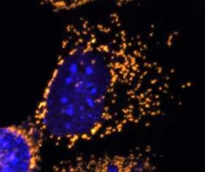
Response: Strictly maternal inheritance of mitochondria, the cellular power stations, and mitochondrial genes that mitochondria harbor, is a major biological paradigm in mammals. Propagation of paternal, sperm-contributed mitochondrial genes, resulting in a condition called heteroplasmy, is seldom observed in mammals, due to post-fertilization elimination sperm mitochondria, referred to as “sperm mitophagy.” Our and others’ recent results suggest that this process is mediated by the synergy of ubiquitin–proteasome system (UPS) pathway that recycles outlived cellular proteins one molecule at a time, and autophagic pathway capable of engulfing and digesting an entire mitochondrion.
Here we demonstrate that the co-inhibition of the ubiquitin-binding autophagy receptor proteins SQSTM1, GABARAP, and UPS, and the UPS protein VCP dependent pathways delayed the digestion of sperm mitochondria inside the fertilized pig egg. By manipulating said proteins, we created heteroplasmic pig embryos with both the paternal and maternal mitochondrial genes. Such animal embryos that could be used as a biomedical model to research and alleviate certain forms of mitochondrial disease.