

03 Sep What an MRI Can See That Other Scans Miss
Most people meet their first MRI the same way. A specialist mentions it almost in passing, a booking gets made and the machine turns out to be far louder than anyone warned you about. MRI is the tool doctors reach for when they need to see soft tissue clearly. Ligaments, nerves, organs and the spinal cord all show up in a level of detail that other scans simply cannot match. According to the NIBIB (National Institute of Biomedical Imaging and Bioengineering), MRI uses a powerful magnetic field and radio waves to produce detailed images of organs and tissues without using ionising radiation, making it particularly valuable for repeated imaging and for visualising soft tissue structures that X-rays and CT scans cannot resolve clearly.
What follows is a practical walkthrough: how MRI differs from an X-ray or CT, what the safety screening is really for, what the scan itself involves and how to approach the report at the end.
Key Takeaways
- MRI uses a strong magnetic field and radio waves rather than ionising radiation
- Its main strength is soft-tissue contrast, which is why it suits brain, spine, joint and organ imaging
- Safety screening exists because metal and the magnet do not mix, so disclose implants and devices honestly
- Most scans run 15 to 30 minutes, with specialised studies taking 40 to 60 minutes
- A qualified MRI technologist performs the scan and a radiologist interprets it
Why MRI Is Not Just a Fancier X-ray
An MRI scanner works on completely different physics from an X-ray or CT machine. Instead of radiation, it uses a powerful magnetic field combined with radio waves to build cross-sectional and three-dimensional images. The practical consequence is soft-tissue contrast. A torn ligament, an inflamed nerve root or a subtle change in brain tissue can be almost invisible on an X-ray and obvious on an MRI. Because there is no ionising radiation involved, MRI also suits repeated imaging. If a condition needs monitoring over years rather than weeks, that matters a great deal.
What Doctors Actually Order MRI For
The list is broad. Neurological symptoms sit near the top, along with spine and nerve conditions. Musculoskeletal injuries involving tendons, ligaments, cartilage and joints are just as common. Abdominal and pelvic organs are the next big group, covering the liver, kidneys, bowel, uterus and prostate. MRI is also used to assess the heart and blood vessels. Tumours, infections and inflammation round out the common referrals. Vascular imaging deserves a separate mention. Using MRA techniques, the same scanner can image blood vessels as part of the same radiation-free examination.
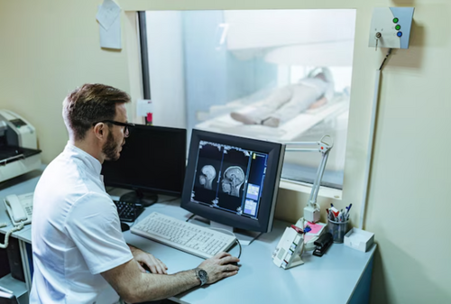
Providers vary in how much of that range they cover under one roof. Advicon Imaging MRI in Sydney spans brain, spine, musculoskeletal, breast, prostate, liver, renal, small bowel and whole body studies, with every scan performed by a qualified MRI technologist and reviewed by an experienced radiologist.
The Safety Questionnaire Is Not a Formality
This is the part patients rush through, and it is the part that matters most. In a clinical MRI suite the magnetic field is present at all times, not only during your scan, and unsecured metal objects can be pulled toward it with significant force. Tell staff before your appointment if you have any of the following:
- A pacemaker or cardiac implant
- A cochlear implant
- Surgical or aneurysm clips
- A neurostimulator
- Metal fragments in your eye
- An insulin pump or drug infusion device
- Magnetic dental implants
Some of these can be safely worked around, and some change whether or how the scan goes ahead. That decision belongs to the imaging team, which is why they need the information in advance rather than on the table. On the day, you remove everything metallic. Keys, jewellery, credit cards, hairpins, piercings and watches all come off, and loose clothing without metal zips makes that process quicker. You may still be asked to change into a gown, so bringing a spare set of clothes is a sensible backup. One detail catches people out: some makeup and hairspray contain metallic particles, so it is worth skipping both on scan day. Also flag it if you are pregnant or breastfeeding, and ask about fasting when you book, since some studies require it and others do not.
What the Scan Itself Is Like
You lie on a padded table that slides into the scanner. Staff hand you a buzzer so you can alert them at any point, which does more for nerves than most people expect. Earplugs or headphones are provided, and you will want them. MRI is loud, though modern equipment is designed to cut noise and improve comfort. The scan runs as a series of sequences, each lasting from 30 seconds to a few minutes. Staying still is the single most useful thing you can do, and some sequences ask for a short breath-hold. Most scans take 15 to 30 minutes. More specialised studies can run 40 to 60 minutes, and you can usually head home straight afterwards unless sedation was used. If you know you are claustrophobic, raise it early. Sedation can be arranged, though you will need someone to drive you home afterwards.
When Contrast Comes Into It
Some MRI examinations use an intravenous contrast agent called gadolinium. It highlights blood vessels, inflammation and abnormal tissue that might otherwise blend into the surrounding anatomy. Contrast is generally very safe. The two things to raise beforehand are any kidney problems and any history of reactions to contrast agents. You will not always know in advance whether your particular scan needs it. The clinic can tell you when you confirm your booking, which is worth asking if you would rather not be surprised on the day.
Bring Your Old Scans With You
This is the most underused piece of advice in imaging. Bring your referral, whatever identification or health card your clinic asks for, and any previous X-ray, CT, ultrasound or MRI studies you have of the same area. Comparison changes interpretation. A finding that looks ambiguous in isolation often becomes clearly stable, or clearly new, the moment a radiologist can hold it against an older scan. If those studies live on a disc in a drawer somewhere, dig them out before the appointment rather than after.
Making Sense of the Report
Your radiologist’s report goes to the doctor who referred you, and that consultation is where the findings get translated into a plan. Reading the report cold, without that context, is where anxiety usually creeps in. Radiology language is precise rather than reassuring, which is a different thing. Phrases like “unremarkable” and “no acute findings” are good news dressed in clinical vocabulary. If you want a head start before your follow-up, a guide to understanding MRI results can break down the structure of a report and the terms that come up most often.
The Bottom Line
MRI earns its place by showing what other scans cannot, without radiation and with a strong safety record when screening is done properly. Your job is simple enough. Answer the safety questions carefully, bring your old imaging, stay still for the sequences and let your doctor interpret the report with you.
Frequently Asked Questions
Is an MRI safe?
Yes. MRI does not use ionising radiation and there are no known long-term side effects when proper safety screening is completed. The main risks relate to metal implants, rare allergic reactions to contrast and claustrophobia.
How long does an MRI take?
Most scans take 15 to 30 minutes. More specialised studies may take 40 to 60 minutes.
Is MRI safe for children?
Yes. It is radiation-free, which makes it suitable for children and for long-term monitoring.
Why is an MRI so noisy?
The noise comes from the scanner’s gradient coils vibrating as currents switch rapidly inside the strong magnetic field, and those vibrations carry through the air as sound. Earplugs or headphones are provided, and newer equipment is built to reduce it.
Do I need to fast before an MRI?
Some scans require fasting and many do not. You will be advised when your appointment is booked, so ask if you have not been told.
What happens if I have a pacemaker or another implant?
Tell the imaging team well before your appointment. Many implants can be worked around safely, but the scan needs to be planned with that information in hand.
For a broader overview of how different medical imaging modalities compare and when MRI, CT, or ultrasound is most appropriate for specific clinical questions, see this MedicalResearch.com overview of choosing the right medical imaging — MRI, CT, ultrasound, and X-ray compared.
Disclaimer: The information on MedicalResearch.com is provided for educational purposes only, and is in no way intended to diagnose, cure, or treat any medical or other condition. Some links are sponsored. MedicalResearch.com and Eminent Domains Inc. do not warrant or endorse products or claims made by third party links. Always seek the advice of your physician or other qualified health provider and ask your doctor any questions you may have regarding a medical condition. In addition to all other limitations and disclaimers in this agreement, service provider and its third party providers disclaim any liability or loss in connection with the content provided on this website.
Last Updated on September 3, 2026 by Marie Benz MD FAAD