
10 Nov FDG-PET Scans of Lung Nodules Should Be Interpreted With Caution
Posted at 21:00h
in Author Interviews, JAMA, Lung Cancer, Medical Imaging, Surgical Research, Vanderbilt
MedicalResearch.com Interview with:
[caption id="attachment_38163" align="alignleft" width="300"]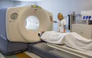 PET Scan Vanderbilt Health[/caption]
Amelia W. Maiga, MD MPH
Vanderbilt General Surgery Resident
VA Quality Scholar, TVHS
MedicalResearch.com: What is the background for this study? What are the main findings?
Response: Positron emission tomography (PET) combined with fludeoxyglucose F18 (FDG) is currently recommended for the noninvasive diagnosis of lung nodules suspicious for lung cancer. Our investigation adds to growing evidence that FDG-PET scans should be interpreted with caution in the diagnosis of lung cancer. Misdiagnosis of lung lesions driven by FDG-PET avidity can lead to unnecessary tests and surgeries for patients, along with potentially additional complications and mortality.
To estimate FDG-PET diagnostic accuracy, we conducted a multi-center retrospective cohort study. The seven cohorts originating from Tennessee, Arizona, Massachusetts and Virginia together comprised 1188 nodules, 81 percent of which were malignant. Smaller nodules were missed by FDG-PET imaging. Surprisingly, negative PET scans were also not reliable indicators of the absence of disease, especially in patients with smaller nodules or who are known to have a high probability of lung cancer prior to the FDG-PET test.
Our study supports a previous meta-analyses that found FDG-PET to be less reliable in regions of the country where fungal lung diseases are endemic. The most common fungal lung diseases in the United States are histoplasmosis, coccidioidomycosis and blastomycosis. All three fungi reside in soils. Histoplasmosis and blastomycosis are common across much of the Mississippi, Ohio and Missouri river valleys and coccidioidomycosis is prevalent in the southwestern U.S. These infections generate inflamed nodules in the lungs (granulomas), which can be mistaken for cancerous lesions by imaging.
PET Scan Vanderbilt Health[/caption]
Amelia W. Maiga, MD MPH
Vanderbilt General Surgery Resident
VA Quality Scholar, TVHS
MedicalResearch.com: What is the background for this study? What are the main findings?
Response: Positron emission tomography (PET) combined with fludeoxyglucose F18 (FDG) is currently recommended for the noninvasive diagnosis of lung nodules suspicious for lung cancer. Our investigation adds to growing evidence that FDG-PET scans should be interpreted with caution in the diagnosis of lung cancer. Misdiagnosis of lung lesions driven by FDG-PET avidity can lead to unnecessary tests and surgeries for patients, along with potentially additional complications and mortality.
To estimate FDG-PET diagnostic accuracy, we conducted a multi-center retrospective cohort study. The seven cohorts originating from Tennessee, Arizona, Massachusetts and Virginia together comprised 1188 nodules, 81 percent of which were malignant. Smaller nodules were missed by FDG-PET imaging. Surprisingly, negative PET scans were also not reliable indicators of the absence of disease, especially in patients with smaller nodules or who are known to have a high probability of lung cancer prior to the FDG-PET test.
Our study supports a previous meta-analyses that found FDG-PET to be less reliable in regions of the country where fungal lung diseases are endemic. The most common fungal lung diseases in the United States are histoplasmosis, coccidioidomycosis and blastomycosis. All three fungi reside in soils. Histoplasmosis and blastomycosis are common across much of the Mississippi, Ohio and Missouri river valleys and coccidioidomycosis is prevalent in the southwestern U.S. These infections generate inflamed nodules in the lungs (granulomas), which can be mistaken for cancerous lesions by imaging.
