 MedicalResearch.com Interview with:
Dr Michael Lee PhD MPhty MChiro BSc
MedicalResearch.com Interview with:
Dr Michael Lee PhD MPhty MChiro BSc
Discipline of Physiotherapy, Faculty of Health Sciences, The University of Sydney
Clinical Neurophysiologist, The Brain & Mind Research Institute, The University of Sydney
Research Affiliate, Neuroscience Research Australia
Neurology Research Fellow, Institute of Neurological Sciences, Prince of Wales Hospital
Medical Research: What is the background for this study?
Dr. Lee: Our research team at the University of Sydney has previously shown that the functioning of peripheral nerves deteriorate following spinal cord injury (SCI). Using novel, non-invasive electrophysiological techniques (nerve excitability testing), we showed in this study that peripheral nerves
below the level of spinal cord injury underwent dramatic functional reorganization. Peripheral nerve dysfunction will not only contribute to a number of undesirable medical complications including peripheral neuropathy and pain, it exacerbates muscle atrophy and can potentially limit the effectiveness of rehabilitative therapies that drive central plasticity. In this study, we were interested to see whether this secondary peripheral nerve dysfunction could be reversed with a short-term targeted peripheral nerve stimulation therapy.
Medical Research: What are the main findings?
Dr. Lee: We studied peripheral nerve function in both the upper (median nerve at the wrist) and lower limbs (peroneal nerve near the fibular head) in 22 patients with acute spinal cord injury (all within 6 months of injury). We then randomly assigned one upper limb and one lower limb nerve to a daily regimen of 30-min peripheral nerve stimulation for 6 week. All study participants continued with standard rehabilitation. The results from our nerve excitability studies showed that 6-weeks of daily stimulation reversed a number of nerve excitability abnormalities secondary to spinal cord injury, and in some cases normalized it to a level comparable to healthy age-matched subjects. The peripheral nerves in the opposite limbs remained dysfunctional over the 6-week period. The results of our study showed convincingly that the addition of peripheral nerve stimulation in the early stages of spinal cord injury is beneficial by ameliorating the downstream effects of
spinal cord injury. Spinal cord injuries can be an unfortunate effect of being a car accident, causing serious issues for those who suffer from it whether financial or physical. Those who find themselves in this type of situation may look into contacting someone like these
car accident injury lawyers near Sacramento who might be able to help them to get compensation for their accident, which could help with phisyotherapy and medical bills.

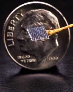 New technique enables rapid calibration of the BrainGate brain-computer interface.[/caption]
David Brandman, MD, PhD
Postdoctoral research associate (neuroengineering), Brown University
Senior neurosurgical resident
Dalhousie University
BrainGate Website
MedicalResearch.com: What is the background for this study?
Response: People with cervical spinal cord injuries, ALS, or brainstem stroke, may lose some or all of their ability to use their arms or hands. In some cases, they may even lose the ability to speak. One approach to restoring neurologic function is by using a brain computer interface (BCI). BCIs record information from the brain, and then translate the recorded brain signals into commands used to control external devices. Our research group and others have shown that intracortical BCIs can provide people with tetraplegia the ability to communicate via a typing interface, to control a robotic limb for self-feeding, and to move their own muscles using functional electrical stimulation. Use of a BCI generally requires the oversight of a trained technician, both for system setup and calibration, before users can begin using the system independently.
An open question with intracortical BCIs is how long it takes people to get up and running before they can communicate independently with 2 dimensional cursor control. The goal of this study was to systematically examine this question in three people with paralysis. As part of the ongoing BrainGate2 clinical trial, each study participant (T5, T8, and T10) had tiny (4x4 mm) arrays of electrodes implanted into a part of their brain that coordinates arm control. Each participant used motor imagery – that is, attempted or imagined moving their body – to control a computer cursor in real time.
New technique enables rapid calibration of the BrainGate brain-computer interface.[/caption]
David Brandman, MD, PhD
Postdoctoral research associate (neuroengineering), Brown University
Senior neurosurgical resident
Dalhousie University
BrainGate Website
MedicalResearch.com: What is the background for this study?
Response: People with cervical spinal cord injuries, ALS, or brainstem stroke, may lose some or all of their ability to use their arms or hands. In some cases, they may even lose the ability to speak. One approach to restoring neurologic function is by using a brain computer interface (BCI). BCIs record information from the brain, and then translate the recorded brain signals into commands used to control external devices. Our research group and others have shown that intracortical BCIs can provide people with tetraplegia the ability to communicate via a typing interface, to control a robotic limb for self-feeding, and to move their own muscles using functional electrical stimulation. Use of a BCI generally requires the oversight of a trained technician, both for system setup and calibration, before users can begin using the system independently.
An open question with intracortical BCIs is how long it takes people to get up and running before they can communicate independently with 2 dimensional cursor control. The goal of this study was to systematically examine this question in three people with paralysis. As part of the ongoing BrainGate2 clinical trial, each study participant (T5, T8, and T10) had tiny (4x4 mm) arrays of electrodes implanted into a part of their brain that coordinates arm control. Each participant used motor imagery – that is, attempted or imagined moving their body – to control a computer cursor in real time.


