Glaucoma, Ophthalmology / 28.10.2024

  glaucoma-opthalmology-eyedoctor.jpg Glaucoma is often called the "sneak thief of sight," and that name fits. Many folks don't know they have it until it's too late. Spotting the earliest sign of glaucoma can help save your vision and get you the care you need. Let's explore four important symptoms to keep an eye out for and discuss why catching glaucoma early is so crucial for keeping your eyes healthy. Please note everyone should be screened for glaucoma as well as other eye diseases including macular degeneration.  Make a regularly scheduled visit to an opthamologist part of your health care routine. Four Key Symptoms Glaucoma is a group of eye conditions that can lead to vision loss and are often associated with increased pressure in the eye. Here are four key symptoms to look out for as earliest sign of glaucoma:
  1. Blurred or Cloudy Vision
One of the most alarming symptoms that could indicate the earliest sign of glaucoma is a subtle change in your vision. Many people notice their vision becoming blurry or cloudy. This problem can get worse as time goes on. It often happens in low-light situations or when moving from bright to dark places. Glaucoma can block fluid from draining out of your eye, causing pressure that makes your vision unclear. If you have trouble focusing, don't ignore these changes. It's important to see an eye doctor. (more…)
Author Interviews, JAMA, Ophthalmology, USPSTF / 02.06.2022

MedicalResearch.com Interview with: Katrina Donahue, M.D., M.P.H. Professor and vice chair of research Department of Family Medicine University of North Carolina at Chapel Hill Dr. Donohue is a family physician and senior research fellow Cecil G. Sheps Center for Health Services Research Dr. Donahue joined the U.S. Preventive Services Task Force in January 2020. MedicalResearch.com:  What is the background for this study?  What are the main findings?  Response: Impaired vision and glaucoma are serious and common conditions facing millions of people nationwide that can affect a person’s independence and quality of life. These recommendations looked at how primary care clinicians can help people who have not noticed any problems with their vision. Unfortunately, there is not enough evidence available to make a recommendation for or against screening adults for glaucoma or older adults for impaired vision in the primary care setting. (more…)
Author Interviews, Genetic Research, Ophthalmology / 08.06.2021

MedicalResearch.com Interview with: Louis Pasquale, MD, FARVO Professor of Ophthalmology Icahn School of Medicine at Mount Sinai. Site Chair of the Department of Ophthalmology Mt. Sinai Hospital and Vice Chair of Translational Ophthalmology Research Mount Sinai Healthcare System  MedicalResearch.com: What is the background for this study? Response: Studies on the relation between caffeine intake and glaucoma have been contradictory, although our work suggested an adverse association amongst people with a self-reported family history of glaucoma. (more…)
Author Interviews, JAMA, Ophthalmology, Rheumatology / 15.12.2019

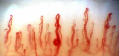
MedicalResearch.com Interview with: Robert Ritch, MD, FACS, FARVO Shelley and Steven Einhorn Distinguished Chair Professor of Ophthalmology, Surgeon Director Emeritus Chief, Glaucoma Services Emeritus The New York Eye and Ear Infirmary of Mount Sinai New York, NY 10003 Founder, Medical Director and Chairman, Scientific Advisory Board The Glaucoma Foundation  MedicalResearch.com: What is the background for this study? Response: Nailfold capillaroscopy (NFC), long used in rheumatology is a new approach to investigation of glaucoma. Posterior to the nailbed and just anterior to the proximal nailfold is the cuticle, which has no structural elements visible to the naked eye. NFC is a non-invasive imaging modality that provides a highly magnified view of the capillaries at the nailfold of digits. It has also been used in ophthalmology to show morphological changes at the nailfold capillaries of POAG and XFG/XFS patients, helping to confirm the systemic nature of these diseases. With nailfold capillaroscopy, an extensive array of capillaries can be seen greatly enlarged on a monitor screen. Capillary loops can be imaged, stored, recorded with videoscopy, and blood flow actively imaged and measured.. The first series of papers on glaucoma were written by Prof Josef Flammer’s group at the turn of the 21st century, looking at vasospasm, blood flow in normal-tension and high-tension glaucoma, and relating ocular blood flow alterations to systemic vascular regulation and relating laser Doppler flowmetry to NFC. Studies from Korea later associated nailbed hemorrhages and loss of nailbed capillaries to the presence of optic disc hemorrhages and investigated correlation of of heart rate variability with visual field defects and nailfold capillaroscopy. Studies by our group began with the publication in 2015 of a paper by Pasquale et al (Nailfold Capillary Abnormalities in Primary Open-Angle Glaucoma: A Multisite Study. IOVS;56:7021) using NFC video microscopy, associating dilated capillaries, avascular zones, and hemorrhages with primary open-angle glaucoma. Successive manuscripts and presentations at conferences have indicated differences between capillary loop patterns in high-tension and normal-tension POAG and exfoliation syndrome/exfoliation glaucoma. Our goal in this paper was to compare nailfold peripheral blood flow in XFG, which had not previously been compared to control subjects using NFC. We explored the peripheral blood flow at the nailfold of patients with high-tension glaucoma, normal-tension glaucoma, exfoliation glaucoma (XFG) and compared it to control subjects further evaluate the possible differences between these glaucoma entities. We examined the morphology and extent of nailfold capillary loops, vascular tortuosity, blood flow, and nailfold hemorrhages. (more…)
Author Interviews, Ophthalmology, Technology / 21.10.2019

MedicalResearch.com Interview with: Louis R. Pasquale, MD Professor of Ophthalmology Icahn School of Medicine at Mount Sinai; Site Chair of the Department of Ophthalmology at The Mount Sinai Hospital and Mount Sinai Queens; Vice Chair of Translational Ophthalmology Research Mount Sinai Health System  MedicalResearch.com: What is the background for this study? What are the main findings? Response: Individual visual field tests provide a 52-point array of functional information about a glaucoma patient but it does not give us a handle on how functionally disabled they might be. A series of visual field tests need to be assessed for functional progression but current conventional algorithms for doing so are governed by ad hoc rules and the various algorithms available for assessing progression do not agree with one another. Finally, in managed care setting where one might be responsible for allocating resources for large numbers of glaucoma patients, it would be valuable to quickly visualize which patients are progressing rapidly and which ones are stable. This could allow for proper allocation of resources and perhaps inquiry into why a subset of patients are doing poorly. We wanted to develop an easy to use tool to quickly visualize how individual glaucoma patients and how groups of glaucoma patients are doing from a functional perspective. (more…)
Author Interviews, Compliance, Electronic Records, JAMA, Ophthalmology / 18.12.2018

MedicalResearch.com Interview with: Michael Vincent Boland, M.D., Ph.D. Glaucoma Center of Excellence Director of Information Technology, Wilmer Eye Institute Associate Professor of Ophthalmology Johns Hopkins University School of Medicine Baltimore, Maryland MedicalResearch.com: What is the background for this study? What are the main findings? Response: Effective medications are available to treat glaucoma and prevent or stop vision loss.
Unfortunately, patients frequently do not use the eye drops as prescribed, oftentimes simply
because they forget to. Since patient medications are now managed via electronic health
records (EHRs), we built a system to deliver automated reminders to patients using the patient
portal to our EHR. We found that the majority (75%, 66 of 88) of participants that received these reminders found them to be useful, and about half (47%, 41 of 88) the participants wanted to
continue using the reminders after the study ended
(more…)
Author Interviews, Health Care Systems, Ophthalmology / 07.11.2018

MedicalResearch.com Interview with: Nathan Radcliffe, MD Senior Faculty, Ophthalmology Glaucoma and Cataract surgeon Mount Sinai Health System MedicalResearch.com: What is the background for this study? Response: Glaucoma is a leading cause of blindness and the mainstay of therapy is to lower the intraocular pressure (IOP) with topical eye drops. Up to 40% of patients may require more than one eye drop to control the disease, and yet taking more than one eye drop bottle can result in higher costs, more eye irritation, worse therapeutic compliance, and possibly worse outcomes, be sure to consult your eye surgeon before increasing any eye treatment to ensure it won't do any further damage to the eye. Compounded therapies (not FDA approved, but made at the physician’s request by a compounding pharmacy) can be created to contain multiple glaucoma therapies in one bottle. We sought to determine if a compounded solution containing three or four drops in one bottle could control glaucoma as well as three or four separate bottles (standard of care) in patients requiring three or four eye drop bottles to control glaucoma. We performed a multi-center, randomized, observer-masked, parallel-group study comparing a compounded therapy containing latanoprost 0.05%, dorzolamide hydrochloride 2%, timolol maleate 0.5%, brimonidine tartrate 0.2% with 0.01% BAK to standard three or four bottle regaimins. We measured IOP and corneal staining (a sign of preservative toxicity), as well as other safety measures at week one, month one, two and three. (more…)
Author Interviews, Emory, Genetic Research, JAMA, Ophthalmology / 24.01.2018

MedicalResearch.com Interview with: Eldon E. Geisert, PhD Professor of Ophthalmology Emory School of Medicine MedicalResearch.com: What is the background for this study? What are the main findings? Response: In the late 1990s a group of doctors began a study of glaucoma patients to determine if there were phenotypes that are predictive for developing glaucoma. In this Ocular Hypertension Treatment Study (OHTS) one of the highly correlated ocular traits was central corneal thickness (CCT). The early clinical studies found that people with thinner corneas were at a higher risk of developing glaucoma. In two large studies, examining thousands of people a number of genes were identified that were risk factors for glaucoma or that controlled CCT in humans. In both cases the identified genes accounted for less than 10% of the genetic risk for glaucoma and less than for 10% of the genetic control for CCT. There was little data linking the genetic control of CCT to the glaucoma risk. Our group has taken an indirect approach to the question, using well-defined mouse genetic system to identify genes modulating CCT and then interrogating human glaucoma data to determine if these genes are associated with glaucoma risk.   (more…)
Author Interviews, JAMA, Ophthalmology / 06.09.2017

MedicalResearch.com Interview with: Andrew Tatham, FRCOphth Consultant Ophthalmologist Princess Alexandra Eye Pavilion and Department of Ophthalmology University of Edinburgh, Scotland  MedicalResearch.com: What is the background for this study? Response: Raised intraocular pressure (IOP) is the major risk factor for the development of glaucoma, the most common cause of irreversible blindness, with lowering IOP the only proven treatment. Until recently the only way to measure IOP was for patients to visit their clinician  meaning it was only possible to obtain a limited number of measurements. This is problematic given that IOP fluctuates and that 75% of individuals have peak IOP outside office hours. If patients could measure their own IOP it would allow far more measurements to be obtained and result in better understanding of the variation and peaks in IOP. This could improve the detection of glaucoma and determine if patients are adequately controlled with medication. Recently, a patient-operated, home IOP monitoring device (iCare HOME) has become available. The patient holds the device close to their eye and the device automatically determines if it is in the correct position to take a measurement. The tonometer then deploys a small probe which gently bounces off the surface of the eye to determine IOP. As the probe is only in contact with the surface of the eye for a few milliseconds no anesthetic is needed. (more…)
Author Interviews, Exercise - Fitness, Ophthalmology, PLoS / 18.01.2016

More on Ophthalmology on MedicalResearch.com MedicalResearch.com Interview with: Robert Ritch, MD, FACS Shelley and Steven Einhorn Distinguished Chair Professor of Ophthalmology Surgeon Director Emeritus and Chief, Glaucoma Services Founder, Medical Director and Chairman, Scientific Advisory Board The Glaucoma Foundation Jessica V. Jasien MEn Einhorn Clinical Research Center The New York Eye and Ear Infirmary of Mount Sinai New York, NY 10003 Medical Research: What is the background for this study? What are the main findings? Response: Glaucoma is the leading cause of irreversible blindness in the United States and elevated intraocular pressure (IOP) is the most common known risk factor for glaucomatous damage. At the current time, IOP is the only modifiable risk factor for which treatment has a proven effect on preventing or slowing the progress of the disease. The story behind this study goes back to 1980, when we saw a 45-year-old woman with severe damage from normal-tension glaucoma, which then was thought to be a disease of the elderly and also thought to be rare, which we now realize was erroneous. The causes of normal-tension glaucoma were also poorly understood. It turned out on questioning that this particular patient had been performing yoga and standing on her head for 20 minutes a day for 20 years. We measured her IOP in this position and it rose from 15 mmHg in the sitting position to 60 mmHg. When measured lying flat, it was 30 mmHg. We measured everyone working in the department standing on their heads and the IOP roughly doubled in each of them. This was our first inkling that marked changes in IOP could result from changes in body position. The background for this study came from the lack of knowledge of IOP rises during yoga inversions, other than the headstand position. We looked at four common inverted yoga positions in glaucoma patients and healthy patients who were all experienced in practicing yoga. The four positions tested were downward facing dog, plow, legs up the wall, and forward bend. Each position showed a direct increase in IOP immediately assuming the yoga position, however the IOP dropped once assuming the seated position after two minutes in the yoga position. The most significant increase in IOP was seen during the downward facing dog position. IOP of each study participant was taken seated (baseline), immediately assuming the yoga position, which was held for two minutes, again at the two minutes of the yoga position, immediately in the seated position following the yoga position, and again after 10 minutes in the seated position. Each position was tested once in this order of IOP measurements. (more…)
Author Interviews, Brigham & Women's - Harvard, JAMA, Nutrition, Ophthalmology / 16.01.2016

More on Ophthalmology on MedicalResearch.com MedicalResearch.com Interview with: Jae Hee Kang, MSc, SC Associate Epidemiologist, Brigham and Women's Hospital Assistant Professor of Medicine, Harvard Medical School Brigham and Women's Hospital Department of Medicine Channing Division of Network Medicine Boston, MA 02115 Medical Research: What is the background for this study? What are the main findings? Dr. Kang: Glaucoma is a leading cause of irreversible blindness worldwide, and primary open-angle glaucoma (POAG) is the most common form of the disease. Little is known on the causes of glaucoma but dysfunction in the regulation of blood flow to the optic nerve, which transmits visual information to the brain, may be involved. Nitric oxide is important for maintenance of blood flow and its signaling may be impaired in glaucoma. We were interested in whether dietary nitrates, an exogenous source of nitric oxide mostly found in green-leafy vegetables, may be related to lower risk of POAG. Medical Research: What is the background for this study? What are the main findings? Dr. Kang: We (Brigham and Women’s Hospital / Harvard Medical School and Massachusetts Eye and Ear researchers) used 25+ years of data from over 100,000 participants in the Nurses' Health Study (63,893 women) and the Health Professionals Follow-up Study (41,094 men). Participants were nurses or other health professionals and were aged 40 years or older and reported eye exams. We collected information on their diet and other health information every two years with questionnaires. During follow-up, 1,483 new cases of primary open-angle glaucoma with visual field loss were identified and confirmed with medical record review. Participants were divided into quintiles (one of five groups) of dietary nitrate intake (quintile 5, approximately 240 mg/day; quintile 1, approximately 80 mg/day) and of green leafy vegetables (quintile 5, approximately 1.5 servings/day; quintile 1, approximately one-third of a serving/day). We observed that greater intake of dietary nitrate and green leafy vegetables (e.g., romaine and iceberg lettuce and kale/chard/mustard greens) was associated with a 20 percent to 30 percent lower POAG risk; the association was particularly strong (40 percent-50 percent lower risk) for POAG with early paracentral visual field loss (a subtype of POAG most linked to dysfunction in blood flow autoregulation). (more…)
Author Interviews, Columbia, Cost of Health Care, JAMA, Ophthalmology / 15.01.2016

More on Health Care Costs on MedicalResearch.com MedicalResearch.com Interview with: Alisa Prager BS Bernard and Shirlee Brown Glaucoma Research Laboratory Department of Ophthalmology Edward S. Harkness Eye Institute Columbia University Medical Center, New York, New York MedicalResearch: What is the background for this study?  Response: The goal of this research was to better understand the impact of glaucoma on non-ophthalmic healthcare use and costs. While there have been other studies assessing costs associated with glaucoma, these studies were primarily derived from either claims data or chart review. Our study used the Medicare Current Beneficiary Survey, which is a dataset that links claims data with survey results. The advantage of this is that the survey data allowed us to assess patient reported outcomes that did not necessarily prompt an encounter with the health care system, such as recent falls or feelings of sadness. The MCBS also provides complete expenditure and source of payment data on health services, including those not covered by Medicare, which allowed us to look at a more full spectrum of both private and public healthcare use and costs among Medicare beneficiaries. MedicalResearch: What are the main findings? Response: We found that Medicare beneficiaries with glaucoma have 27% higher likelihood of inpatient hospitalizations and home health aide visits compared to those without glaucoma, even after adjusting for covariates and excluding individuals who were admitted to the hospital with a diagnosis of glaucoma. When we stratified glaucoma patients based on self-reported visual disability, we found that those with self-reported visual disability were more likely to complain of depression, falls and difficulty walking compared to those without. We also found that glaucoma patients incurred a predicted $2,903 higher mean annual total healthcare costs from all sources compared to those without glaucoma after adjusting for socioeconomic factors and comorbidities. Costs were higher among those who reported visual disability, and remained higher after excluding outpatient payments. (more…)
Author Interviews, Diabetes, JAMA, Ophthalmology, University of Michigan / 06.06.2015

Julia E. Richards, Ph.D. Harold F. Falls Professor of Ophthalmology and Visual Sciences Professor of Epidemiology Director, Glaucoma Research Center The University of MichiganMedicalResearch.com Interview with: Julia E. Richards, Ph.D. Harold F. Falls Professor of Ophthalmology and Visual Sciences Professor of Epidemiology Director, Glaucoma Research Center The University of Michigan Medical Research: What is the background for this study? Response: We have a special interest in how the developmental processes of aging increase the risk of late onset diseases. We wondered whether drugs that target known aging pathways might be able to reduce risk of late onset disease. In the aging field, an emerging area of interest has been the category of drugs called caloric restriction mimetic (CRM) drugs, which have been found to extend life span and to reduce risk or delay onset of some late-onset diseases. These caloric restriction mimetic drugs target a set of pathways that have come to be seen as playing roles in longevity. One of these caloric restriction mimetic drugs, metformin, happens to also be one of the most common drugs used in the treatment of type 2 diabetes. Glaucoma is a leading cause of blindness worldwide and classical open-angle glaucoma shows onset in late middle age or late age, so we hypothesized that a caloric restriction mimetic drug might be able to reduce the risk of open-angle glaucoma. We used data from a large health services database to compare the rate at which open-angle glaucoma developed in individuals with diabetes mellitus who used metformin versus those who did not use metformin. We predicted that metformin would be associated with reduced risk of open-angle glaucoma. Medical Research: What are the main findings? Response: We found that use of metformin was associated with reduced risk of open-angle glaucoma. A 2 gram per day dose of the CRM drug metformin for two years was associated with a 20.8% reduction in risk of developing open-angle glaucoma. When we looked at the highest quartile of drug prescribed (>1,100 grams over a two year period) we found a 25% reduction in risk relative to those taking no metformin. This risk reduction is seen even when we account for glycemic control in the form of glycated hemoglobin, and use of other diabetes drugs was not associated with reduced risk of open-angle glaucoma. A possible explanation for our findings might be that the mechanism of risk reduction is taking place by CRM drug mechanisms that target aging pathways rather than through glycemic control of diabetes. In the long run, the approaches to late onset diseases in general will become much more powerful if we can use parallel approaches that simultaneously target both the aging processes going on and the disease-specific pathways going on. In the literature we see caloric restriction mimetic drugs metformin, rapamycin and resveratrol all being explored for their ability to target points in aging pathways in ways that can impact the risk of a variety of late-onset diseases, so it will be important for those interested in the risk factors affecting late onset diseases to pay attention to how caloric restriction mimetic drugs might be altering risk for those late onset diseases. (more…)
Author Interviews, Cost of Health Care, Medicare, Ophthalmology / 21.04.2015

MedicalResearch.com Interview with: Dan Gong BA Yale University School of MedicineMedicalResearch.com Interview with: Dan Gong BA Yale University School of Medicine ------------ James C. Tsai, M.D., M.B.A. President - New York Eye and Ear Infirmary of Mount Sinai Delafield-Rodgers Professor and Chair Department of Ophthalmology Icahn School of Medicine at Mount Sinai Medical Research: What is the background for this study? What are the main findings?
  • Congress first introduced the Medicare Physician Fee Schedule built on the resource-based relative value scale (RBRVS) in the Omnibus Budget Reconciliation Act of 1989. Until recently, Medicare payments to physicians were adjusted annually based on the sustainable growth rate (SGR) formula.
  • When adjusting physician payments, one controversial belief by policymakers was the assumption that in response to fee reductions, physicians would recuperate one-half of lost revenue by increasing the volume and complexity of services.
  • This study questioned this assumption that this inverse relationship between Medicare payment and procedural volume is uniform across all procedures. In particular, glaucoma procedures have not been studied in the past.
  • Using a fixed effects regression model, we found that for six commonly performed glaucoma procedures, four did not have any significant Medicare payment and procedural volume relationship (laser trabeculoplasty, trabeculectomy with and without previous surgery, aqueous shunt to reservoir). Two procedures, laser iridotomy and scleral reinforcement with graft, did have significant and inverse associations between Medicare payment and procedural volume. (more…)
Author Interviews, Circadian Rhythm, Ophthalmology, Sleep Disorders, UCSD / 09.04.2015

MedicalResearch.com Interview with: Carolina P B Gracitelli, M.D. Ophthalmology - PhD Candidate/ Research Fellow University of California San Diego - Hamilton Glaucoma Center  Medical Research: What is the background for this study? What are the main findings?  Dr. Gracitelli:  Of all the diseases that can lead to blindness, glaucoma is one of the most important diseases; it affects more than 70 million people worldwide, of whom approximately 10 % are bilaterally blind. Different studies have reported that the damage caused by glaucomatous disease lead to retinal ganglion cell (RGC) loss and consequently loss of intrinsically photosensitive retinal ganglion cells (ipRGCs), which is a subtype of RGC. This subpopulation of RGC is clearly related with non-image-forming visual function such as photic synchronization of circadian rhythms  and the pupillary light reflex. However, the true impact of glaucoma on sleep quality, sleep disturbance or circadian rhythm was until nowadays controversial. The main clinical finding of our study was that glaucoma leads to RGC death, including ipRGC death. These cells are connected to several non-image-forming functions, including circadian photoentrainment and pupillary reflexes. Therefore, the image-forming and non-image-forming visual systems are associated with glaucoma. Circadian function has not been well investigated in clinical daily practice, but it can interfere with the quality of life of these patients. Concerns about sleep disturbances in glaucoma patients should be incorporated into clinical evaluations.   Medical Research: What should clinicians and patients take away from your report? Dr. Gracitelli:  Our data support the concept that glaucoma is associated with a loss of ipRGCs that mediate the pupillary light response, particularly to the sustained component of the blue flash with a luminance of 250 cd/m2. Additionally, glaucoma patients had significant sleep disturbances that were inversely correlated with a measure of ipRGC function, the pupillary light reflex. These results suggest that the loss of ipRGCs in glaucoma may also lead to sleep disturbances. Both non-visual functions of ipRGCs are correlated, indicating that attention should be paid to the non-image forming visual functions in glaucoma patients.   Medical Research: What recommendations do you have for future research as a result of this study?  Dr. Gracitelli:  Sleep disorders is a complex system, therefore, some conclusions in this study should be carefully evaluated. Further studies with larger cohorts could also help to elucidate the association between the pupillary reflex and the polysomnography parameters. And longitudinal studies can better explain the associations between sleep disorders and glaucoma progression.  In addition, we know that there are several types of ipRGCs and they have specific functions (pupillary reflex or circadian rhythms), therefore, evaluations would also need to include a more thorough assessment to understand better the specific role of ipRGCs in sleep disturbances. However, it is true that these ipRGCs functions are impaired in glaucoma, affecting the quality of life of these patients.   Citation:   Intrinsically Photosensitive Retinal Ganglion Cell Activity Is Associated with Decreased Sleep Quality in Patients with Glaucoma  Gracitelli, Carolina P.B. et al. Ophthalmology Published Online: April 06, 2015 DOI: http://dx.doi.org/10.1016/j.ophtha.2015.02.030MedicalResearch.com Interview with: Carolina P B Gracitelli, M.D. Ophthalmology - PhD Candidate/ Research Fellow University of California San Diego - Hamilton Glaucoma Center Medical Research: What is the background for this study? What are the main findings? Dr. Gracitelli: Of all the diseases that can lead to blindness, glaucoma is one of the most important diseases; it affects more than 70 million people worldwide, of whom approximately 10 % are bilaterally blind. Different studies have reported that the damage caused by glaucomatous disease lead to retinal ganglion cell (RGC) loss and consequently loss of intrinsically photosensitive retinal ganglion cells (ipRGCs), which is a subtype of retinal ganglion cell. This subpopulation of RGC is clearly related with non-image-forming visual function such as photic synchronization of circadian rhythms and the pupillary light reflex. However, the true impact of glaucoma on sleep quality, sleep disturbance or circadian rhythm was until nowadays controversial. The main clinical finding of our study was that glaucoma leads to retinal ganglion cell death, including ipRGC death. These cells are connected to several non-image-forming functions, including circadian photoentrainment and pupillary reflexes. Therefore, the image-forming and non-image-forming visual systems are associated with glaucoma. Circadian function has not been well investigated in clinical daily practice, but it can interfere with the quality of life of these patients. Concerns about sleep disturbances in glaucoma patients should be incorporated into clinical evaluations. (more…)
Author Interviews, Blood Pressure - Hypertension, Ophthalmology / 12.01.2015

MedicalResearch.com Interview with: Zheng He and Bang V. Bui Department of Optometry & Vision Sciences University of Melbourne, Parkville, Australia Medical Research: What is the background for this study? What are the main findings? Response: Glaucoma, the second leading cause of blindness in the world, is a condition that occurs when too much pressure builds up inside the eye. This excess pressure injures the optic nerve (the wire that transmits visual information to the brain) resulting in vision loss. Many risk factors for glaucoma are not well understood. High blood pressure (> 140/90 mmHg) is probably the most common comorbidity in patients presenting to optometry clinics. The overall prevalence of hypertension worldwide is over 26%, and increases dramatically with advancing age. Long-term hypertension leads to remodeling of the heart and blood vessels, promoting the risk of multiple end organ damage. Whilst chronic hypertension is a well-documented risk factor for stroke, the link between hypertension and glaucoma remains unclear. Previously, it was thought that high blood pressure could counteract high intraocular pressure, which is a clear risk factor for glaucoma. However, this issue may be more complicated than first thought. The Baltimore Eye Survey compared the prevalence of glaucoma in young and older patients with hypertension. They found that young patients with high blood pressure were at lower risk of glaucoma compared to the entire cohort. This outcome is consistent with the idea that higher blood pressure provides better perfusion pressure to the eye. Paradoxically systemic hypertension in older subjects actually increased the risk of glaucoma. Its seems that longer durations of systemic hypertension impact glaucoma risk negatively. One explanation for this is that any benefit from high blood pressure counteracting high eye pressure is lost as damage to blood vessels — a consequence of hypertension — becomes more dominant. This hypothesis was tested by comparing the effect of acute (one hour) and chronic (four weeks) hypertension in lab rats with elevated eye pressure. When blood pressure was raised for four weeks, there was less functional protection against eye pressure elevation compared with the one-hour case. This shows that having high blood pressure for a longer time compromises the eye's capacity to cope with high eye pressure. This impairment was associated with thicker and narrower blood vessels and a reduced capacity for the eye to maintain blood flow at normal levels in response to eye pressure elevation (this process is known as autoregulation). Thus in chronic hypertension, smaller reduction in ocular perfusion pressure can result in blood flow deficiency. (more…)