MedicalResearch.com Interview with:
[caption id="attachment_41105" align="alignleft" width="133"]

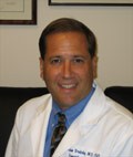
Dr. Mitchell[/caption]
Aaron Mitchell, MD
Division of Hematology/Oncology, Department of Medicine,
Lineberger Comprehensive Cancer Center
The Cecil G. Sheps Center for Health Services Research
The University of North Carolina at Chapel Hill
MedicalResearch.com: What is the background for this study? What are the main findings?
Response: Financial relationships between physicians and the pharmaceutical industry are very common. However, we are just beginning to figure out whether these relationships may lead to potentially concerning changes in physician behavior - whether physicians tend to prescribe more of the drugs made by a company that has given them money. We decided to ask whether oncologists who receive money from drugmakers are more likely to use the cancer drugs made by companies that have given them money in the past.
In studying two specific groups of cancer drugs, one for kidney cancer and one for chronic myeloid leukemia (CML), we found that oncologists who had received payments such as meals, consulting fees, travel & lodging expenses from the manufacturer of one of these drugs tended to use that drug more. When looking at oncologists who received payments for research, we found increased prescribing among the kidney cancer drugs but not the CML drugs.