MedicalResearch.com Interview with:
[caption id="attachment_52467" align="alignleft" width="130"]

Dr. Ritch[/caption]
Robert Ritch, MD, FACS, FARVO
Shelley and Steven Einhorn Distinguished Chair
Professor of Ophthalmology, Surgeon Director Emeritus
Chief, Glaucoma Services Emeritus
The New York Eye and Ear Infirmary of Mount Sinai
New York, NY 10003
Founder, Medical Director and
Chairman, Scientific Advisory Board
The Glaucoma Foundation
MedicalResearch.com: What is the background for this study?
Response: Nailfold capillaroscopy (NFC), long used in rheumatology is a new approach to investigation of glaucoma.
Posterior to the nailbed and just anterior to the proximal nailfold is the cuticle, which has no structural elements visible to the naked eye. NFC is a non-invasive imaging modality that provides a highly magnified view of the capillaries at the nailfold of digits. It has also been used in ophthalmology to show morphological changes at the nailfold capillaries of POAG and XFG/XFS patients, helping to confirm the systemic nature of these diseases.
[caption id="attachment_52469" align="alignleft" width="400"]
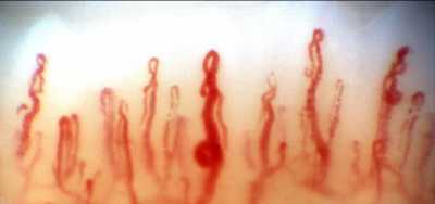
Nailfold Capillaroscopy / Dr. Ritch[/caption]
With nailfold capillaroscopy, an extensive array of capillaries can be seen greatly enlarged on a monitor screen. Capillary loops can be imaged, stored, recorded with videoscopy, and blood flow actively imaged and measured..
The first series of papers on glaucoma were written by Prof Josef Flammer’s group at the turn of the 21
st century, looking at vasospasm, blood flow in normal-tension and high-tension glaucoma, and relating ocular blood flow alterations to systemic vascular regulation and relating laser Doppler flowmetry to NFC.
Studies from Korea later associated nailbed hemorrhages and loss of nailbed capillaries to the presence of optic disc hemorrhages and investigated correlation of of heart rate variability with visual field defects and nailfold capillaroscopy.
Studies by our group began with the publication in 2015 of a paper by Pasquale et al (Nailfold Capillary Abnormalities in Primary Open-Angle Glaucoma: A Multisite Study. IOVS;56:7021) using NFC video microscopy, associating dilated capillaries, avascular zones, and hemorrhages with primary open-angle glaucoma. Successive manuscripts and presentations at conferences have indicated differences between capillary loop patterns in high-tension and normal-tension POAG and exfoliation syndrome/exfoliation glaucoma.
Our goal in this paper was to compare nailfold peripheral blood flow in XFG, which had not previously been compared to control subjects using NFC. We explored the peripheral blood flow at the nailfold of patients with high-tension glaucoma, normal-tension glaucoma, exfoliation glaucoma (XFG) and compared it to control subjects further evaluate the possible difference
s between these glaucoma entities. We examined the morphology and extent of nailfold capillary loops, vascular tortuosity, blood flow, and nailfold hemorrhages.


 Dr. Christina Weng[/caption]
MedicalResearch.com Interview with:
Christina Y. Weng, MD, MBA
Assistant Professor-Vitreoretinal Diseases & Surgery
Baylor College of Medicine-Cullen Eye Institute
Medical Research: What is the background for this study? What are the main findings?
Dr. Weng: Telemedicine has been around for a long time, but only recently have technological advances solidified its utility as a reliable, effective, and cost-efficient method of healthcare provision. The application of telemedicine in the field of ophthalmology has been propelled by the development of high-quality non-mydriatic cameras, HIPAA-compliant servers for the storage and transfer of patient data, and the growing demand for ophthalmological care despite the relatively stagnant supply of eye care specialists. The global epidemic of diabetes mellitus has contributed significantly to this growing demand, as the majority of patients with diabetes will develop diabetic retinopathy in their lifetime.
Today, there are over 29 million Americans with diabetes, and diabetic retinopathy is the leading cause of blindness in working age adults in the United States. The American Academy of Ophthalmology’s and American Diabetes Association’s formal screening guidelines recommend that all diabetic patients receive an annual dilated funduscopic examination. Unfortunately, the compliance rate with this recommendation is quite dismal at an estimated 50-65%. It is even lower amongst minority populations which comprise the demographic majority of those served by the Harris Health System in Harris County, Texas, the third most populous county in the United States.
In 2013, the Harris Health System initiated a teleretinal screening program housed by eight of the district’s primary care clinics. In this system, patients with diabetes are identified by their primary care provider (PCP) during their appointments, immediately directed to receive funduscopic photographs by trained on-site personnel operating non-mydriatic cameras, and provided a follow-up recommendation (e.g., referral for in-clinic examination versus repeat imaging in 1 year) depending on the interpretation of their images. The images included in our study were interpreted via two different ways—once by the IRISTM (Intelligent Retinal Imaging Systems) proprietary auto-reader and then again by a trained ophthalmic specialist from the IRISTM reading center. The primary aim of this study was to evaluate the utility of the auto-reader by comparing its results to those of the reading center.
Data for 15,015 screened diabetic patients (30,030 eyes) were included. The sensitivity of the auto-reader in detecting severe non-proliferative diabetic retinopathy or worse, deemed sight threatening diabetic eye disease (STDED), compared to the reading center interpretation of the same images was 66.4% (95% confidence interval [CI] 62.8% - 69.9%) with a false negative rate of 2%. In a population where 15.8% of diabetics have STDED, the negative predictive value of the auto-reader was 97.8% (CI 96.8% - 98.6%).
Dr. Christina Weng[/caption]
MedicalResearch.com Interview with:
Christina Y. Weng, MD, MBA
Assistant Professor-Vitreoretinal Diseases & Surgery
Baylor College of Medicine-Cullen Eye Institute
Medical Research: What is the background for this study? What are the main findings?
Dr. Weng: Telemedicine has been around for a long time, but only recently have technological advances solidified its utility as a reliable, effective, and cost-efficient method of healthcare provision. The application of telemedicine in the field of ophthalmology has been propelled by the development of high-quality non-mydriatic cameras, HIPAA-compliant servers for the storage and transfer of patient data, and the growing demand for ophthalmological care despite the relatively stagnant supply of eye care specialists. The global epidemic of diabetes mellitus has contributed significantly to this growing demand, as the majority of patients with diabetes will develop diabetic retinopathy in their lifetime.
Today, there are over 29 million Americans with diabetes, and diabetic retinopathy is the leading cause of blindness in working age adults in the United States. The American Academy of Ophthalmology’s and American Diabetes Association’s formal screening guidelines recommend that all diabetic patients receive an annual dilated funduscopic examination. Unfortunately, the compliance rate with this recommendation is quite dismal at an estimated 50-65%. It is even lower amongst minority populations which comprise the demographic majority of those served by the Harris Health System in Harris County, Texas, the third most populous county in the United States.
In 2013, the Harris Health System initiated a teleretinal screening program housed by eight of the district’s primary care clinics. In this system, patients with diabetes are identified by their primary care provider (PCP) during their appointments, immediately directed to receive funduscopic photographs by trained on-site personnel operating non-mydriatic cameras, and provided a follow-up recommendation (e.g., referral for in-clinic examination versus repeat imaging in 1 year) depending on the interpretation of their images. The images included in our study were interpreted via two different ways—once by the IRISTM (Intelligent Retinal Imaging Systems) proprietary auto-reader and then again by a trained ophthalmic specialist from the IRISTM reading center. The primary aim of this study was to evaluate the utility of the auto-reader by comparing its results to those of the reading center.
Data for 15,015 screened diabetic patients (30,030 eyes) were included. The sensitivity of the auto-reader in detecting severe non-proliferative diabetic retinopathy or worse, deemed sight threatening diabetic eye disease (STDED), compared to the reading center interpretation of the same images was 66.4% (95% confidence interval [CI] 62.8% - 69.9%) with a false negative rate of 2%. In a population where 15.8% of diabetics have STDED, the negative predictive value of the auto-reader was 97.8% (CI 96.8% - 98.6%).


