08 Jun Hebrew University: First WWOX Gene Replacement Therapy Administered to Child With Hereditary Seizures
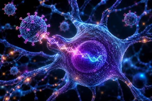
[caption id="attachment_74143" align="aligncenter" width="500"] Conceptual illustration of AAV9-mediated delivery of the WWOX gene to neurons, representing the first clinical use of a gene replacement therapy designed to restore WWOX function in the brain of an infant with WOREE syndrome.Credit: Hebrew University of Jerusalem / AI-generated illustration[/caption]
MedicalResearch.com Interview with:
Prof. Rami Aqeilan
Jacob M. Eisenberg and Thomas W. Baylek Chair for Medical Research in the field of Genetic Engineering
Lautenberg Center for Immunology and Cancer Research
Faculty of Medicine
Hebrew University of Jerusalem
Jerusalem, Israel
This therapy is based on more than a decade of research led by Prof. Rami Aqeilan, brought together with scientists, clinicians, and biotechnology leaders from Israel and the United States, including Dr. Naama Orenstein and Dr. Dror Kraus of Schneider Children's Medical Center and Dr. Yael Weiss, CEO of Mahzi Therapeutics.
MedicalResearch.com: What is the background for this study? Would you briefly explain the functions of the WWOX gene? Response: WWOX (WW domain-containing oxidoreductase) is a highly conserved gene that plays essential roles in brain development, neuronal function, and cellular stress responses. Nearly two decades ago, our laboratory and others began studying WWOX because of its involvement in cancer biology. However, over the past decade it became increasingly clear that WWOX is also critical for normal brain development. Inherited loss-of-function mutations in WWOX cause a devastating neurological disorder known as WOREE syndrome (WWOX-Related Epileptic Encephalopathy). Affected children typically develop severe, treatment-resistant epilepsy during infancy, profound developmental delay, intellectual disability, and significant motor impairment. Unfortunately, there has been no disease-modifying therapy available for these patients. The foundation for this therapeutic approach came from years of fundamental research in our laboratory aimed at understanding the biological role of WWOX in the nervous system. Using genetically engineered mouse models, we discovered that deleting WWOX specifically in neurons was sufficient to reproduce the major neurological features observed in mice lacking WWOX throughout the entire body. This finding demonstrated that neuronal WWOX deficiency is a primary driver of the disease and suggested that restoring WWOX function in neurons might be sufficient to achieve therapeutic benefit. Based on this insight, we developed a gene replacement strategy designed to restore WWOX expression selectively in neurons using an adeno-associated viral (AAV) vector. In preclinical studies, delivery of this vector into the brains of WWOX-deficient mice resulted in remarkable rescue of the disease phenotype. Treated animals exhibited normal behavior, elimination of seizures, and substantial correction of the neurological abnormalities associated with WWOX deficiency. These findings provided the critical proof-of-concept that neuronal gene replacement could effectively reverse key features of the disease and laid the scientific foundation for translating this approach toward clinical application in patients with WOREE syndrome.





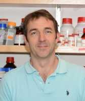






 Amanda Sierra, PhD
Research Professor and Group Leader
Ramón y Cajal Fellow
Achucarro Basque Center for Neuroscience
Laida Bidea
Bizkaia Science and Technology Park
Zamudio, Bizkaia, Spain
MedicalResearch.com: What is the background for this study? What are the main findings?
Dr. Sierra: Microglia phagocytosis of apoptotic cells is at the core of the brain regenerative response to recover the homeostasis of the brain parenchyma after damage because it prevents the spillover of toxic intracellular contents and is actively anti-inflammatory. However, while neuronal death is widespread in neurodegenerative diseases (Alzheimer´s, Parkinson´s, multiple sclerosis) and well as in ischemic and traumatic brain injuries, we have a complete lack of knowledge of the efficiency of microglial phagocytosis in the diseased brain.
In this paper we have discovered that microglia have a generalized response to apoptotic challenges: when confronted to a rise in the number of newborn cells, microglia display a combination of different strategies to boost their phagocytic output: increase the phagocytic capacity of each cell, recruit more cells to become phagocytic, or increase the total number of microglia (Abiega et al., PLoS Biol 2015). Thus, microglia have a very large potential for phagocytosis that could be summoned when needed.
To our surprise, however, in pathological conditions such as epilepsy (mouse and human), microglial phagocytosis was blocked. We have made use of the adult neurogenic cascade, where newborn cells undergo apoptosis naturally and are engulfed by “unchallenged microglia” (Sierra et al. Cell Stem Cell 2010), to establish the baseline of microglial phagocytosis efficiency. Whereas in physiological conditions microglia phagocytose over 90% of the apoptotic cells and remove them in under 1.5h, soon after the seizures it only engulfed 10% of the apoptotic cells and took up to 6h to digest them. This is the first quantification of microglial phagocytosis efficiency in the diseased mouse and human brain..
The block in phagocytosis was a rather complex phenomenon related to an impaired recognition (reduction of phagocytosis receptors) as well as impaired motility and targeting (reduced basal motility). We have also shown that the impairment is mediated at least partially by altered ATP microgradients: ATP is not only a neuro- and gliotransmitter widely released during seizures but is also a well-known “find-me” signal released by apoptotic cells. Thus, during seizures microglia became “blinded” by the neuronal hyperactivity and could not find the apoptotic cells.
In addition, we have shown that impairing phagocytosis releases the break on the inflammatory response. In fact, the impaired microglia were in a pro-inflammatory state and produced more cytokines such as tumor necrosis factor alfa (TNFa) or interleukin-1beta (IL-1b), which are well known neurotoxic and epileptogenic factors.
Amanda Sierra, PhD
Research Professor and Group Leader
Ramón y Cajal Fellow
Achucarro Basque Center for Neuroscience
Laida Bidea
Bizkaia Science and Technology Park
Zamudio, Bizkaia, Spain
MedicalResearch.com: What is the background for this study? What are the main findings?
Dr. Sierra: Microglia phagocytosis of apoptotic cells is at the core of the brain regenerative response to recover the homeostasis of the brain parenchyma after damage because it prevents the spillover of toxic intracellular contents and is actively anti-inflammatory. However, while neuronal death is widespread in neurodegenerative diseases (Alzheimer´s, Parkinson´s, multiple sclerosis) and well as in ischemic and traumatic brain injuries, we have a complete lack of knowledge of the efficiency of microglial phagocytosis in the diseased brain.
In this paper we have discovered that microglia have a generalized response to apoptotic challenges: when confronted to a rise in the number of newborn cells, microglia display a combination of different strategies to boost their phagocytic output: increase the phagocytic capacity of each cell, recruit more cells to become phagocytic, or increase the total number of microglia (Abiega et al., PLoS Biol 2015). Thus, microglia have a very large potential for phagocytosis that could be summoned when needed.
To our surprise, however, in pathological conditions such as epilepsy (mouse and human), microglial phagocytosis was blocked. We have made use of the adult neurogenic cascade, where newborn cells undergo apoptosis naturally and are engulfed by “unchallenged microglia” (Sierra et al. Cell Stem Cell 2010), to establish the baseline of microglial phagocytosis efficiency. Whereas in physiological conditions microglia phagocytose over 90% of the apoptotic cells and remove them in under 1.5h, soon after the seizures it only engulfed 10% of the apoptotic cells and took up to 6h to digest them. This is the first quantification of microglial phagocytosis efficiency in the diseased mouse and human brain..
The block in phagocytosis was a rather complex phenomenon related to an impaired recognition (reduction of phagocytosis receptors) as well as impaired motility and targeting (reduced basal motility). We have also shown that the impairment is mediated at least partially by altered ATP microgradients: ATP is not only a neuro- and gliotransmitter widely released during seizures but is also a well-known “find-me” signal released by apoptotic cells. Thus, during seizures microglia became “blinded” by the neuronal hyperactivity and could not find the apoptotic cells.
In addition, we have shown that impairing phagocytosis releases the break on the inflammatory response. In fact, the impaired microglia were in a pro-inflammatory state and produced more cytokines such as tumor necrosis factor alfa (TNFa) or interleukin-1beta (IL-1b), which are well known neurotoxic and epileptogenic factors.