MedicalResearch.com Interview with:
[caption id="attachment_23557" align="alignleft" width="200"]
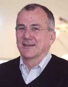
Dr. Ezequiel Morsella[/caption]
Ezequiel Morsella, Ph.D.
Associate Professor of Neuroscience
Department of Psychology
San Francisco State University
Assistant Adjunct Professor
Department of Neurology
University of California, San Francisco
Boardmember, Scientific Advisory Board
Institute of Cognitive Neurology (INECO),
Buenos Aires
MedicalResearch.com: What is the background for this study? What are the main findings?
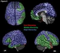
Dr. Morsella: The study is based on Passive Frame Theory, which I discuss below in brief, and on
ironic processing, in which one is more likely to think about something (e.g., white bears) when instructed to not think about that thing. Based on this research, the Reflexive Imagery Task (RIT) reveals that, following the activation of certain "action sets" (i.e., dispositions to act one way or another), conscious thoughts can arise involuntarily and systematically when one is presented with certain stimuli. In the most basic version of the RIT, subjects are presented with visual objects and instructed to not think of the names of the objects, which is challenging. In the new study, we show that the effect arises not only for automatic processes (e.g., forms of cued-memory retrieval) but also for processes involving more, in a sense, moving parts (e.g., symbol manipulation, in which symbols are mentally manipulated). In the study, subjects were first trained to perform a word-manipulation task similar to the game of Pig Latin (e.g., “CAR” becomes “AR-CAY”). This task involves complex symbol manipulations. After training, though participants were instructed to no longer transform stimulus words in this way, the RIT effect still arose on roughly 40% of the trials.
The present experiment provides additional evidence for Passive Frame Theory, a new, comprehensive and internally coherent framework that illuminates the role of conscious processing in the brain. Click here for more information about Passive Frame Theory:
https://www.psychologytoday.com/blog/consciousness-and-the-brain/201604/passive-frame-theory-new-synthesis
Although consciousness is not "epiphenomenal" (meaning that it serves no function) or omnipresent (e.g., as in panpsychism, which states that consciousness is a property of all matter), in Passive Frame Theory, the role of consciousness is much more passive and less teleological (i.e., less purposeful) than that of other theoretical accounts. The framework reveals that consciousness has few moving parts and no memory, no reasoning, or symbol manipulation, which is relevant to the present study. Consciousness does the same thing, over and over, for various processes, making it seem that it does more than it does. Hence, consciousness, over time, seems to be more flexible than it actually is.

 MedicalResearch.com Interview with:
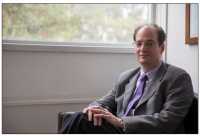
Mary Kay Lobo, PhD
Associate Professor
University of Maryland School of Medicine
Department of Anatomy and Neurobiology
Baltimore, MD 21201
MedicalResearch.com: What is the background for this study? What are the main findings?
Response: Altered energy balance has been studied in drug abuse but the fundamental source of energy, mitochondria, has not been well examined. In this study we found that a molecular regulator of mitochondrial fission (division) is increased in the nucleus accumbens, a major brain reward region, of rodents exposed to repeated cocaine and postmortem samples of cocaine dependent individuals. We further found that mitochondrial fission is increased in a nucleus accumbens neuron subtype in rodents that self-administer cocaine. Pharmacological blockade of mitochondrial fission can prevent physiological responses to cocaine in this neuron subtype while reducing cocaine-mediated behaviors. Finally, genetic reduction of mitochondrial fission in this neuron subtype in the nucleus accumbens can reduce drug (cocaine) seeking in rodents previously exposed to cocaine. In contrast, increasing mitochondrial fission, in this neuron subtype, enhances cocaine seeking behavior.
MedicalResearch.com Interview with:
Mary Kay Lobo, PhD
Associate Professor
University of Maryland School of Medicine
Department of Anatomy and Neurobiology
Baltimore, MD 21201
MedicalResearch.com: What is the background for this study? What are the main findings?
Response: Altered energy balance has been studied in drug abuse but the fundamental source of energy, mitochondria, has not been well examined. In this study we found that a molecular regulator of mitochondrial fission (division) is increased in the nucleus accumbens, a major brain reward region, of rodents exposed to repeated cocaine and postmortem samples of cocaine dependent individuals. We further found that mitochondrial fission is increased in a nucleus accumbens neuron subtype in rodents that self-administer cocaine. Pharmacological blockade of mitochondrial fission can prevent physiological responses to cocaine in this neuron subtype while reducing cocaine-mediated behaviors. Finally, genetic reduction of mitochondrial fission in this neuron subtype in the nucleus accumbens can reduce drug (cocaine) seeking in rodents previously exposed to cocaine. In contrast, increasing mitochondrial fission, in this neuron subtype, enhances cocaine seeking behavior.
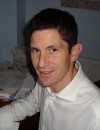


 Dr. Susanne Asu Wolf[/caption]
Susanne Asu Wolf PhD
Max-Delbrueck-Center for Molecular Medicine
Berlin, Germany
MedicalResearch.com: What inspired you to research this link between Ly6Chi monocytes, antibiotics and neurogenesis?
Dr. Wolf: As a neuroimmunologist I research the communication between the immune system and the brain. Amongst other research groups we found almost 10 years ago that T cells are needed to maintain brain homeostasis and plasticity, namely neurogenesis. Since only activated T cells enter the brain, we were looking for a mouse model, where immune cells are not activated. My former supervisor Polly Matzinger (NIH), a well-known immunologist, suggested to use germ free mice, born and raised in an isolator without any contact to a pathogen or any bacteria. I did a pilot experiment with the germ free mice, but wanted to get closer to possible applications in humans. Since humans are rarely born and raised in a sterile environment, I was looking for another model. By chance I met with the group of Bereswill and Heimesaat (Berlin, Charite) who provided me with a model, where due to prolonged treatment with an antibiotic cocktail, the microbiota are below detection level and the mice are also virtually germ free. They got me into contact with the second senior author of the paper Ildiko Dunay (University of Magdeburg). Her expertise is the function of Ly6Chi monocytes during infection with malaria or toxoplasmosis.
Now we were ready to investigate the gut-immune-brain axis with the focus on neurogenesis and cognition. Meanwhile the impact of the microbiome on behavior was reported by several research groups using “sterile” germ free mice and I was also curious if we could see similar differences in our antibiotic treated mice.
Dr. Susanne Asu Wolf[/caption]
Susanne Asu Wolf PhD
Max-Delbrueck-Center for Molecular Medicine
Berlin, Germany
MedicalResearch.com: What inspired you to research this link between Ly6Chi monocytes, antibiotics and neurogenesis?
Dr. Wolf: As a neuroimmunologist I research the communication between the immune system and the brain. Amongst other research groups we found almost 10 years ago that T cells are needed to maintain brain homeostasis and plasticity, namely neurogenesis. Since only activated T cells enter the brain, we were looking for a mouse model, where immune cells are not activated. My former supervisor Polly Matzinger (NIH), a well-known immunologist, suggested to use germ free mice, born and raised in an isolator without any contact to a pathogen or any bacteria. I did a pilot experiment with the germ free mice, but wanted to get closer to possible applications in humans. Since humans are rarely born and raised in a sterile environment, I was looking for another model. By chance I met with the group of Bereswill and Heimesaat (Berlin, Charite) who provided me with a model, where due to prolonged treatment with an antibiotic cocktail, the microbiota are below detection level and the mice are also virtually germ free. They got me into contact with the second senior author of the paper Ildiko Dunay (University of Magdeburg). Her expertise is the function of Ly6Chi monocytes during infection with malaria or toxoplasmosis.
Now we were ready to investigate the gut-immune-brain axis with the focus on neurogenesis and cognition. Meanwhile the impact of the microbiome on behavior was reported by several research groups using “sterile” germ free mice and I was also curious if we could see similar differences in our antibiotic treated mice.