

 Dr. Ledoux[/caption]
Andrée-Anne Ledoux, PhD
Children’s Hospital of Eastern Ontario Research Institute
Ottawa, Ontario, Canada
MedicalResearch.com: What is the background for this study?
Response: The natural recovery processes from a pediatric concussion remains poorly characterized throughout childhood. Children’s brains go through many phases of growth during development and sex differences exist. Therefore a 6-year-old child may not have the same recovery trajectory as an adolescent because of biopsychosocial differences. Thus, this study explored symptom improvement after concussion while considering these two key demographic factors. Understanding symptom improvement at different stages of development is important in order to provide the best possible care.
The study examined data from 2,716 children and adolescents who had presented at nine emergency departments across Canada and were diagnosed with concussion. We examined the natural progression of self-reported symptom recovery following pediatric concussion over the initial three months after injury. Participants in the study were aged 5 to 18 years old with acute concussion, enrolled from August 1, 2013, to May 31, 2015. We examined different age cohorts – 5 to 7 years of age, 8 to 12 years of age, and 13 to 18 years of age, and investigated how sex is associated with recovery.
Our study represents the largest study to evaluate symptom improvement trajectories in concussed pediatric population.
 Dr. Moseby-Knappe[/caption]
Marion Moseby-Knappe, MD
Neurologist and Researcher
Center for Cardiac Arrest at Lund University and
Skane University Hospital
Lund, Sweden
MedicalResearch.com: What is the background for this study? What are the main findings?
Response: Our research focuses on improving methods for examining unconscious patients treated on intensive care units after cardiac arrest. If a patient does not wake up within the first days after cardiac arrest, physicians need to evaluate how likely it is that the patient will awaken at all and to which extent there is brain injury.
According to European and American guidelines, decisions on further medical treatment of cardiac arrest patients should always be based on a combination of examinations and not only one single method. Various methods are combined when assessing the patient such as examining different neurologic reflexes, head scans (computed tomography or magnetic resonance imaging), other specialist examinations (electroencephalogram or somatosensory evoked potentials) or blood markers.
Our research focuses on patients included in the largest cardiac arrest trial to date, the Targeted Temperature Management after Out-of-Hospital Cardiac Arrest (TTM) Trial.
 Sean C. Rose, MD
Pediatric sports neurologist and co-director
Complex Concussion Clinic
Nationwide Children’s Hospital
Assistant professor of Pediatrics
The Ohio State University
MedicalResearch.com: What is the background for this study? What are the main findings?
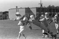
Response: The link between sub-concussive head impacts and declines in neurocognitive function has been reported by some studies, yet refuted by others. There is very little evidence that has been collected in children as they are sustaining these head impacts.
We initiated a multi-year study of youth football players to provide a more in-depth look at the question. We measured head impacts using helmet sensors during the 2016 football season. 112 players age 9-18 completed a battery of neurocognitive tests before and after the football season.
We found that neither the total burden of head impacts nor the intensity of individual impacts were associated with changes in testing performance from pre to post-season.
 Prof. Cooper[/caption]
Jamie Cooper AO
BMBS MD FRACP FCICM FAHMS
Professor of Intensive Care Medicine
Monash University
Deputy Director & Head of Research,
Intensive Care & Hyperbaric Medicine
The Alfred, Melbourne
MedicalResearch.com: What is the background for this study?
Response: 50-60 million people each year suffer a traumatic brain injury (TBI) . When the injury is severe only one half are able to live independently afterwards.
Cooling the brain (hypothermia) is often used in intensive care units for decades to decrease inflammation and brain swelling and hopefully to improve outcomes, but clinical staff have had uncertainty whether benefits outweigh complications.
We conducted the largest randomised trial of hypothermia in TBI, in 500 patients, in 6 countries, called POLAR. We started cooling by ambulance staff, to give hypothermia the best chance to benefit patients. We continued for 3-7 days in hospital ind ICU. We measured functional outcomes at 6 months.
 Dr. Ewing-Cobbs[/caption]
Dr. Linda Ewing-Cobbs PhD
Professor in the Department of Pediatrics
McGovern Medical School
University of Texas Health Science Center at Houston
Harriet and Joe Foster Chair in Cognitive Neuroscience
MedicalResearch.com: What is the background for this study? What are the main findings?
Response: Children may have long-lasting psychological and physical symptoms after an injury. Post-concussive symptoms (PCS) are nonspecific cognitive, physical, and mood symptoms such as difficulty concentrating, headache, and irritability. These symptoms occur in approximately 15 to 30% children after mild traumatic brain injury (TBI). Although PCS often resolve within one month, some children experience symptoms for longer periods of time.
 Sasank Chilamkurthy
AI Scientist,
Qure.ai
MedicalResearch.com: What is the background for this study?
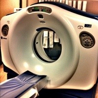
Response: Head CT scan is one of the most commonly used imaging protocols besides chest x-ray. They are used for patients with symptoms suggesting stroke, rise in intracranial pressure or head trauma. These manifest in findings like intracranial haemorrhage, midline shift or fracture.
Scans with these critical findings need to be read immediately. But radiologists evaluate the scans on first-come-first-serve basis or based on stat/routine markers set by clinicians. If the scans with critical findings are somehow pushed to the top of radiologists’ work list, it could substantially decrease time to diagnosis and therefore decrease mortality and morbidity associated with stroke/head trauma.

 Dr. Haarbauer-Krupa[/caption]
Juliet Haarbauer-Krupa, PhD
Senior Health Scientist
Division of Unintentional Injury Prevention
CDC
MedicalResearch.com: What is the background for this study?
 Dr. Hess[/caption]
Erik P. Hess MD MSc
Professor and Vice Chair for Research
Department of Emergency Medicine
UAB Medicine
he University of Alabama at Birmingham
Birmingham Alabama 35249
MedicalResearch.com: What is the background for this study? What are the main findings?
Response: 450,000 children present to U.S emergency departments each year for evaluation of head trauma. Physicians obtain head computed tomography (CT) scans in 37%-50% of these patients, with less than 10% showing evidence of traumatic brain injury and only 0.2% that require neurosurgical treatment.
In order to avoid unnecessary CT scans and to limit radiation exposure, the Pediatric Emergency Care Applied Research Network (PECARN) developed clinical prediction rules that consist of 6 readily available factors that can be assessed from the history and physical examination. If none of these risk factors are present, a CT scan is not indicated.
If either of 2 high risk factors such as signs of a skull fracture are present, CT scanning is indicated.
If 1 or 2 non-high risk factors are present, then either CT scanning or observation are recommended, depending on considerations such as parental preference, clinician experience and/or symptom progression.
In this study we designed a parent decision aid, “Head CT Choice” to educate the parent about the difference between a concussion – which does not show up on a CT scan – and a more serious brain injury causing bleeding in or around the brain. The decision aid also shows parents their child’s risk for a serious brain injury – less than 1% risk in the majority of patients in our trial – what to observe their child at home for should they opt not to obtain a CT scan, and the advantages and disadvantages of CT scanning versus home observation.
In our trial, we did not observe a difference in the rate of head CT scans obtained in the ED but did find that parents who were engaged in shared decision-making using Head CT Choice were more knowledgeable about their child’s risk for serious brain injury, has less difficulty making the decision because they were clearer about the advantages and disadvantages of the diagnostic options, and were more involved in decision-making by their physician. Parents also less frequently sought additional testing for their child within 1 week of the emergency department visit.

 Dr. Breiding[/caption]
Matt Breiding, PhD
Team Lead, Division of Unintentional Injury Prevention
Center for Disease Control and Prevention
MedicalResearch.com: What is the background for this study? What are the main findings?
Response: Caused by a bump, blow or jolt to the head or body, a mild traumatic brain injury (mTBI) can lead to short- or long-term problems that can affect how a child thinks, acts, feels, and learns. CDC’s Pediatric mTBI Guideline is based on the most comprehensive review of the science on pediatric mTBI diagnosis and management to date—covering 25 years of research.
The guideline consists of 19 sets of clinical recommendations that cover diagnosis, prognosis, and management and treatment. These recommendations are applicable to healthcare providers who care for pediatric patients with mTBI in all practice settings and outline actions healthcare providers can take to improve the health of their patients with this injury.
The CDC Pediatric mTBI Guideline outlines specific actions healthcare providers can take to help young patients and includes 5 key recommendations. Specifically, they recommend that physicians:
 Susanna Rosi, PhD
Director of Neurocognitive Research
Brain and Spinal Injury Center
Professor in the departments of Physical Therapy and Rehabilitation Science and of Neurological Surgery
UCSF
MedicalResearch.com: What is the background for this study? What are the main findings?
Response: NASA and private space companies like SpaceX plan to send humans to the red planet within the next 15 years — but among the major challenges facing future crewed space missions is how to protect astronauts from the dangerous cosmic radiation of deep space.
In this study we identified the first potential treatment for the brain damage caused by exposure to cosmic rays — a treatment can be given after exposure and that prevents memory impairment in mice exposed to simulated space radiation.
 Deborah E. Barnes, PhD, MPH
Professor, UCSF Weill Institute for Neurosciences
Departments of Psychiatry and Epidemiology & Biostatistics
University of California, San Francisco: http://profiles.ucsf.edu/deborah.barnes
Research Health Sciences Specialist
San Francisco VA Medical Center
MedicalResearch.com: What is the background for this study? What are the main findings?
 Dr. Jeff Russell[/caption]
Jeffrey A. (Jeff) Russell, PhD, AT, FIADMS
Science and Health in Artistic Performance
Division of Athletic Training, School of Applied Health Sciences and Wellness
Ohio University
Athens, OH 45701
MedicalResearch.com: What is the background for this study? What are the main findings?
Response: Everyone knows that enormous attention is given to concussions in sports today. Those involved in performing arts experience many head impacts, too; yet, they receive neither the attention nor the specialized care for concussions that athletes do.
At Ohio University’s Clinic for Science and Health in Artistic Performance (SHAPe Clinic) that I direct, we were seeing a number of theater students suffer concussions. So, Brooke Daniell and I decided explore this trend more closely. This is the first known published research to evaluate the prevalence of head impacts in theater personnel. In the sample we studied, which comprised predominantly those involved in various aspects of theater production, the prevalence of receiving at least one head impact in a theater career was 67%. Of those who sustained at least one head impact from theater, 77% reported three or more head impacts, and 39% reported more than five impacts. More troubling, of those who said they had received a head impact that was accompanied by concussion-like symptoms, 70% indicated that they continued their work, and half of those did not report the incident to anyone.
 Dr. Christopher Abeare[/caption]
Christopher Abeare, Ph.D.
Associate Professor
Clinical Neuropsychology
Department of Psychology
University of Windsor
Windsor, Ontario
MedicalResearch.com: What is the background for this study? What are the main findings?
Response: In this study, we examined the prevalence of invalid performance on baseline neurocognitive testing in sport concussion. Baseline testing is a commonly employed practice in which the cognitive abilities of athletes are assessed pre-season. These baseline test results are then used as a point of comparison against which post-injury neurocognitive test results can be compared, thereby creating a more individualized approach to the assessment of neurocognitive functioning.
However, there has been growing concern about the validity of baseline test results, meaning that there is concern over the degree to which the scores on these baseline tests actually reflect an athlete’s true cognitive ability. There are many reasons why their test scores might not reflect their actual ability, ranging from inattentiveness during testing and lack of appreciation of the importance of doing their best on testing to intentional underperformance (aka “sandbagging” or malingering).
As a result of these concerns, 4 different validity measures have been developed. We compared these 4 validity measures, head to head, in a sample of 7897 athletes aged 10 to 21 years.
We found that 56% of athletes failed at least 1 of these validity measures, suggesting that as many as 56% of athletes have scores that may not reflect their true ability level. We then tested the hypothesis that age would be related to the proportion of athletes with invalid performance. Our findings supported this hypothesis in that nearly 84% of 10-year-olds failed at least one validity measure and 29% of 21-year-olds failed at least one.
 Dr Chris Gale
Clinical Senior Lecturer in Neonatal Medicine
Imperial College London and
Consultant Neonatologist at Chelsea and Westminster Hospital
NHS Foundation Trust
MedicalResearch.com: What is the background for this study? What are the main findings?
Response: As part of a drive to make England a safer place to give birth, the Department of Health in England has set a target of reducing the number of babies that incur brain injury during or soon after birth by 20% by 2020 and to halve them by 2030.
Before now United Kingdom health services did not have a standard definition of brain injury in babies and there has been no systematic collection of data for this purpose. With colleagues and in collaboration with the Department of Health, we have devised a practical way to measure the incidence rate of brain injury in babies using routinely recorded data held in the National Neonatal Research Database.
The research estimated that 3,418 babies suffered conditions linked to brain injury at or soon after birth in 2015, which equates to an incidence rate of 5.14 per 1,000 live births. For preterm births (babies born at or less than 37 weeks) the rate was 25.88 per 1,000 live births in 2015, almost six times greater than the rate for full-term births, which was 3.47 per 1,000 live births.
Overall, the research found that the most common type of condition that contributed brain injuries was damage caused by lack of oxygen to the brain, called hypoxic ischaemic encephalopathy; this is seen mainly in term babies. For preterm babies, the largest contributor to brain injuries is from bleeding into and around the ventricles of the brain, a condition called periventricular haemorrhage.
It is also the first time that brain injuries in babies have been measured using data gathered routinely during day to day clinical care on NHS neonatal units. The use of routine data required no additional work for clinical staff and provides a valuable way to measure the effectiveness of interventions to reduce brain injury.
 Dr. Hicks[/caption]
Dr. Steven D. Hicks, M.D., Ph.D
Penn State Health
MedicalResearch.com: What is the background for this study? What are the main findings?
Response: Previous research has shown that small epigenetic molecules called microRNAs are altered in the blood after a traumatic brain injury. Our own pilot research showed that microRNAs were also changed in the saliva after brain injury and that some of these changes mirrored changes in cerebrospinal fluid. In this study we investigated whether salivary microRNA patterns after a concussion could be used to predict the duration and character of symptoms one month after injury.
We found that levels of five microRNAs predicted presence of symptoms one month later with greater accuracy (~85%) than standard surveys of symptom burden (~65%). Interestingly, one of the predictive salivary microRNAs (miR-320c) targets pathways involved in synaptic plasticity and was significantly correlated with attention difficulties one month after concussive injury.
 Prof J van der Naalt[/caption]
Prof.dr. J van der Naalt PhD
Department of Neurology University Medical Center Groningen
Groningen, The Netherlands
MedicalResearch.com: What is the background for this study? What are the main findings?
Response: Mild traumatic brain injury occurs frequently and is one of the leading cause of morbidity in adults worldwide. It is a major social-economic problem with one in three patients had persistent complaints several months after injury that interfere with resumption of daily activities and work.
One of the most important questions concerns the finding that some patients recover without complaints and others do not after sustaining a mild traumatic brain injury. In a follow-up study with more than 1000 participants we found that personality factors are a major factor in the recovery process. In particular coping, that is the way patients adapt to persistent complaints, is important next to emotional distress and impact of the injury.
In an add-on study with fMRI we found that in the early phase after injury, the interaction between specific brain networks was temporarily changed. However, when regarding persistent posttraumatic complaints , specific personality characteristics significantly determine long term outcome.
 Dr. Cusimano[/caption]
Michael D. Cusimano MD, FRCSC, DABNS, FACS, PhD, MHPE
Adjunct Scientist in the Keenan Research Centre of the Li Ka Shing Knowledge Institute of St. Michael's Hospital
Division of Neurosurgery, St. Michael\'s Hospital
Professor of Neurosurgery, Education and Public Health
University of Toronto
MedicalResearch.com: What is the background for this study?
Response: Baseball is played by millions annually and is traditionally seen as a low risk sport for head injury when compared to sports like American Football, Ice Hockey and Rugby. Over 6 million children and youth are enrolled in formal baseball or softball leagues annually.
 Dr. Yang[/caption]
Jingzhen (Ginger) Yang, PhD, MPH
Principal Investigator
Associate Professor, Center for Injury Research and Policy
The Research Institute at Nationwide Children’s Hospital
Dept. of Pediatrics, College of Medicine, The Ohio State University
Columbus, Ohio 43205
MedicalResearch.com: What is the background for this study?
Response: From 2009-2014, all 50 states and the District of Columbia passed their state TBI laws, more commonly known as concussion laws, to mitigate severe consequences of concussions.
These laws often include 3 core components:
(1) mandatory removal from play following actual or suspected concussions,
(2) requirements to receive clearance to return to play from a licensed health professional, and
(3) education of coaches, parents, and athletes regarding concussion symptoms and signs.
Our study aimed to evaluate whether the laws achieve the intended impact.
MedicalResearch.com: What are the main findings?
Response: The main findings showed that:
 Dr. Adrian Harel[/caption]
Dr. Adrian Harel, PhD
Chief Executive Officer
Medicortex Finland
MedicalResearch.com: What is the background for this study? What are the main findings?
Response: Every 15 seconds, someone in the United States suffers a new head injury. Of the 2.5M people treated in hospital emergency rooms each year, 80,000 become permanently disabled because of TBI. Currently, there are no reliable diagnostic tests to assess the presence or severity of an injury on-site, nor are there any pharmaceutical therapies that could stop the secondary injury from spreading. Accurate diagnostics would benefit especially mild cases of TBI (concussions), which, if occurring repeatedly, may cause neurodegenerative conditions such as Chronic Traumatic Encephalopathy (which is typical for athletes in NFL and Ice-hockey).
We have performed extensive preclinical research comparing fluid biopsies from normal and injured lab animals. The results showed some unique biomarkers released as a biodegradation products after head injury. The data served as the basis and confirmation for our patent applications to protect the biomarker concept.
Medicortex has completed a clinical proof-of-concept trial in collaboration with Turku University Hospital (Tyks). Samples from 12 TBI patients and 12 healthy volunteers were collected and analyzed for the presence and for the level of the biomarker in state-of-the-art laboratories. The study demonstrated the diagnostic potential of the new biomarker in humans and it confirmed the prior preclinical findings. This was a significant milestone for Medicortex.
 Dr. Virendra Mishra[/caption]
Virendra Mishra, Ph.D.
Department of Imaging Research
Cleveland Clinic Lou Ruvo Center for Brain Health in Las Vegas
MedicalResearch.com: What is the background for this study? What are the main findings?
Response: Repetitive head trauma has been shown to be a risk factor for various neurodegenerative disorders, mood swings, depression and chronic traumatic encephalopathy. There has been a significant amount of research into identifying an imaging biomarker of mild traumatic brain injury (mTBI) due to repetitive head trauma. Unfortunately, most of the biomarkers have not been able to find a successful translation to clinics. Additionally, the quest for the mTBI imaging biomarker especially using Magnetic Resonance Imaging (MRI) techniques has been done by looking at either the gray matter (T1-weighted) or the white matter (Diffusion Tensor Imaging) independently; and both have shown changes that are associated with repetitive head trauma.
Hence in this study, we wanted to investigate if combining gray matter and white matter information enables us to better predict the fighters who are more vulnerable to cognitive decline due to repetitive head trauma. Our method found seven imaging biomarkers that when combined together in a multivariate sense were able to predict with greater than 73% accuracy those fighters who are vulnerable to cognitive decline both at baseline and follow-up. The imaging biomarkers were indeed a combination of gray and white matter measures of regions reported previously in the literature. A key point in our study was we found the regions predicting cognitive decline without enforcing any assumptions on the regions previously reported.
 Dr. Daneshvar[/caption]
Daniel H. Daneshvar, M.D., Ph.D.
Chronic Traumatic Encephalopathy Center
Team Up Against Concussions | Founder
Boston University
MedicalResearch.com: What is the background for this study? What are the main findings?
Response: Chronic traumatic encephalopathy (CTE) is a neurodegenerative disease associated with repetitive head impacts. CTE was first described in JAMA in 1928. In the 99 years since, just over 100 cases of CTE have been described in the world’s literature.
This study nearly doubles the number of reported cases of CTE, with 177 cases of CTE in football players. Of note, 110 of the 111 athletes who played in the NFL had CTE. This study represents the largest and the most methodologically rigorous description of a series of patients with CTE ever published. Such a richness of data regarding the clinical and pathological features of CTE has never been previously compiled. As such, this study represents an important advance to the medical literature and an enormous scientific advance in our understanding of chronic traumatic encephalopathy.
 Dr. Hill[/caption]
Dr Lisa J Hill PhD
Institute of Inflammation and Ageing
Research Fellow
Neuroscience and Ophthalmology
Institute of Inflammation and Ageing
College of Medical and Dental Sciences
University of Birmingham UK
MedicalResearch.com: What is the background for this study? What are the main findings?
Response: Traumatic brain injury (TBI) is the leading cause of death and disability among young adults and, according to the World Health Organization, by 2020 TBI will become the world’s leading cause of neurological disability across all age groups. Early and correct diagnosis of traumatic brain injury is one of the most challenging aspects faced by clinicians. Being able to detect compounds in the blood that help to determine how severe the brain injury is would be of great benefit to patients and aid in their treatment. Inflammatory markers are particularly suited for biomarker discovery as TBI leads to very early alterations in inflammatory proteins. The discovery of reliable biomarkers for the management of TBI would improve clinical interventions.
We collected blood samples from 30 injured patients within the first hour of injury prior to the patient arriving at hospital and analysed them. Analysis of protein biomarkers from blood taken within the first hour of injury has never been carried out until now. We used a panel of 92 inflammation-associated human proteins when analysing the blood samples. The analysis identified three inflammatory proteins, known as CST5, AXIN1 and TRAIL, as novel biomarkers of TBI.
 Dr. Hicks[/caption]
Steven Daniel Hicks, MD, PhD
Assistant Professor, Division of Academic General Pediatrics
College of Medicine
Penn State Health
MedicalResearch.com: What is the background for this study? What are the main findings?
Response: There are about 3 million concussions in the US each year and the majority occur in children. Parents of children with concussions commonly cite length of recovery as a major concern, but pediatricians have no objective or accurate tests for addressing this concern.
Our research group previously identified small regulatory molecules called microRNAs that were altered in both the spinal fluid and saliva in children with traumatic brain injuries. In this study we investigated whether those microRNAs could predict duration of concussion symptoms. In 52 children with concussion we found a set of microRNAs that predict whether concussion symptoms would last beyond one month with over 80% accuracy. This was significantly more accurate than survey based tools such as the sports concussion assessment tool or a modified concussion clinical risk score. Interestingly, the microRNAs with predictive accuracy targeted pathways involved in brain repair and showed correlations with specific concussion symptoms.
 Dr. Sergey Dryga[/caption]
Sergey A. Dryga, PhD, MBA
Chief Scientific Officer
BioDirection, Inc.
MedicalResearch.com: What is the background for this study? What are the main findings?
Response: When patients have suffered a head injury, they typically undergo a series of subjective cognitive tests to confirm a diagnosis of a concussion or other traumatic brain injury. In many cases these tests are inaccurate and inconsistent, increasing the risk of misdiagnosis. In other cases, patients may undergo an unnecessary CT scan, which is costly and exposes them to radiation. Early, objective diagnostic testing of patients who have experienced a head injury can support more rapid and appropriate treatment decisions while potentially reducing the use of unnecessary CT scans or other forms of intervention.
We know that protein biomarkers, including S100 calcium binding protein beta (S100β) and glial fibrillary acidic protein (GFAP), are released from the brain into the bloodstream immediately following a concussion or other traumatic brain injury. The Tbit™ System is a new medical device based on a nanotechnology biosensor that rapidly detects and accurately measures these protein biomarkers. The system includes a disposable cartridge and portable analyzer designed for testing using a single drop of blood at the earliest stages of a concussion.
This pre-clinical study was designed to evaluate the ability of the Tbit System to screen traumatic brain injury patients for a CT positive or CT negative test. Frozen plasma samples were collected from a total of 100 patients who had undergone CT scans post hospital admission. The Tbit System demonstrated 100% sensitivity with no false negative results, and a 41% specificity level.
 Emily Dennis[/caption]
Emily Dennis PhD
Postdoctoral Scholar
Imaging Genetics Center
Mark and Mary Stevens Neuroimaging and Informatics Institute
USC
MedicalResearch.com: What is the background for this study? What are the main findings?
Response: We know that there is heterogeneity in outcome post-traumatic brain injury (TBI), but we generally think of this as a continuous variable - with most patients falling in the middle and only a few at the extremes in terms of recovery process and outcome.
Our main finding was that interhemispheric transfer time (IHTT - the time it takes for information to move from one hemisphere of the brain to the other) identified 2 subgroups of TBI patients - those with slow IHTT and those with normal IHTT. These two groups show differences in cognitive function and brain structure, with the IHTT slow group showing structural disruptions that become progressively worse while the IHTT normal group seems to be recovering from the injury.
 Dr. Hsu[/caption]
Wellington K. Hsu, MD
Clifford C. Raisbeck, MD, Professor of Orthopaedic Surgery
Associate Professor of Orthopaedic Surgery
Northwestern University
Chicago, IL
MedicalResearch.com: What is the background for this study? What are the main findings?
Response: Concussions remains a significant problem in youth sports. The recent enactment of Traumatic brain injury laws have certainly heightened awareness regarding this problem. Our study looked at publicly available data regarding diagnosis of concussion in high school athletes. We found that females are more likely to be diagnosed with a concussion than males. We also concluded that girl soccer players and boys football players are at highest risk for a diagnosis of concussion. Since the neck meant of the Traumatic brain injury state laws, the diagnosis of concussion in this patient group increased significantly past decade.
 Dr. Ujwal Chaudhary[/caption]
Dr. Ujwal Chaudhary, PhD
Institute of Medical Psychology and Behavioral Neurobiology
University of Tübingen
Tübingen, Germany
MedicalResearch.com: What is the background for this study?
Response: Amyotrophic Lateral Sclerosis (ALS) is a progressive neurodegenerative disorder which causes an Individual to be in Locked-in state (LIS), i.e. the patients have control of their vertical eye movement and blinking, and ultimately in Completely Locked-in state (CLIS), i.e, no control over their eye muscle. There are several assistive and augmentative (AAC) technology along with EEG based BCI which can be used be by the patients in LIS for communication but once they are in CLIS they do not have any means of communication. Hence, there was a need to find an alternative learning paradigm and probably another neuroimaging technique to design a more effective BCI to help ALS patient in CLIS with communication.
 Dr. Julianne Schmidt[/caption]
Julianne Schmidt, PhD, ATC
Assistant Professor
Department of Kinesiology
The University of Georgia
Athens GA
MedicalResearch.com: What is the background for this study? What are the main findings?
Response: Less than half of all people with a concussion intend to reduce their driving at any point.
Current recommendations surrounding concussion focus on when it is safe to return to sport or return to the classroom, but return to driving is usually ignored and has not been studied.