
infections Tag
15 Nov High Treatment Failure Rates Among Elderly With Purulent Skin Infections
MedicalResearch.com Interview with: [caption id="attachment_29656" align="alignleft" width="150"] Dr. John P. Haran[/caption]
John P. Haran, MD
Assistant Professor
Department of Emergency Medicine
University of Massachusetts Medical School
UMass Memorial Medical Group
Worcester, MA
MedicalResearch.com: What is the background for this study?
Response: In 2014, the Infectious Disease Society of America (IDSA) updated their guidelines for the management of skin and soft tissue infection in response to high MRSA infection rates as well as high treatment failure rates for skin and soft tissue infections. Greater than 1 in 5 patients treated for a skin abscess will fail initial treatment.
Historically antibiotics have been shown to be unnecessary in the treatment of uncomplicated purulent infections. This notion has been recently challenges when authors published a randomized control trial using trimethoprim-sulfamethoxazone in the NEJM that demonstrated a minimal increase in cure rates for outpatient treatment of uncomplicated skin purulent skin infections. In this study they did not follow IDSA-guidelines nor model or stratify their analysis. It is possible their findings may be due to at-risk patient groups that did not receive antibiotics. Many widely used clinical decision rules incorporate age into their decision algorithms, however the IDSA did not do this with their recent guidelines.
Dr. John P. Haran[/caption]
John P. Haran, MD
Assistant Professor
Department of Emergency Medicine
University of Massachusetts Medical School
UMass Memorial Medical Group
Worcester, MA
MedicalResearch.com: What is the background for this study?
Response: In 2014, the Infectious Disease Society of America (IDSA) updated their guidelines for the management of skin and soft tissue infection in response to high MRSA infection rates as well as high treatment failure rates for skin and soft tissue infections. Greater than 1 in 5 patients treated for a skin abscess will fail initial treatment.
Historically antibiotics have been shown to be unnecessary in the treatment of uncomplicated purulent infections. This notion has been recently challenges when authors published a randomized control trial using trimethoprim-sulfamethoxazone in the NEJM that demonstrated a minimal increase in cure rates for outpatient treatment of uncomplicated skin purulent skin infections. In this study they did not follow IDSA-guidelines nor model or stratify their analysis. It is possible their findings may be due to at-risk patient groups that did not receive antibiotics. Many widely used clinical decision rules incorporate age into their decision algorithms, however the IDSA did not do this with their recent guidelines.
17 Oct Travelers Often Bring Home Resistant Bacteria From Endemic Areas
MedicalResearch.com Interview with: [caption id="attachment_28922" align="alignleft" width="180"] Dr. John Penders[/caption]
John Penders PhD
Assistant professor
Dept. of Medical Microbiology
Maastricht University, Maastricht
MedicalResearch.com: What is the background for this study?
Response: Antimicrobial resistance (AMR) constitutes an increasingly important human health hazard worldwide. Especially, the rapid emergence and global spread of multidrug resistant Enterobacteriaceae is worrisome. These bacteria often produce enzymes like extended-spectrum beta-lactamases (ESBLs) and carbapenemases, which inactivate most beta-lactam antibiotics, and are often co-resistant to multiple other antibiotic classes. Consequently, treatment options for infections with multidrug resistant Enterobacteriaceae are limited.
The prevalence of antimicrobial-resistant bacteria is generally higher in low and middle income countries as a result of inappropriate use of antimicrobial agents, overcrowding and lack of hygiene and infection control measures. The exponential increase of international travel to such endemic areas may substantially contribute to the emergence and spread of AMR as it allows resistant bacteria to be rapidly transported between regions.
Indeed several previous studies had already indicated that international travel is a major risk factor for colonisation with ESBL-producing Enterobacteriaceae. However, many questions remained unanswered, such as the travel destinations and potential risk-behaviour that provide the highest risk for colonisation, how long travellers remain colonised after they return and whether they can transmit these resistant bacteria to other people within their household once they returned from their travel.
That is why we initiated the largest prospective study on the acquisition and spread of multidrug resistant bacteria in returning travellers. This multicenter study, conducted by Maastricht University Medical Center, Erasmus University Medical Center, Academic Medical Center Amsterdam, Havenziekenhuis and Utrecht University, included 2,001 travellers and 215 household members. Fecal samples of these participants were collected before and immediately after travel as well as 1, 3, 6 and 12 months after travel return and screened for the prescence of multidrug resistant Enterobacteriaceae. Moreover, extensive information on demographics, travel details, illnesses and behaviour were collected at all follow-up moments by repeated questionnaires.
Dr. John Penders[/caption]
John Penders PhD
Assistant professor
Dept. of Medical Microbiology
Maastricht University, Maastricht
MedicalResearch.com: What is the background for this study?
Response: Antimicrobial resistance (AMR) constitutes an increasingly important human health hazard worldwide. Especially, the rapid emergence and global spread of multidrug resistant Enterobacteriaceae is worrisome. These bacteria often produce enzymes like extended-spectrum beta-lactamases (ESBLs) and carbapenemases, which inactivate most beta-lactam antibiotics, and are often co-resistant to multiple other antibiotic classes. Consequently, treatment options for infections with multidrug resistant Enterobacteriaceae are limited.
The prevalence of antimicrobial-resistant bacteria is generally higher in low and middle income countries as a result of inappropriate use of antimicrobial agents, overcrowding and lack of hygiene and infection control measures. The exponential increase of international travel to such endemic areas may substantially contribute to the emergence and spread of AMR as it allows resistant bacteria to be rapidly transported between regions.
Indeed several previous studies had already indicated that international travel is a major risk factor for colonisation with ESBL-producing Enterobacteriaceae. However, many questions remained unanswered, such as the travel destinations and potential risk-behaviour that provide the highest risk for colonisation, how long travellers remain colonised after they return and whether they can transmit these resistant bacteria to other people within their household once they returned from their travel.
That is why we initiated the largest prospective study on the acquisition and spread of multidrug resistant bacteria in returning travellers. This multicenter study, conducted by Maastricht University Medical Center, Erasmus University Medical Center, Academic Medical Center Amsterdam, Havenziekenhuis and Utrecht University, included 2,001 travellers and 215 household members. Fecal samples of these participants were collected before and immediately after travel as well as 1, 3, 6 and 12 months after travel return and screened for the prescence of multidrug resistant Enterobacteriaceae. Moreover, extensive information on demographics, travel details, illnesses and behaviour were collected at all follow-up moments by repeated questionnaires.
12 Oct Screening Programs For Congenital CMV Found To Be Cost-Effective
MedicalResearch.com Interview with: [caption id="attachment_28765" align="alignleft" width="97"] Dr. Soren Gantt[/caption]
Dr. Soren Gantt MD, PhD, MPH
Investigator, BC Children's Hospital
Associate Professor, Department of Infectious and Immunological Diseases (Pediatrics)
Faculty of Medicine, University of British Columbia
MedicalResearch.com: What is the background for this study? What are the main findings?
Response: Cytomegalovirus (CMV) is a common virus that is usually transmitted through bodily fluids such as saliva, urine, blood, and breast milk, but it can also cause congenital infection (from a pregnant woman to her fetus). While it doesn’t usually cause problems for most children or adults, congenital CMV often causes serious problems. Congenital CMV causes 25 per cent of all childhood hearing loss and it’s the second most common cause of intellectual disability. Without screening, most infected newborns are not diagnosed in time to treat them with antivirals or provide other care that can make a big difference to improving their life-long outcomes.
Our study showed that screening programs for congenital CMV infection are cost-effective. We found that the cost of identifying one case of congenital CMV ranges from US$2000 to US$10,000 through universal screening, or US$566 to $2833 through a targeted screening approach. Our model showed that screening programs resulted in a net savings for the health care system of approximately USD$21 to $32 per newborn for universal screening or USD$11-$27 per newborn for targeted screening by reducing lifetime costs for therapies and lost productivity due to CMV-related health problems.
This finding addresses a major barrier to implementing CMV screening programs, as costs have often been viewed as an issue.
Dr. Soren Gantt[/caption]
Dr. Soren Gantt MD, PhD, MPH
Investigator, BC Children's Hospital
Associate Professor, Department of Infectious and Immunological Diseases (Pediatrics)
Faculty of Medicine, University of British Columbia
MedicalResearch.com: What is the background for this study? What are the main findings?
Response: Cytomegalovirus (CMV) is a common virus that is usually transmitted through bodily fluids such as saliva, urine, blood, and breast milk, but it can also cause congenital infection (from a pregnant woman to her fetus). While it doesn’t usually cause problems for most children or adults, congenital CMV often causes serious problems. Congenital CMV causes 25 per cent of all childhood hearing loss and it’s the second most common cause of intellectual disability. Without screening, most infected newborns are not diagnosed in time to treat them with antivirals or provide other care that can make a big difference to improving their life-long outcomes.
Our study showed that screening programs for congenital CMV infection are cost-effective. We found that the cost of identifying one case of congenital CMV ranges from US$2000 to US$10,000 through universal screening, or US$566 to $2833 through a targeted screening approach. Our model showed that screening programs resulted in a net savings for the health care system of approximately USD$21 to $32 per newborn for universal screening or USD$11-$27 per newborn for targeted screening by reducing lifetime costs for therapies and lost productivity due to CMV-related health problems.
This finding addresses a major barrier to implementing CMV screening programs, as costs have often been viewed as an issue.
30 Sep Triclosan-Containing Sutures Reduced Surgical Infections in Children
MedicalResearch.com Interview with: Dr Marjo Renko MD PEDEGO Research Unit University of Oulu Oulu, Finland MedicalResearch.com: What is the background for this study? What are the main findings? Response: For over a decade there has been suture materials containing antiseptic agent in the market. Trials in adults have shown some possible benefits from these sutures as in some studies they have reduced occurrence of surgical site infections. Only one small study had so far been published in children and thus we decided to carry out a large trial comparing sutures containing triclosan with ordinary ones. Our trial included over 1500 children who came to Oulu University Hospital for surgery. Surgical site infections were carefully monitored. Surgical site infections occurred in 2.6 % of the children who received absorbing sutures containing triclosan while that occurred in 5,4 % of the children who received ordinary sutures.28 Sep Excess Zinc May Predispose to C.diff By Altering Gut Microbiome
MedicalResearch.com Interview with: [caption id="attachment_28403" align="alignleft" width="147"] Dr. Eric P Skaar,[/caption]
Eric P Skaar, Ph.D., MPH
Director, Division of Molecular Pathogenesis
Ernest W. Goodpasture Professor of Pathology
Vice Chair for Basic Research, Department of Pathology, Microbiology, and Immunology
Vanderbilt University School of Medicine
MedicalResearch.com: What is the background for this study? What are the main findings?
Response: Nutrient metals are known to be a critical driver of the outcome of host-pathogen interactions, and C. difficile is the most common cause of hospital-acquired infections. C. difficile infection typically occurs following antibiotic-mediated disruption of the healthy microbiome. We were interested in learning how nutrient metals can shape the microbiome and impact the outcome of Clostridium difficile infection.
We found that excess zinc alters the structure of the microbiome and increases the severity of C. difficile infection in mice.
Dr. Eric P Skaar,[/caption]
Eric P Skaar, Ph.D., MPH
Director, Division of Molecular Pathogenesis
Ernest W. Goodpasture Professor of Pathology
Vice Chair for Basic Research, Department of Pathology, Microbiology, and Immunology
Vanderbilt University School of Medicine
MedicalResearch.com: What is the background for this study? What are the main findings?
Response: Nutrient metals are known to be a critical driver of the outcome of host-pathogen interactions, and C. difficile is the most common cause of hospital-acquired infections. C. difficile infection typically occurs following antibiotic-mediated disruption of the healthy microbiome. We were interested in learning how nutrient metals can shape the microbiome and impact the outcome of Clostridium difficile infection.
We found that excess zinc alters the structure of the microbiome and increases the severity of C. difficile infection in mice.
17 Sep Resistant E. Coli Produces Slimy Coating To Prevent Removal By Immune System
MedicalResearch.com Interview with: [caption id="attachment_28049" align="alignleft" width="133"] Dr. Daria Van Tyne[/caption]
Dr. Daria Van Tyne, PhD
The Gilmore Lab
Department of Ophthalmology
Harvard Medical School
Massachusetts Eye and Ear Infirmary
Boston, Massachusetts
MedicalResearch.com: What is the background for this study? What are the main findings?
Response: A specific clone of E. coli, type ST131, which produces an extended-spectrum beta-lactamase (ESBL – an enzyme that inactivates many penicillin-type antibiotics), has rapidly spread around the globe to become the leading cause of multidrug-resistant, non-intestinal E. coli infection. Despite this, E. coli is a rare cause of infection of the cornea. A patient was recently seen at the Massachusetts Eye and Ear Infirmary with a severe E. coli infection of the cornea, and the large number of antibiotic resistances of this strain tipped us off to the possibility that it might be the highly virulent ST131 ESBL type. By sequencing the DNA of its genome, we found that it was indeed ST131 ESBL E. coli. Moreover, we discovered a new mutation in this strain that allows it to produce a slimy outer coating on its surface. This slime layer, or capsule, makes the bacteria more resistant to removal by phagocytic cells of the immune system. The slime layer also makes these particular colonies appear different on a special type of agar that contains the dye Congo Red.
Dr. Daria Van Tyne[/caption]
Dr. Daria Van Tyne, PhD
The Gilmore Lab
Department of Ophthalmology
Harvard Medical School
Massachusetts Eye and Ear Infirmary
Boston, Massachusetts
MedicalResearch.com: What is the background for this study? What are the main findings?
Response: A specific clone of E. coli, type ST131, which produces an extended-spectrum beta-lactamase (ESBL – an enzyme that inactivates many penicillin-type antibiotics), has rapidly spread around the globe to become the leading cause of multidrug-resistant, non-intestinal E. coli infection. Despite this, E. coli is a rare cause of infection of the cornea. A patient was recently seen at the Massachusetts Eye and Ear Infirmary with a severe E. coli infection of the cornea, and the large number of antibiotic resistances of this strain tipped us off to the possibility that it might be the highly virulent ST131 ESBL type. By sequencing the DNA of its genome, we found that it was indeed ST131 ESBL E. coli. Moreover, we discovered a new mutation in this strain that allows it to produce a slimy outer coating on its surface. This slime layer, or capsule, makes the bacteria more resistant to removal by phagocytic cells of the immune system. The slime layer also makes these particular colonies appear different on a special type of agar that contains the dye Congo Red.
12 Sep Roundworm Infection Transmitted By Raccoons Has Serious Neurologic Complications
MedicalResearch.com Interview with: [caption id="attachment_27834" align="alignleft" width="193"] Dr. Anita Sircar[/caption]
Anita D. Sircar, MD
Epidemic Intelligence Service
Division of Parasitic Diseases and Malaria
Center for Global Health
CDC
MedicalResearch.com: What is the background for this study?
Response: Baylisascaris procyonis is a roundworm commonly found in raccoons. It can be found anywhere in the United States where raccoons live. People, especially children, can be infected by this roundworm when they accidentally ingest contaminated raccoon feces. Infection with Baylisascaris procyonis can have severe outcomes in people such as blindness and even death if not treated promptly.
Despite expansion of the geographic distribution of Baylisascaris procyonis in the last 14 years and probable increasing human exposure, baylisascariasis is likely an underreported disease: only 22 documented cases were reported in the United States during 1973–2010.
During May 2013–December 2015, seven additional cases of baylisascariasis were identified among patients in the United States through testing at CDC, including six cases of central nervous system disease and one of ocular disease. Laboratory and clinical information for each patient was gathered and reviewed in a case series to contribute to knowledge about Baylisascaris procyonis infection. All seven patients survived, although approximately half had residual neurologic sequelae.
Dr. Anita Sircar[/caption]
Anita D. Sircar, MD
Epidemic Intelligence Service
Division of Parasitic Diseases and Malaria
Center for Global Health
CDC
MedicalResearch.com: What is the background for this study?
Response: Baylisascaris procyonis is a roundworm commonly found in raccoons. It can be found anywhere in the United States where raccoons live. People, especially children, can be infected by this roundworm when they accidentally ingest contaminated raccoon feces. Infection with Baylisascaris procyonis can have severe outcomes in people such as blindness and even death if not treated promptly.
Despite expansion of the geographic distribution of Baylisascaris procyonis in the last 14 years and probable increasing human exposure, baylisascariasis is likely an underreported disease: only 22 documented cases were reported in the United States during 1973–2010.
During May 2013–December 2015, seven additional cases of baylisascariasis were identified among patients in the United States through testing at CDC, including six cases of central nervous system disease and one of ocular disease. Laboratory and clinical information for each patient was gathered and reviewed in a case series to contribute to knowledge about Baylisascaris procyonis infection. All seven patients survived, although approximately half had residual neurologic sequelae.
07 Sep Inflammatory Molecule Links Metabolic Environment and Innate Immunity
MedicalResearch.com Interview with: [caption id="attachment_27723" align="alignleft" width="142"] Dr. David Underhill[/caption]
David Underhill, PhD
Professor of Biomedical Sciences
Research scientist, F. Widjaja Foundation Inflammatory Bowel and Immunobiology Research Institute
Cedars-Sinai
Los Angeles, CA
MedicalResearch.com: What is the background for this study?
Response: “Innate immunity” is the body’s natural resistance to microbial infection and stands in contrast to “adaptive immunity,” which is the body’s learned response to infection (e.g. antibodies and vaccines). In the standard model of innate immunity that has emerged over the last several decades, scientists have come to understand that the human genome encodes many “receptors” that have evolved as sensors for specific common microbial molecules, such as bacterial or viral DNA or components of bacterial or fungal cell walls. The job of these receptors is to survey the environment (skin, blood, etc.) for potentially dangerous microbes and initiate inflammatory responses if they are found. These activities are essential for defense against infection, and people and animals with defects in these sensors or the responses they trigger can be susceptible to infection.
My laboratory has been interested for more than a decade in identifying these innate sensors and the microbial targets that they recognize. In this study, we were looking for the sensor that allows white blood cells (e.g. macrophages and dendritic cells) to detect Gram-positive bacterial cell walls and trigger a specific inflammatory response: secretion of the potent inflammatory mediator interleukin-1β (IL-1β).
Dr. David Underhill[/caption]
David Underhill, PhD
Professor of Biomedical Sciences
Research scientist, F. Widjaja Foundation Inflammatory Bowel and Immunobiology Research Institute
Cedars-Sinai
Los Angeles, CA
MedicalResearch.com: What is the background for this study?
Response: “Innate immunity” is the body’s natural resistance to microbial infection and stands in contrast to “adaptive immunity,” which is the body’s learned response to infection (e.g. antibodies and vaccines). In the standard model of innate immunity that has emerged over the last several decades, scientists have come to understand that the human genome encodes many “receptors” that have evolved as sensors for specific common microbial molecules, such as bacterial or viral DNA or components of bacterial or fungal cell walls. The job of these receptors is to survey the environment (skin, blood, etc.) for potentially dangerous microbes and initiate inflammatory responses if they are found. These activities are essential for defense against infection, and people and animals with defects in these sensors or the responses they trigger can be susceptible to infection.
My laboratory has been interested for more than a decade in identifying these innate sensors and the microbial targets that they recognize. In this study, we were looking for the sensor that allows white blood cells (e.g. macrophages and dendritic cells) to detect Gram-positive bacterial cell walls and trigger a specific inflammatory response: secretion of the potent inflammatory mediator interleukin-1β (IL-1β).
07 Sep Over the Counter Lice Remedies Unlikely to Be Effective
MedicalResearch.com Interview with: [caption id="attachment_27707" align="alignleft" width="120"]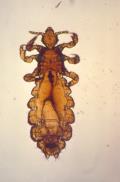 Head Louse[/caption]
William Ryan B.V.Sc.
Ryan Mitchell Associates LLC
Westfield, NJ and
Bernard Cohen, MD
Professor Dermatology and
Ellen Koch, MD
Division of Pediatric Dermatology
Johns Hopkins Medicine, Baltimore, Maryland
MedicalResearch.com: What is the background for this study? What are the main findings?
Response: As a group we were concerned about the misinformation that continues to be promulgated on the internet and through other sites. Importantly, the group consisted of experts with specific experience in the management of head louse infestations, from pediatric dermatology, pediatrics, school nursing and head louse research fields. Even information sources that we would have expected to be credible are outdated, unreliable or both, often continuing myths about head louse infestations and how they can be controlled. We wanted to provide a balanced and informed perspective that would help physicians and parents recognize that head louse infestations do not present a serious problem, and can be well managed with an informed approach to treatment.
The main findings are that over the counder products (permethrin/pyrethrins) are unlikely to be effective, and that that there are safe and effective products that are available by prescription.
Interestingly, head lice do affect Indian and African children in their home countries, but virtually nonexistent in African Americans in North America. There has been speculation about hair grooming regimen or structure of African American hair but the cause is unknown. In a study we performed assessing resistance to over the counter pediculicide components over a decade ago in Baltimore, we were not able to find a single African American child with head lice. We were not able to recruit any patients from the Baltimore City Schools.
Head Louse[/caption]
William Ryan B.V.Sc.
Ryan Mitchell Associates LLC
Westfield, NJ and
Bernard Cohen, MD
Professor Dermatology and
Ellen Koch, MD
Division of Pediatric Dermatology
Johns Hopkins Medicine, Baltimore, Maryland
MedicalResearch.com: What is the background for this study? What are the main findings?
Response: As a group we were concerned about the misinformation that continues to be promulgated on the internet and through other sites. Importantly, the group consisted of experts with specific experience in the management of head louse infestations, from pediatric dermatology, pediatrics, school nursing and head louse research fields. Even information sources that we would have expected to be credible are outdated, unreliable or both, often continuing myths about head louse infestations and how they can be controlled. We wanted to provide a balanced and informed perspective that would help physicians and parents recognize that head louse infestations do not present a serious problem, and can be well managed with an informed approach to treatment.
The main findings are that over the counder products (permethrin/pyrethrins) are unlikely to be effective, and that that there are safe and effective products that are available by prescription.
Interestingly, head lice do affect Indian and African children in their home countries, but virtually nonexistent in African Americans in North America. There has been speculation about hair grooming regimen or structure of African American hair but the cause is unknown. In a study we performed assessing resistance to over the counter pediculicide components over a decade ago in Baltimore, we were not able to find a single African American child with head lice. We were not able to recruit any patients from the Baltimore City Schools.
07 Sep Which Come First? Atopic Dermatitis or Staph Aureus Colonisation?
MedicalResearch.com Interview with: [caption id="attachment_27696" align="alignleft" width="125"] Prof. Alan Irvine[/caption]
Prof. Alan Irvine DSc
Consultant Dermatologist Our Lady’s Children’s Hospital
Associate Professor of Dermatology
Trinity College Dublin
MedicalResearch.com: What is the background for this study? What are the main findings?
Response: The background is that atopic dermatitis (AD) has a close relationship with staphylococcus aureus (SA) colonisation, and this is known to drive flares or exacerbations of AD but before our report it was not known which came first-AD colonisation or atopic dermatitis?
By following a cohort pf patients very carefully over a 1 year period and regularly sampling their skin microbiome we were able to show that SA colonisation did not precede development of AD and in fact that several non SA species of staphylococcus actually appeared to be protective for developing atopic dermatitis.
This is an important new finding in the complex relationship between the microbiome and skin inflammation, suggesting that some commensal bacterial are anti-inflammatory or protective.
Prof. Alan Irvine[/caption]
Prof. Alan Irvine DSc
Consultant Dermatologist Our Lady’s Children’s Hospital
Associate Professor of Dermatology
Trinity College Dublin
MedicalResearch.com: What is the background for this study? What are the main findings?
Response: The background is that atopic dermatitis (AD) has a close relationship with staphylococcus aureus (SA) colonisation, and this is known to drive flares or exacerbations of AD but before our report it was not known which came first-AD colonisation or atopic dermatitis?
By following a cohort pf patients very carefully over a 1 year period and regularly sampling their skin microbiome we were able to show that SA colonisation did not precede development of AD and in fact that several non SA species of staphylococcus actually appeared to be protective for developing atopic dermatitis.
This is an important new finding in the complex relationship between the microbiome and skin inflammation, suggesting that some commensal bacterial are anti-inflammatory or protective.
29 Aug ICUs Might Consider Avoiding Tap Water To Limit Pseudomonas Infections from Faucets
MedicalResearch.com Interview with: [caption id="attachment_27441" align="alignleft" width="200"] Dr. Cohen Regev[/caption]
Dr. Cohen Regev, M.D
Head of the infectious diseases and infection control units
Sanz Medical Center, Laniado hospital
Netanya, Israel
MedicalResearch.com: What is the background for this study?
Response: During 3 months in 2012 we had a number of clinical isolates of Pseudomonas aeruginosa (PA) in our neonatal intensive care unit (NICU) and a high incidence of colonization among ventilated patients in our medical-surgical intensive care unit (MSICU). The origin of PA may be from various environmental sources (‘exogenous’), from the patients’ own microbiome (‘endogenous’), or from both. Since in NICUs the origin is usually exogenous, we investigated the sources of the bacteria, focusing on the faucets of these units, as they were previously incriminated as causes of outbreaks in ICUs.
The study was conducted in Sanz medical center, a 400-bed community hospital located in central Israel. In the NICU we obtained several environmental cultures from faucets using a bacterial swab by rubbing the tip into the distal part of the faucet. Aerators were dismantled from all faucets, cultured from their inner part using a swab and were not repositioned. Contaminated faucets were occasionally replaced or treated with enzymatic fluid and sterilization by Ethylene Oxide. During the intervention and since, neonates were bathed only with warmed sterile water, and tap water was allowed only for hand hygiene practices.
In the MSICU tap water was used only for bathing the patients. All other uses of tap water, such as drinking, moistening and mouth treatments, were allowed using only sterile water. The units' faucets were sampled on two different days concurrently with surveillance cultures of pharyngeal, sputum and urine from the patients.
Bacteria were identified with VITEK 2 (Biomerieux®) and typing was done by Enterobacterial Repetitive Intergenic Consensus (ERIC) PCR.
Dr. Cohen Regev[/caption]
Dr. Cohen Regev, M.D
Head of the infectious diseases and infection control units
Sanz Medical Center, Laniado hospital
Netanya, Israel
MedicalResearch.com: What is the background for this study?
Response: During 3 months in 2012 we had a number of clinical isolates of Pseudomonas aeruginosa (PA) in our neonatal intensive care unit (NICU) and a high incidence of colonization among ventilated patients in our medical-surgical intensive care unit (MSICU). The origin of PA may be from various environmental sources (‘exogenous’), from the patients’ own microbiome (‘endogenous’), or from both. Since in NICUs the origin is usually exogenous, we investigated the sources of the bacteria, focusing on the faucets of these units, as they were previously incriminated as causes of outbreaks in ICUs.
The study was conducted in Sanz medical center, a 400-bed community hospital located in central Israel. In the NICU we obtained several environmental cultures from faucets using a bacterial swab by rubbing the tip into the distal part of the faucet. Aerators were dismantled from all faucets, cultured from their inner part using a swab and were not repositioned. Contaminated faucets were occasionally replaced or treated with enzymatic fluid and sterilization by Ethylene Oxide. During the intervention and since, neonates were bathed only with warmed sterile water, and tap water was allowed only for hand hygiene practices.
In the MSICU tap water was used only for bathing the patients. All other uses of tap water, such as drinking, moistening and mouth treatments, were allowed using only sterile water. The units' faucets were sampled on two different days concurrently with surveillance cultures of pharyngeal, sputum and urine from the patients.
Bacteria were identified with VITEK 2 (Biomerieux®) and typing was done by Enterobacterial Repetitive Intergenic Consensus (ERIC) PCR.
29 Aug Baylor Scientists Grow Noroviruses In Lab For First Time
MedicalResearch.com Interview with: [caption id="attachment_27424" align="alignleft" width="132"] Dr. Mary Estes[/caption]
Mary K. Estes, Ph.D.
Distinguished Service Professor
Cullen Endowed Chair of Human and Molecular Virology
Department of Molecular Virology and Microbiology
Baylor College of Medicine
Houston, TX 77030
MedicalResearch.com: What is the background for this study?
Response: Noroviruses are the most common cause of acute gastroenteritis (vomiting and diarrhea) worldwide and the leading cause of food-borne gastroenteritis. They also can cause chronic (long-lasting) illness in immunocompromised patients. These viruses are highly contagious and spread rapidly among people. The first report of an outbreak caused by a norovirus was in an elementary school in Norwalk, Ohio in 1968. Since that time, it became known that the virus damaged cells in the small intestine of infected people but attempts by many research groups to grow human noroviruses in the laboratory in a variety of intestinal cancer cells lines failed. This inability to grow human norovirus has been considered the single greatest barrier to norovirus research because it limited studies to understand how the virus makes people sick and how to inactivate the virus to prevent infection.
Dr. Mary Estes[/caption]
Mary K. Estes, Ph.D.
Distinguished Service Professor
Cullen Endowed Chair of Human and Molecular Virology
Department of Molecular Virology and Microbiology
Baylor College of Medicine
Houston, TX 77030
MedicalResearch.com: What is the background for this study?
Response: Noroviruses are the most common cause of acute gastroenteritis (vomiting and diarrhea) worldwide and the leading cause of food-borne gastroenteritis. They also can cause chronic (long-lasting) illness in immunocompromised patients. These viruses are highly contagious and spread rapidly among people. The first report of an outbreak caused by a norovirus was in an elementary school in Norwalk, Ohio in 1968. Since that time, it became known that the virus damaged cells in the small intestine of infected people but attempts by many research groups to grow human noroviruses in the laboratory in a variety of intestinal cancer cells lines failed. This inability to grow human norovirus has been considered the single greatest barrier to norovirus research because it limited studies to understand how the virus makes people sick and how to inactivate the virus to prevent infection.
19 Aug Genes That Enable C. diff Toxin Production Identified
MedicalResearch.com Interview with: [caption id="attachment_27186" align="alignleft" width="141"] Dr. Charles Darkoh[/caption]
Charles Darkoh, Ph.D., MS., MSc.
Assistant Professor
University of Texas Health Science Center at Houston
School of Public Health
Department of Epidemiology, Human Genetics & Environmental Sciences
Center for Infectious Diseases
Houston, Texas 77030
MedicalResearch.com: What is the background for this study? What are the main findings?
Response: Clostridium difficile (Cdiff) is a multidrug-resistant pathogen that takes over the colon after the good bacteria in the colon have been wiped out by antibiotic therapy. As a result, antibiotic treatment is a major risk factor for C. diff infections. Because of the ability of C. diff to inactivate the majority of the antibiotics currently available, it has become necessary to urgently develop a non-antibiotic therapy for this life-threatening infection. We know that C. diff causes disease by producing toxins, designated toxin A and B. During infection, the toxins are released into the colon resulting in diarrhea and inflammation of the colon as well as other diarrhea-associated illnesses. We also know that C. diff strains that are unable to produce toxins cannot cause disease. Therefore, the toxins are promising targets for a non-antibiotic therapy.
We reported last year that C. difficile regulates toxin production using quorum sensing — a system that allows bacteria to coordinate their biological activities as a group. Two sets of quorum-sensing genes (agr1 and agr2) were identified. These genes form part of a signaling communication system that makes a small peptide, which serves as a cue for the infecting bacterial population to turn on their toxin genes.
In this study we used genetic analysis to identify which of these two sets of genes is responsible for regulating the toxins. Our results demonstrates that agr1 is the culprit. This is because Cdiff agr1 mutant cannot produce toxins and unable to cause disease in mice, whereas the agr2 mutant can cause disease just like the wild type C.diff.
Dr. Charles Darkoh[/caption]
Charles Darkoh, Ph.D., MS., MSc.
Assistant Professor
University of Texas Health Science Center at Houston
School of Public Health
Department of Epidemiology, Human Genetics & Environmental Sciences
Center for Infectious Diseases
Houston, Texas 77030
MedicalResearch.com: What is the background for this study? What are the main findings?
Response: Clostridium difficile (Cdiff) is a multidrug-resistant pathogen that takes over the colon after the good bacteria in the colon have been wiped out by antibiotic therapy. As a result, antibiotic treatment is a major risk factor for C. diff infections. Because of the ability of C. diff to inactivate the majority of the antibiotics currently available, it has become necessary to urgently develop a non-antibiotic therapy for this life-threatening infection. We know that C. diff causes disease by producing toxins, designated toxin A and B. During infection, the toxins are released into the colon resulting in diarrhea and inflammation of the colon as well as other diarrhea-associated illnesses. We also know that C. diff strains that are unable to produce toxins cannot cause disease. Therefore, the toxins are promising targets for a non-antibiotic therapy.
We reported last year that C. difficile regulates toxin production using quorum sensing — a system that allows bacteria to coordinate their biological activities as a group. Two sets of quorum-sensing genes (agr1 and agr2) were identified. These genes form part of a signaling communication system that makes a small peptide, which serves as a cue for the infecting bacterial population to turn on their toxin genes.
In this study we used genetic analysis to identify which of these two sets of genes is responsible for regulating the toxins. Our results demonstrates that agr1 is the culprit. This is because Cdiff agr1 mutant cannot produce toxins and unable to cause disease in mice, whereas the agr2 mutant can cause disease just like the wild type C.diff.
12 Aug Early Onset of Pediatric Acute Otitis Media Raises Risk of More Frequent Episodes
MedicalResearch.com Interview with: [caption id="attachment_26952" align="alignleft" width="162"] Marieke de Hoog[/caption]
Marieke de Hoog
Assistant Professor
Julius Centrum voor Gezondheidswetenschappen en Eerstelijnsgeneeskunde
UMC Utrecht
The Netherlands
MedicalResearch.com: What is the background for this study?
Response: Acute otitis media (AOM) is a prime reason for doctor consultations and antibiotic use in children. Although symptoms of AOM may resolve spontaneously, these infections have a significant impact on child and family life and carry a considerable health care and economic burden. Acute otitis media occurring early in life, also called early-onset AOM, has been suggested as a risk factor for subsequent Acute otitis media episodes during childhood and could therefore also impact health care resource use. Identifying the critical age-period and quantifying the long-term consequences of early-onset AOM is important to guide future management and prevention programs aiming to reduce the burden of AOM.
Marieke de Hoog[/caption]
Marieke de Hoog
Assistant Professor
Julius Centrum voor Gezondheidswetenschappen en Eerstelijnsgeneeskunde
UMC Utrecht
The Netherlands
MedicalResearch.com: What is the background for this study?
Response: Acute otitis media (AOM) is a prime reason for doctor consultations and antibiotic use in children. Although symptoms of AOM may resolve spontaneously, these infections have a significant impact on child and family life and carry a considerable health care and economic burden. Acute otitis media occurring early in life, also called early-onset AOM, has been suggested as a risk factor for subsequent Acute otitis media episodes during childhood and could therefore also impact health care resource use. Identifying the critical age-period and quantifying the long-term consequences of early-onset AOM is important to guide future management and prevention programs aiming to reduce the burden of AOM.
11 Aug Serious Infections Associated With Increased Risk of Suicide
Posted at 22:01h
in Author Interviews, Critical Care - Intensive Care - ICUs, Infections, JAMA, Mental Health Research
MedicalResearch.com Interview with:
Helene Lund-Sørensen BM
Department of Biomedical Sciences
Section of Cellular and Metabolic Research
University of Copenhagen
MedicalResearch.com: What is the background for this study? What are the main findings?
Response: Accumulating research has shown that inflammation and infections are associated with psychiatric diagnoses and interactions between infectious agents, known to affect the brain, and suicidal behavior have been reported.
We find an increased risk of death by suicide among individuals hospitalized with infections. The risk of suicide increased in a dose-response relationship with the number of hospitalizations with infections and with the number of days hospitalized with infections. We also examined the risk of suicide association with the time since the last hospitalization with infection and found that infection was linked to an elevated risk with the strongest effect after 1 and 2 years compared with those without infections.
05 Aug Preventing Bacteria From Sticking Would Eliminate Need For Antibiotics
MedicalResearch.com Interview with: [caption id="attachment_26775" align="alignleft" width="153"] Dr. Peter Monk[/caption]
Dr Peter Monk BSc PhD
Faculty Director of International Affairs
Reader in Immunology
Department of Infection, Immunity and Cardiovascular Disease
Sheffield University Medical School
MedicalResearch.com: What is the background for this study? What are the main findings?
Response: The tetraspanin proteins are found on the surface of all mammalian cells. The cell surface is the place where cells 'socialise': they talk to each other to coordinate activities, stick to each other to form tissues and sometimes crawl across each other to get to where they need to go. Tetraspanins have an important job to do in the organisation of the cell surface, amongst other things enabling the formation of 'sticky patches' (tetraspanin-enriched microdomains or TEM) that cause cells to adhere together or provide traction to allow movement. Some bacteria have evolved ways of hijacking the TEM for their own ends, adhering to tightly to these structures so that the normal things that sweep bacteria away (such as blood, sweat and tears!) are no longer effective. At this point, infection begins.
We have found that the TEM can be partly disrupted, by adding small parts of tetraspanins (peptides) to cells. The peptides seem to work by weakening the tetraspanin glue that holds the TEM together and causing the other components that give the 'stickiness' to the TEM to become more spaced out. We use the analogy of Velcro(TM), where the fabric hooks and loops are held together in woven material; loosen the weave and the hooks and loops fall apart, no longer able to engage strongly with the loops in the opposing piece of fabric.
Using reconstructed human skin, we were able to show that the tetraspanin peptides were both safe and effective; they did not affect wound healing in burned skin, but they could lower the bacterial load in the wound by 50%. This would allow the immune system (including the fluid that 'weeps' from wounds) to deal with the remaining bacteria more easily. Unlike conventional antibiotics that tend to kill bacteria, our peptides simply cause them to get washed away, so not invoking the evolutionary selective mechanisms that lead to resistance.
Dr. Peter Monk[/caption]
Dr Peter Monk BSc PhD
Faculty Director of International Affairs
Reader in Immunology
Department of Infection, Immunity and Cardiovascular Disease
Sheffield University Medical School
MedicalResearch.com: What is the background for this study? What are the main findings?
Response: The tetraspanin proteins are found on the surface of all mammalian cells. The cell surface is the place where cells 'socialise': they talk to each other to coordinate activities, stick to each other to form tissues and sometimes crawl across each other to get to where they need to go. Tetraspanins have an important job to do in the organisation of the cell surface, amongst other things enabling the formation of 'sticky patches' (tetraspanin-enriched microdomains or TEM) that cause cells to adhere together or provide traction to allow movement. Some bacteria have evolved ways of hijacking the TEM for their own ends, adhering to tightly to these structures so that the normal things that sweep bacteria away (such as blood, sweat and tears!) are no longer effective. At this point, infection begins.
We have found that the TEM can be partly disrupted, by adding small parts of tetraspanins (peptides) to cells. The peptides seem to work by weakening the tetraspanin glue that holds the TEM together and causing the other components that give the 'stickiness' to the TEM to become more spaced out. We use the analogy of Velcro(TM), where the fabric hooks and loops are held together in woven material; loosen the weave and the hooks and loops fall apart, no longer able to engage strongly with the loops in the opposing piece of fabric.
Using reconstructed human skin, we were able to show that the tetraspanin peptides were both safe and effective; they did not affect wound healing in burned skin, but they could lower the bacterial load in the wound by 50%. This would allow the immune system (including the fluid that 'weeps' from wounds) to deal with the remaining bacteria more easily. Unlike conventional antibiotics that tend to kill bacteria, our peptides simply cause them to get washed away, so not invoking the evolutionary selective mechanisms that lead to resistance.
05 Aug Bacteria Causing Dental Caries Transmitted From Family and Friends
MedicalResearch.com Interview with: [caption id="attachment_26712" align="alignleft" width="149"] Dr. David Macintyre[/caption]
Stephanie S. Momeni, MS, MBA
Doctoral Candidate, Department of Biology
DART Trainee, Department of Pediatric Dentistry & IOHR
UAB School of Dentistry
Birmingham, Alabama 35294-0007
MedicalResearch.com: What is the background for this study? What are the main findings?
Response: This study was a small part of a large scale of S. mutans in a group of high-caries risk children and their household family members in Perry County, Alabama, USA. Overall dental caries is a dietary and infectious disease that we seek to understand better. We found only 34 rep-PCR genotypes for over 13,000 bacterial isolates from over 594 individual subjects. With so much commonality we wanted to determine if any conclusions could be made about transmission.
The key findings are:
• Children having multiple S. mutans genotypes were 2.3 times more likely to have dental caries.
• Analysis for transmission performed from two perspectives (by child and by genotype) indicating 63% of children shared at least 1 genotype with their mother, but 72% of children had at least 1 genotype not shared with any household family members.
• Child-to-child transmission of some genotypes is highly probable.
• About 1/3 of isolates observed were transient, and may confound the search for strains associated with tooth decay.
Dr. David Macintyre[/caption]
Stephanie S. Momeni, MS, MBA
Doctoral Candidate, Department of Biology
DART Trainee, Department of Pediatric Dentistry & IOHR
UAB School of Dentistry
Birmingham, Alabama 35294-0007
MedicalResearch.com: What is the background for this study? What are the main findings?
Response: This study was a small part of a large scale of S. mutans in a group of high-caries risk children and their household family members in Perry County, Alabama, USA. Overall dental caries is a dietary and infectious disease that we seek to understand better. We found only 34 rep-PCR genotypes for over 13,000 bacterial isolates from over 594 individual subjects. With so much commonality we wanted to determine if any conclusions could be made about transmission.
The key findings are:
• Children having multiple S. mutans genotypes were 2.3 times more likely to have dental caries.
• Analysis for transmission performed from two perspectives (by child and by genotype) indicating 63% of children shared at least 1 genotype with their mother, but 72% of children had at least 1 genotype not shared with any household family members.
• Child-to-child transmission of some genotypes is highly probable.
• About 1/3 of isolates observed were transient, and may confound the search for strains associated with tooth decay.
27 Jul Fewer Childhood Infections Do Not Explain Marked Increase In Human Longevity
MedicalResearch.com Interview with: [caption id="attachment_26526" align="alignleft" width="119"] Dr. Adam Hayward[/caption]
Adam Hayward PhD
Impact Research Fellow
University of Stirling
MedicalResearch.com: What is the background for this study?
Response: Adult life expectancies in industrialized countries have increased dramatically in the last 150 years, even once we’ve accounted for the fact that previously common deaths in childhood and now very rare. One hypothesis seeking to explain this increase is that childhood infections cause chronic inflammation, which are then linked with heart disease and stroke in later life, reducing lifespan.
Since such childhood infections were previously common but are now, thanks to vaccine and sanitation, much rarer, chronic inflammation should be lower and people should live longer and be less likely to die from early-onset heart disease. If this hypothesis is correct, we should see that higher exposure to infections in early life leads to increased adult mortality and deaths from heart disease and stroke.
Dr. Adam Hayward[/caption]
Adam Hayward PhD
Impact Research Fellow
University of Stirling
MedicalResearch.com: What is the background for this study?
Response: Adult life expectancies in industrialized countries have increased dramatically in the last 150 years, even once we’ve accounted for the fact that previously common deaths in childhood and now very rare. One hypothesis seeking to explain this increase is that childhood infections cause chronic inflammation, which are then linked with heart disease and stroke in later life, reducing lifespan.
Since such childhood infections were previously common but are now, thanks to vaccine and sanitation, much rarer, chronic inflammation should be lower and people should live longer and be less likely to die from early-onset heart disease. If this hypothesis is correct, we should see that higher exposure to infections in early life leads to increased adult mortality and deaths from heart disease and stroke.
25 Jul Shorter Course of Antibiotics May Be Enough For Community Acquired Pneumonia
MedicalResearch.com Interview with: Dr. Ane Uranga MD Department of Pneumology, Galdakao-Usansolo Hospital Galdakao, Bizkaia, Spain MedicalResearch.com: What is the background for this study? What are the main findings? Response: Despite clear benefits of shorter antibiotic treatments, reducing the duration of treatment remains challenging in daily clinical practice. Actually, IDSA/ATS recommendations for Community Acquired Pneumonia (CAP) suggested a minimum of 5 days of treatment based on clinical stability criteria. However, in our study the median of duration of antibiotic treatment in the control group was as high as 10 days. The main finding is that receiving 5 days antibiotic treatment in hospitalized patients suffering from CAP is not inferior to arbitrary treatment schedules in terms of clinical success.20 Jul Travelers Are Importing Intestinal Antibiotic-Resistant Superbugs
MedicalResearch.com Interview with: [caption id="attachment_26331" align="alignleft" width="100"] Herr Dr. Endimiani[/caption]
Prof. Andrea Endimiani, MD, PhD
Institute for Infectious Diseases
University of Bern
MedicalResearch.com: What is the background for this study?
Response: The spread of multidrug-resistant Gram-negative bacteria represents a serious issue for the healthcare system worldwide because our antibiotic armamentarium is becoming too limited. These «superbugs» may cause serious infections with high morbidity and mortality rates – there are already 700,000 estimated deaths per year worldwide because common antimicrobial therapies have become ineffective. In this scenario, colistin has represented the last active antibiotic option able to cure many infected people.
Unfortunately, in November 2015 a new mechanism of resistance against colistin was found with a high prevalence in Escherichia coli and Klebsiella pneumoniae strains detected in China among humans, food animals, and chicken meat; more recently, it has also been found in other countries.
This mechanism is encoded by a gene (named mcr-1) that is plasmid-mediated, thus assuring its great ability to mobilize and spread between different enterobacteria, including those normally present in the human and animal intestinal tracts.
Herr Dr. Endimiani[/caption]
Prof. Andrea Endimiani, MD, PhD
Institute for Infectious Diseases
University of Bern
MedicalResearch.com: What is the background for this study?
Response: The spread of multidrug-resistant Gram-negative bacteria represents a serious issue for the healthcare system worldwide because our antibiotic armamentarium is becoming too limited. These «superbugs» may cause serious infections with high morbidity and mortality rates – there are already 700,000 estimated deaths per year worldwide because common antimicrobial therapies have become ineffective. In this scenario, colistin has represented the last active antibiotic option able to cure many infected people.
Unfortunately, in November 2015 a new mechanism of resistance against colistin was found with a high prevalence in Escherichia coli and Klebsiella pneumoniae strains detected in China among humans, food animals, and chicken meat; more recently, it has also been found in other countries.
This mechanism is encoded by a gene (named mcr-1) that is plasmid-mediated, thus assuring its great ability to mobilize and spread between different enterobacteria, including those normally present in the human and animal intestinal tracts.
12 Jul Malignant Bacteria Infects Brain and Spinal Cord Through Nasal Inhalation
MedicalResearch.com Interview with: Jenny A. K. Ekberg Eskitis Institute for Drug Discovery Faculty of Health Sciences and Medicine, Bond University Institute for Health and Biomedical Innovation, Queensland University of Technology, School of Medical Science, Griffith University Queensland, Australia MedicalResearch.com: What is the background for this study? What are the main findings? Response: Melioidosis is a tropical bacterial infection that causes around 90,000 deaths world-wide each year. It is caused by the bacteria Burkholderia pseudomallei which can cause pneumonia and a serious flu-like illness which can cause death, however if the brain is infected, which happens in particular with the Australian variant of the disease, the mortality is particularly high (~25 %). The route by which the bacteria invade and progress through the central nervous system is to date largely unknown. We have now shown in an animal model that the bacteria can penetrate the trigeminal nerve within the nasal cavity and then rapidly invade the brainstem and spinal cord only 24 hours after intranasal inoculation. By migrating along the trigeminal nerve, the bacteria bypasses the blood-brain barrier.This study constitutes the first characterization of the path by which B. pseudomallei bacteria migrate all the way from the nasal cavity into the spinal cord.05 Jul Risk of Staph Bacteremia Can Run in Families
MedicalResearch.com Interview with: Louise Bruun Østergaard MD. Ph.D student Faculty of Medicine and Faculty of Engineering and Science Aalborg University Department of Cardiology, Gentofte Hospital Hellerup MedicalResearch.com: What is the background for this study? What are the main findings? Response: Staphylococcus aureus bacteremia strikes people of all ages resulting in devastating consequence even in young and healthy individuals. Animal studies have shown that the susceptibility to Staphylococcus aureus differs among different genetic strains in mice, suggesting that genetic differences could influence the susceptibility to Staphylococcus aureus in other spices. As a first step in determining whether genetics influence risk of Staphylococcus aureus infections we aimed to study whether a family history of Staphylococcus aureus bacteremia in first-degree relatives was associated with risk of the disease.25 Jun Phase 2 Study of New Combination Antibiotics Successfully Treats Complicated UTIs
MedicalResearch.com Interview with: [caption id="attachment_25517" align="alignleft" width="162"] Dr. Amanda Paschke[/caption]
Amanda Paschke, MD
Director, Infectious Disease Clinical Research
Merck Research Laboratories
MedicalResearch.com: What is the background for this study? What are the main findings?
Response: Relebactam is an investigational beta-lactamase inhibitor being developed as a fixed-dose combination with imipenem/cilastatin, which is a broad-spectrum antibiotic in the carbapenem class. In preclinical studies, this combination demonstrated antibacterial activity against a broad range of multidrug-resistant Gram-negative pathogens, including those producing extended-spectrum beta-lactamases such as Klebsiella pneumoniae carbapenemase (KPC)-producing Enterobacteriaceae and AmpC-producing Pseudomonas aeruginosa. Many of the most concerning infections caused by “superbugs” are caused by Gram-negative bacteria. These bacteria have evolved to be resistant to commonly used antibacterials, and even to antibacterials used as “last resort” treatment, which is why finding ways to treat them has become urgent. The addition of relebactam to imipenem is designed to restore activity of imipenem against certain imipenem-resistant strains of Gram-negative bacteria known to cause serious infections among people who often have other underlying medical conditions, which complicates treatment.
This was a Phase 2, multicenter, randomized, double-blind, non-inferiority study. The study looked at the use of relebactam plus imipenem versus imipenem alone for the treatment of adult patients with complicated urinary tract infections. The primary endpoint for the trial was microbiological response at the completion of IV study therapy. The study met its primary endpoint, demonstrating that the combination of relebactam with imipenem was as at least as effective as imipenem alone for the treatment of complicated urinary tract infections. The trial also demonstrated that the combination of relebactam plus imipenem is well-tolerated, with a safety profile similar to that of imipenem alone in this patient.
Dr. Amanda Paschke[/caption]
Amanda Paschke, MD
Director, Infectious Disease Clinical Research
Merck Research Laboratories
MedicalResearch.com: What is the background for this study? What are the main findings?
Response: Relebactam is an investigational beta-lactamase inhibitor being developed as a fixed-dose combination with imipenem/cilastatin, which is a broad-spectrum antibiotic in the carbapenem class. In preclinical studies, this combination demonstrated antibacterial activity against a broad range of multidrug-resistant Gram-negative pathogens, including those producing extended-spectrum beta-lactamases such as Klebsiella pneumoniae carbapenemase (KPC)-producing Enterobacteriaceae and AmpC-producing Pseudomonas aeruginosa. Many of the most concerning infections caused by “superbugs” are caused by Gram-negative bacteria. These bacteria have evolved to be resistant to commonly used antibacterials, and even to antibacterials used as “last resort” treatment, which is why finding ways to treat them has become urgent. The addition of relebactam to imipenem is designed to restore activity of imipenem against certain imipenem-resistant strains of Gram-negative bacteria known to cause serious infections among people who often have other underlying medical conditions, which complicates treatment.
This was a Phase 2, multicenter, randomized, double-blind, non-inferiority study. The study looked at the use of relebactam plus imipenem versus imipenem alone for the treatment of adult patients with complicated urinary tract infections. The primary endpoint for the trial was microbiological response at the completion of IV study therapy. The study met its primary endpoint, demonstrating that the combination of relebactam with imipenem was as at least as effective as imipenem alone for the treatment of complicated urinary tract infections. The trial also demonstrated that the combination of relebactam plus imipenem is well-tolerated, with a safety profile similar to that of imipenem alone in this patient.
21 Jun Psoriasis Patients At Higher Risk of Serious Infections
MedicalResearch.com Interview with: [caption id="attachment_24142" align="alignleft" width="128"] Dr. Jonathan Silverberg[/caption]
Dr. Jonathan L. Silverberg MD PhD MPH
Assistant Professor in Dermatology
Medical Social Sciences and Preventive Medicine
Northwestern University, Chicago, Illinois
MedicalResearch.com: What is the background for this study? What are the main findings?
Response: Psoriasis is associated with a number of potential risk factors for developing serious infections, including impaired skin-barrier function, immune dysregulation, use of systemic immunosuppressant and biologic treatments. We hypothesized that adults with psoriasis have higher rates of serious infections.
We examined data from the 2002-2012 National Inpatient Sample, which contains a representative 20% sample of all hospitalizations in the United States. We found that psoriasis was associated with multiple serious infections, including methicillin-resistant Staphylococcus aureus, cellulitis, herpes simplex virus infection, infectious arthritis, osteomyelitis, meningitis, encephalitis and tuberculosis. Rates of serious infections increased over all time.
Significant predictors of serious infections in patients with psoriasis included non-white race, lower estimated income quartile, and Medicaid, Medicare, or self-pay insurance status. These findings suggest that poor access to adequate dermatologic care may be associated with higher rates of infections.
Dr. Jonathan Silverberg[/caption]
Dr. Jonathan L. Silverberg MD PhD MPH
Assistant Professor in Dermatology
Medical Social Sciences and Preventive Medicine
Northwestern University, Chicago, Illinois
MedicalResearch.com: What is the background for this study? What are the main findings?
Response: Psoriasis is associated with a number of potential risk factors for developing serious infections, including impaired skin-barrier function, immune dysregulation, use of systemic immunosuppressant and biologic treatments. We hypothesized that adults with psoriasis have higher rates of serious infections.
We examined data from the 2002-2012 National Inpatient Sample, which contains a representative 20% sample of all hospitalizations in the United States. We found that psoriasis was associated with multiple serious infections, including methicillin-resistant Staphylococcus aureus, cellulitis, herpes simplex virus infection, infectious arthritis, osteomyelitis, meningitis, encephalitis and tuberculosis. Rates of serious infections increased over all time.
Significant predictors of serious infections in patients with psoriasis included non-white race, lower estimated income quartile, and Medicaid, Medicare, or self-pay insurance status. These findings suggest that poor access to adequate dermatologic care may be associated with higher rates of infections.
13 Jun ‘Epi-Chip’ Aims To Reconstruct Entire Infection Exposure History From Drop of Blood
Posted at 19:43h
in Author Interviews, Biomarkers, Cancer Research, Infections, Technology, University of Pittsburgh
MedicalResearch.com Interview with:
[caption id="attachment_25161" align="alignleft" width="160"] Dr. Donald Burke[/caption]
Donald S. Burke, M.D.
Dean of the University of Pittsburgh Graduate School of Public Health
Director of the University of Pittsburgh Center for Vaccine Research
MedicalResearch.com: What is the background for this study? What are the main findings?
Dr. Burke: At the University of Pittsburgh we developed a unique method for detecting antibodies in the blood of patients in a proof-of-principle study that opens the door to development of simple diagnostic tests for diseases for which no microbial cause is known, including auto-immune diseases, cancers and other conditions.
We used a technique pioneered by co-author Thomas Kodadek, Ph.D., of the Scripps Research Institute, that synthesizes random molecular shapes called “peptoids” hooked onto microscopic plastic beads. The technique can produce millions of molecular shapes. The peptoids are not organic, but if they match to the corresponding shape on an antibody, that antibody will connect to them, allowing the scientist to pull out that bead and examine that peptoid and its corresponding antibody.
My team chemically generated a huge library of random molecular shapes. Then, using blood from HIV-infected patients and from non-infected people, we screened a million of these random molecular shapes to find the ones that bound only to antibodies present in the blood of HIV-infected patients, but not the healthy controls. No HIV proteins or structures were used to construct or select the peptoids, but the approach, nonetheless, successfully led to selection of the best molecular shapes to use in screening for HIV antibodies.
We then resynthesized that HIV-antibody-targeting peptoid in mass and tested it by screening hundreds of samples from the Multicenter AIDS Cohort Study (MACS), a confidential research study of the natural history of treated and untreated HIV/AIDS in men who have sex with men (supported by the National Institutes of Health). Study co-author Charles Rinaldo, Ph.D., chair of Pitt Public Health’s Department of Infectious Diseases and Microbiology and director of the Pittsburgh arm of the MACS, selected the samples, but blinded the testers to which samples were HIV-positive or -negative. The test distinguished between the samples of HIV-positive blood and HIV-negative blood with a high degree of accuracy.
Dr. Donald Burke[/caption]
Donald S. Burke, M.D.
Dean of the University of Pittsburgh Graduate School of Public Health
Director of the University of Pittsburgh Center for Vaccine Research
MedicalResearch.com: What is the background for this study? What are the main findings?
Dr. Burke: At the University of Pittsburgh we developed a unique method for detecting antibodies in the blood of patients in a proof-of-principle study that opens the door to development of simple diagnostic tests for diseases for which no microbial cause is known, including auto-immune diseases, cancers and other conditions.
We used a technique pioneered by co-author Thomas Kodadek, Ph.D., of the Scripps Research Institute, that synthesizes random molecular shapes called “peptoids” hooked onto microscopic plastic beads. The technique can produce millions of molecular shapes. The peptoids are not organic, but if they match to the corresponding shape on an antibody, that antibody will connect to them, allowing the scientist to pull out that bead and examine that peptoid and its corresponding antibody.
My team chemically generated a huge library of random molecular shapes. Then, using blood from HIV-infected patients and from non-infected people, we screened a million of these random molecular shapes to find the ones that bound only to antibodies present in the blood of HIV-infected patients, but not the healthy controls. No HIV proteins or structures were used to construct or select the peptoids, but the approach, nonetheless, successfully led to selection of the best molecular shapes to use in screening for HIV antibodies.
We then resynthesized that HIV-antibody-targeting peptoid in mass and tested it by screening hundreds of samples from the Multicenter AIDS Cohort Study (MACS), a confidential research study of the natural history of treated and untreated HIV/AIDS in men who have sex with men (supported by the National Institutes of Health). Study co-author Charles Rinaldo, Ph.D., chair of Pitt Public Health’s Department of Infectious Diseases and Microbiology and director of the Pittsburgh arm of the MACS, selected the samples, but blinded the testers to which samples were HIV-positive or -negative. The test distinguished between the samples of HIV-positive blood and HIV-negative blood with a high degree of accuracy.
10 Jun Staph Blood Infections Doubled in Patients on Steroids
MedicalResearch.com Interview with: [caption id="attachment_25120" align="alignleft" width="142"] Dr. Jesper Smit[/caption]
Jesper Smit, MD
Department of Clinical Microbiology
Aalborg University Hospital
Aalborg, Denmark
MedicalResearch.com: What is the background for this study? What are the main findings?
Dr. Smit: Use of glucocorticoids have been suggested to be associated with increased risk of blood infections by Staphylococcus aureus, but the existing evidence is sparse. Therefore, we conducted a large case-control study to investigate this topic in detail.
We found that the risk of staphylococcal blood infections was more than doubled in users of systemic glucocorticoids compared with non-users and that the risk of infection escalated with increasing dose.
Dr. Jesper Smit[/caption]
Jesper Smit, MD
Department of Clinical Microbiology
Aalborg University Hospital
Aalborg, Denmark
MedicalResearch.com: What is the background for this study? What are the main findings?
Dr. Smit: Use of glucocorticoids have been suggested to be associated with increased risk of blood infections by Staphylococcus aureus, but the existing evidence is sparse. Therefore, we conducted a large case-control study to investigate this topic in detail.
We found that the risk of staphylococcal blood infections was more than doubled in users of systemic glucocorticoids compared with non-users and that the risk of infection escalated with increasing dose.
01 Jun Childhood Infections Linked to Later Life Obesity and Impaired Vascular Function
Posted at 01:46h
in Author Interviews, Heart Disease, Infections, Pediatrics, Pediatrics, Weight Research
MedicalResearch.com Interview with:
[caption id="attachment_24810" align="alignleft" width="180"] Dr. Markus Juonala[/caption]
Markus Juonala, MD, PhD
Murdoch Childrens Research Institute, Parkville
Victoria, Australia
MedicalResearch.com: What is the background for this study? What are the main findings?
Dr. Juonala: This is an epidemiological follow-up study investigating whether childhood infections and socieconomic status are associated with cardiovasular risk factor and early chances in vasculature.
The main finding was that childhood infections were associated with obesity and impaired vascular function in adulthood among those individuals with low socioeconomic status.
Dr. Markus Juonala[/caption]
Markus Juonala, MD, PhD
Murdoch Childrens Research Institute, Parkville
Victoria, Australia
MedicalResearch.com: What is the background for this study? What are the main findings?
Dr. Juonala: This is an epidemiological follow-up study investigating whether childhood infections and socieconomic status are associated with cardiovasular risk factor and early chances in vasculature.
The main finding was that childhood infections were associated with obesity and impaired vascular function in adulthood among those individuals with low socioeconomic status.
22 May Respiratory and Urinary Tract Infections Common in TAVR Patients
Posted at 11:39h
in Author Interviews, Heart Disease, Hospital Acquired, Infections, Surgical Research
MedicalResearch.com Interview with:
[caption id="attachment_24564" align="alignleft" width="180"] Dr. Nombela Franco[/caption]
Luis Nombela-Franco, MD, PhD
Structural cardiology program.
Interventional Cardiology department.
Hospital Clínico San Carlos, Cardiovascular Institute
Madrid, Spain
(Dr. Nombela-Franco, has a special interest in interest on percutaneous treatment of structural heart disease and coronary interventions with special focus on chronic total occlusion)
MedicalResearch.com: What is the background for this study?
Dr. Nombela-Franco: In-hospital infections are one of the most common complications that may occur following medical and surgical admissions, significantly impacted length of hospital stay, costs and clinical outcomes. In addition, approximately one third of hospital-acquired infections are preventable.
Transcatheter aortic valve replacement (TAVR) is currently the standard of care for symptomatic patients with severe aortic stenosis deemed at high surgical risk or inoperable. Patients undergoing TAVR have several comorbidities and the invasive (although less invasive the surgical treatment) nature of the procedure and peri-operative care confers a high likelihood in-hospital infections in such patients. This study analyzed the incidence, predictive factors and impact of in-hospital infections in patients undergoing transcatheter aortic valve implantation.
Dr. Nombela Franco[/caption]
Luis Nombela-Franco, MD, PhD
Structural cardiology program.
Interventional Cardiology department.
Hospital Clínico San Carlos, Cardiovascular Institute
Madrid, Spain
(Dr. Nombela-Franco, has a special interest in interest on percutaneous treatment of structural heart disease and coronary interventions with special focus on chronic total occlusion)
MedicalResearch.com: What is the background for this study?
Dr. Nombela-Franco: In-hospital infections are one of the most common complications that may occur following medical and surgical admissions, significantly impacted length of hospital stay, costs and clinical outcomes. In addition, approximately one third of hospital-acquired infections are preventable.
Transcatheter aortic valve replacement (TAVR) is currently the standard of care for symptomatic patients with severe aortic stenosis deemed at high surgical risk or inoperable. Patients undergoing TAVR have several comorbidities and the invasive (although less invasive the surgical treatment) nature of the procedure and peri-operative care confers a high likelihood in-hospital infections in such patients. This study analyzed the incidence, predictive factors and impact of in-hospital infections in patients undergoing transcatheter aortic valve implantation.
18 May Simple Test Kit Can Detect E. Coli In Contaminated Water Samples
MedicalResearch.com Interview with: [caption id="attachment_24460" align="alignleft" width="200"] Dr. Sushanta Mitra[/caption]
Sushanta K. Mitra, PhD, PEng
Associate Vice-President Research
Kaneff Professor in Micro & Nanotechnology for Social Innovation
FCSME, FASME, FEIC, FRSC, FCAE, FAAAS Y
York University Toronto
MedicalResearch.com: What is the background for this study? What are the main findings?
Dr. Mitra: As a mechanical engineer I got interested in the water problem when I had discussions with Tata Consultancy Services (TCS), India and the tertiary public health centre doctors near Mumbai, where the doctors had to deal with large number of patients with water-borne diseases. This was hugely a challenge from resource point of view as the doctors would much preferred to have their attention focused on more pressing diseases. They approached me about developing tools for rapid detection of water-borne pathogen in drinking water. Hence, my journey started on water quality monitoring.
MedicalResearch.com: What is the background for this study? What are the main findings?
Dr. Mitra: Here, we have developed a low-cost compact E. coli and total coliform detection system, which uses commercially available plunger-tube assembly. We incorporate a hydrogel (porous matrix) inside the tube so that the plunger-tube assembly act as a concentrator and a detector at the same time. Specially formulated enzymatic substrates are caged inside the hydrogel so that an E. coli cell trapped within the hydrogel will be lysed and react with the enzymatic substrates to produce a red color.
Dr. Sushanta Mitra[/caption]
Sushanta K. Mitra, PhD, PEng
Associate Vice-President Research
Kaneff Professor in Micro & Nanotechnology for Social Innovation
FCSME, FASME, FEIC, FRSC, FCAE, FAAAS Y
York University Toronto
MedicalResearch.com: What is the background for this study? What are the main findings?
Dr. Mitra: As a mechanical engineer I got interested in the water problem when I had discussions with Tata Consultancy Services (TCS), India and the tertiary public health centre doctors near Mumbai, where the doctors had to deal with large number of patients with water-borne diseases. This was hugely a challenge from resource point of view as the doctors would much preferred to have their attention focused on more pressing diseases. They approached me about developing tools for rapid detection of water-borne pathogen in drinking water. Hence, my journey started on water quality monitoring.
MedicalResearch.com: What is the background for this study? What are the main findings?
Dr. Mitra: Here, we have developed a low-cost compact E. coli and total coliform detection system, which uses commercially available plunger-tube assembly. We incorporate a hydrogel (porous matrix) inside the tube so that the plunger-tube assembly act as a concentrator and a detector at the same time. Specially formulated enzymatic substrates are caged inside the hydrogel so that an E. coli cell trapped within the hydrogel will be lysed and react with the enzymatic substrates to produce a red color.