
Mental Health Research
12 Dec Access To Two Different Health Care Systems Can Lead To Dangerous Presciption Combinations
Posted at 22:06h
in Alzheimer's - Dementia, Annals Internal Medicine, Author Interviews, Critical Care - Intensive Care - ICUs, Health Care Systems, University of Pittsburgh
MedicalResearch.com Interview with:
[caption id="attachment_30462" align="alignleft" width="144"] Dr. Joshua Thorpe[/caption]
Joshua M. Thorpe, PhD, MPH
From the Center for Health Equity Research and Promotion
Veterans Affairs Pittsburgh Healthcare System
Pittsburgh Pennsylvania, and
Center for Health Services Research in Primary Care
Department of Pharmacy and Therapeutics
University of Pittsburgh School of Pharmacy
MedicalResearch.com: What is the background for this study?
Response: Care coordination for persons with dementia is challenging for health care systems under the best of circumstances. These coordination challenges are exacerbated in Medicare-eligible veterans who receive care through both Medicare and the Department of Veterans Affairs (VA). Recent Medicare and VA policy changes (e.g., Medicare Part D, Veteran’s Choice Act) expand veterans’ access to providers outside the VA. While access to care may be improved, seeking care across multiple health systems may disrupt care coordination and increase the risk of unsafe prescribing - particularly in veterans with dementia. To see how expanded access to care outside the VA might influence medication safety for veterans with dementia, we studied prescribing safety in Veterans who qualified for prescriptions through the VA as well as through the Medicare Part D drug benefit.
Dr. Joshua Thorpe[/caption]
Joshua M. Thorpe, PhD, MPH
From the Center for Health Equity Research and Promotion
Veterans Affairs Pittsburgh Healthcare System
Pittsburgh Pennsylvania, and
Center for Health Services Research in Primary Care
Department of Pharmacy and Therapeutics
University of Pittsburgh School of Pharmacy
MedicalResearch.com: What is the background for this study?
Response: Care coordination for persons with dementia is challenging for health care systems under the best of circumstances. These coordination challenges are exacerbated in Medicare-eligible veterans who receive care through both Medicare and the Department of Veterans Affairs (VA). Recent Medicare and VA policy changes (e.g., Medicare Part D, Veteran’s Choice Act) expand veterans’ access to providers outside the VA. While access to care may be improved, seeking care across multiple health systems may disrupt care coordination and increase the risk of unsafe prescribing - particularly in veterans with dementia. To see how expanded access to care outside the VA might influence medication safety for veterans with dementia, we studied prescribing safety in Veterans who qualified for prescriptions through the VA as well as through the Medicare Part D drug benefit.
09 Dec Computerized Working Memory Training in Pediatric Sickle Cell Disease
MedicalResearch.com Interview with: [caption id="attachment_30413" align="alignleft" width="200"] Dr. Steven J. Hardy[/caption]
Steven J. Hardy, Phd
Licensed Clinical Psychologist
Divisions of Hematology and Oncology
Children’s National Health System
Assistant Professor of Pediatrics and Psychiatry & Behavioral Sciences
George Washington School of Medicine and Health Sciences Washington, DC
MedicalResearch.com: What is the background for this study? What are the main findings?
Response: Children with sickle cell disease exhibit neurocognitive deficits as a consequence of either silent or overt cerebral infarction or disease-related non-infarct central nervous system effects (likely resulting from chronic anemia and hypoxic events). These complications often lead to impairment in executive functioning (e.g., working memory, attention, inhibition, cognitive flexibility), which can make it difficult to focus in class, plan for long-term school projects, remember and carry out multi-step tasks or assignments, and stay organized. The literature on interventions to reduce neurocognitive sequelae of sickle cell disease is extremely limited.
Our research team investigated a promising home-based, computerized cognitive training program (Cogmed) involving repeated practice on performance-adapted exercises targeting working memory with a sample of youth (ages 7 – 16) with sickle cell disease. Of the participants who have enrolled in the study (n = 70), 49% exhibited working memory deficits (<25% in the general population have a working memory deficit) and were randomized to an eight-week waitlist or to begin Cogmed immediately. Participants who used Cogmed demonstrated significant improvements on multiple measures of working memory, while those randomized to the waitlist group only exhibited such improvements after receiving Cogmed. Approximately 25% of participants completed the recommended number of Cogmed sessions (20 – 25 sessions). However, analyses revealed that participants who completed at least 10 sessions (about 50% of the participants) showed comparable levels of working memory improvement.
Dr. Steven J. Hardy[/caption]
Steven J. Hardy, Phd
Licensed Clinical Psychologist
Divisions of Hematology and Oncology
Children’s National Health System
Assistant Professor of Pediatrics and Psychiatry & Behavioral Sciences
George Washington School of Medicine and Health Sciences Washington, DC
MedicalResearch.com: What is the background for this study? What are the main findings?
Response: Children with sickle cell disease exhibit neurocognitive deficits as a consequence of either silent or overt cerebral infarction or disease-related non-infarct central nervous system effects (likely resulting from chronic anemia and hypoxic events). These complications often lead to impairment in executive functioning (e.g., working memory, attention, inhibition, cognitive flexibility), which can make it difficult to focus in class, plan for long-term school projects, remember and carry out multi-step tasks or assignments, and stay organized. The literature on interventions to reduce neurocognitive sequelae of sickle cell disease is extremely limited.
Our research team investigated a promising home-based, computerized cognitive training program (Cogmed) involving repeated practice on performance-adapted exercises targeting working memory with a sample of youth (ages 7 – 16) with sickle cell disease. Of the participants who have enrolled in the study (n = 70), 49% exhibited working memory deficits (<25% in the general population have a working memory deficit) and were randomized to an eight-week waitlist or to begin Cogmed immediately. Participants who used Cogmed demonstrated significant improvements on multiple measures of working memory, while those randomized to the waitlist group only exhibited such improvements after receiving Cogmed. Approximately 25% of participants completed the recommended number of Cogmed sessions (20 – 25 sessions). However, analyses revealed that participants who completed at least 10 sessions (about 50% of the participants) showed comparable levels of working memory improvement.
09 Dec Cerebral Perfusion Is Perturbed by Preterm Birth and Brain Injury
MedicalResearch.com Interview with: Eman S. Mahdi, MD, MBChB Pediatric Radiology Fellow [caption id="attachment_30368" align="alignleft" width="133"] Dr. Catherine Limperopoulos[/caption]
Catherine Limperopoulos, PhD
Director, Developing Brain Research Laboratory
Co-Director of Research, Division of Neonatology
Diagnostic Imaging and Radiology
Children’s National Health System
Washington, DC
MedicalResearch.com: What is the background for this study? What are the main findings?
Response: Premature birth is a major public health concern in the United States affecting 1 in 10 infants each year. Prematurity-related brain injury is very common and associated with a high prevalence of brain injury and accompanying lifelong neurodevelopmental morbidities.
Early disturbances in systemic and cerebral hemodynamics are thought to mediate prematurity-related brain injury. The extent to which cerebral blood flow (CBF) is disturbed in preterm birth is poorly understood, in large part because of the lack of monitoring techniques that can directly and non-invasively measure cerebral blood flow.
We report for the first time early disturbances in global and regional cerebral blood flow in preterm infants following brain injury on conventional magnetic resonance imaging (MRI) over the third trimester of ex-uterine life using arterial spin labelling images. In terms of regional differences, we saw a marked decrease in blood flow to the thalamus and the pons, regions known to be metabolically active during this time.
Dr. Catherine Limperopoulos[/caption]
Catherine Limperopoulos, PhD
Director, Developing Brain Research Laboratory
Co-Director of Research, Division of Neonatology
Diagnostic Imaging and Radiology
Children’s National Health System
Washington, DC
MedicalResearch.com: What is the background for this study? What are the main findings?
Response: Premature birth is a major public health concern in the United States affecting 1 in 10 infants each year. Prematurity-related brain injury is very common and associated with a high prevalence of brain injury and accompanying lifelong neurodevelopmental morbidities.
Early disturbances in systemic and cerebral hemodynamics are thought to mediate prematurity-related brain injury. The extent to which cerebral blood flow (CBF) is disturbed in preterm birth is poorly understood, in large part because of the lack of monitoring techniques that can directly and non-invasively measure cerebral blood flow.
We report for the first time early disturbances in global and regional cerebral blood flow in preterm infants following brain injury on conventional magnetic resonance imaging (MRI) over the third trimester of ex-uterine life using arterial spin labelling images. In terms of regional differences, we saw a marked decrease in blood flow to the thalamus and the pons, regions known to be metabolically active during this time.
09 Dec Strong SSRIs Linked To Increase Risk of Intracranial Hemorrhage
MedicalResearch.com Interview with: Christel Renoux, MD, PhD Assistant Professor, Dept. of Neurology & Neurosurgery McGill University Centre For Clinical Epidemiology Jewish General Hospital - Lady Davis Research Institute Montreal Canada MedicalResearch.com: What is the background for this study? Response: Selective serotonin reuptake inhibitors (SSRIs) increase the risk for abnormal bleeding, in particular, gastrointestinal tract bleeding. Previous studies also suggested an increased risk for intracranial hemorrhage (ICH) in patients treated with SSRIs compared to non users. However, even if this risk exists, the comparison with a non-treated group may exaggerate the strength of a potential association and the comparison with a group of patients treated with other antidepressants may help better delineate the risk. The potential bleeding effect of antidepressants is linked to the strength of serotonin inhibition reuptake, and antidepressants that are strong inhibitors of serotonin reuptake have been associated with the risk for gastrointestinal or abnormal bleeding compared with weak inhibitors but the risk of ICH is unclear.08 Dec More Medical Students May Have Non-Apparent Disabilities
MedicalResearch.com Interview with:
Lisa Meeks , PhD Director, Medical Student Disability UCSF Medical Center
[caption id="attachment_30304" align="alignnone" width="171"] Dr. Lisa Meeks[/caption]
MedicalResearch.com: What is the background for this study? What are the main findings? Response: This was the first study to include students with AD/HD, learning, psychological, and chronic health conditions. This study found that the prevalence of students with disabilities is up to four times higher than previous studies indicated.
AD/HD, learning, and psychological disabilities were the most prevalent, suggesting that most students with disabilities in medicine have non-apparent disabilities. Within MD granting programs, the number of students self-reporting disability varied between 0% and 12%. Explanations for the high variability between programs are unknown, however, anecdotal reports suggest the degree to which programs have dedicated resources and inclusive practices for students with disabilities influence student disclosure.03 Dec Is Depression in Mild Cognitive Impairment a Precursor to Dementia?
MedicalResearch.com Interview with: Zahinoor Ismail MD FRCPC Clinical Associate Professor, Hotchkiss Brain Institute University of Calgary MedicalResearch.com: What is the background for this study? What are the main findings? Response: Depression and depressive symptoms are common in mild cognitive impairment (MCI). Evidence suggests that depression in MCI increases the likelihood of progression from MCI to dementia, compared to non-depressed people with MCI. In the newer construct of mild behavioural impairment (MBI), which describes the relationship between later life onset of sustained and impactful neuropsychiatric symptoms and the risk of cognitive decline and dementia, depression is an important subdomain (in addition to apathy, impulse control, social cognition and psychotic symptoms). Thus, depression and depressive symptoms are a significant risk factor for cognitive, behavioural and functional outcomes in older adults who have at most mild cognitive impairment. As the importance of neuropsychiatric symptoms in older adults emerges, good prevalence estimates are required to inform clinicians and researchers as well as public health policy and decision makers. We performed a systematic review and meta-analysis to determine the best estimate of prevalence of depression in mild cognitive impairment. We included 57 studies, representing 20,892 participants in the analysis. While we determined that the omnibus prevalence estimate was 32%, there was significant heterogeneity in this sample based on setting. In community samples, the rate was 25%, but in clinical samples this was higher at 40%. Additionally, different case ascertainment methods for depression (self report, clinician administered or caregiver report) and different MCI criteria didn't change the prevalence estimates.02 Dec Mental and Physical Disorders Linked at Early Age
MedicalResearch.com Interview with: Prof. Dr. Gunther Meinlschmidt, Psych
University of Basel, Department of Psychology, Division of Clinical Psychology and Epidemiology
Faculty of Medicine
Switzerland
MedicalResearch.com: What is the background for this study?
Response: Physical diseases and mental disorders affect a person’s quality of life. Further, they present a huge challenge for the healthcare system. It has been reported that physical and mental disorders systematically co-occur already early in life. What we wanted to know is whether there are certain temporal patterns between mental disorders and physical diseases during childhood and adolescence. A better understanding of such patterns may help to reveal processes that could be relevant both to the origins of physical diseases and mental disorders and to their treatment.
Prof. Dr. Gunther Meinlschmidt, Psych
University of Basel, Department of Psychology, Division of Clinical Psychology and Epidemiology
Faculty of Medicine
Switzerland
MedicalResearch.com: What is the background for this study?
Response: Physical diseases and mental disorders affect a person’s quality of life. Further, they present a huge challenge for the healthcare system. It has been reported that physical and mental disorders systematically co-occur already early in life. What we wanted to know is whether there are certain temporal patterns between mental disorders and physical diseases during childhood and adolescence. A better understanding of such patterns may help to reveal processes that could be relevant both to the origins of physical diseases and mental disorders and to their treatment.
02 Dec Longitudinal Study of Post-Concussion Syndrome: Not Everyone Recovers
MedicalResearch.com Interview with: Dr. Charles Tator, Neurosurgeon Toronto Western Hospital and Director Canadian Concussion Centre MedicalResearch.com: What is the background for this study? What are the main findings? Response: The main findings of this study are that the number of symptoms of post-concussion syndrome (PCS) are related to how long PCS lasts. Furthermore, physicians need to be more vigorous in their treatment of PCS symptoms and use the treatments that exist for each symptom where possible such as headache, vertigo, anxiety. The sooner sufferers of PCS receive treatment for their symptoms, the better.02 Dec Can Probiotics in Yogurt Protect Against Stress and Anxiety?
Posted at 14:32h
in Author Interviews, Gastrointestinal Disease, Mental Health Research, Nature, Probiotics
MedicalResearch.com Interview with:
[caption id="attachment_30115" align="alignleft" width="200"]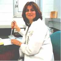 Dr. Elizabeth Bryda[/caption]
Elizabeth Bryda, PhD
Professor, Director, Rat Resource and Research Center
Veterinary Pathobiology
University of Missouri
Columbia, Missouri
MedicalResearch.com: What is the background for this study? What are the main findings?
Response: A number of groups have demonstrated the ability of probiotics to benefit digestive health and there is a growing body of evidence to suggest an association between mental health and “gut health”. We were interested to see if probiotic bacteria could decrease anxiety- or stress-related behavior in a controlled setting using zebrafish as our model organism of choice for these studies.
We were able to show that Lactobacillus plantarum decreased overall anxiety-related behavior and protected against stress-induced dysbiosis (microbial imbalance). The fact that administration of probiotic bacteria also protected other resident gut bacteria from the dramatic changes seen in “stressed” fish not receiving the probiotic was unexpected and suggested that these bacteria may be working at the level of the GI tract and the central nervous system.
Dr. Elizabeth Bryda[/caption]
Elizabeth Bryda, PhD
Professor, Director, Rat Resource and Research Center
Veterinary Pathobiology
University of Missouri
Columbia, Missouri
MedicalResearch.com: What is the background for this study? What are the main findings?
Response: A number of groups have demonstrated the ability of probiotics to benefit digestive health and there is a growing body of evidence to suggest an association between mental health and “gut health”. We were interested to see if probiotic bacteria could decrease anxiety- or stress-related behavior in a controlled setting using zebrafish as our model organism of choice for these studies.
We were able to show that Lactobacillus plantarum decreased overall anxiety-related behavior and protected against stress-induced dysbiosis (microbial imbalance). The fact that administration of probiotic bacteria also protected other resident gut bacteria from the dramatic changes seen in “stressed” fish not receiving the probiotic was unexpected and suggested that these bacteria may be working at the level of the GI tract and the central nervous system.
25 Nov Children With Autism Miss Social Cues of Eye Contact
MedicalResearch.com Interview with: [caption id="attachment_29960" align="alignleft" width="126"] Dr. Warren Jones[/caption]
Warren Jones, PhD
Director of Research, Marcus Autism Center
Children's Healthcare of Atlanta
CHOA Distinguished Chair in Autism
Asst. Professor, Dept. of Pediatrics
Emory University School of Medicine
Atlanta, Georgia 30329
MedicalResearch.com: What is the background for this study? What are the main findings?
Response: These results help clarify an important and longstanding question in autism: why do children with autism look less at other people’s eyes?
Two ideas for reduced eye contact in autism have been proposed:
- One idea is that children with autism avoid eye contact because they find it stressful and negative.
- The other idea is that children with autism look less at other people’s eyes because the social cues from the eyes are not perceived as particularly meaningful or important.
This study is important because each idea reflects a very different understanding of what autism is. And maybe even more importantly, each idea reflects a very different view about the right treatment approach to autism and to reduced eye contact in autism.
To answer this question, we used eye-tracking technology to study how 86 children with and without autism paid attention to other people’s eyes.
Children were tested when they were just two years old, at their time of initial diagnosis.
Dr. Warren Jones[/caption]
Warren Jones, PhD
Director of Research, Marcus Autism Center
Children's Healthcare of Atlanta
CHOA Distinguished Chair in Autism
Asst. Professor, Dept. of Pediatrics
Emory University School of Medicine
Atlanta, Georgia 30329
MedicalResearch.com: What is the background for this study? What are the main findings?
Response: These results help clarify an important and longstanding question in autism: why do children with autism look less at other people’s eyes?
Two ideas for reduced eye contact in autism have been proposed:
- One idea is that children with autism avoid eye contact because they find it stressful and negative.
- The other idea is that children with autism look less at other people’s eyes because the social cues from the eyes are not perceived as particularly meaningful or important.
This study is important because each idea reflects a very different understanding of what autism is. And maybe even more importantly, each idea reflects a very different view about the right treatment approach to autism and to reduced eye contact in autism.
To answer this question, we used eye-tracking technology to study how 86 children with and without autism paid attention to other people’s eyes.
Children were tested when they were just two years old, at their time of initial diagnosis.
22 Nov ADHD: Heritability of Structural and Functional Brain Connectivity in Families
MedicalResearch.com Interview with: [caption id="attachment_29904" align="alignleft" width="200"] Dr. Gustavo Sudre[/caption]
Gustavo Sudre, PhD
Section on Neurobehavioral Clinical Research, Social and Behavioral Research Branch
National Human Genome Research Institute
Bethesda, Maryland
MedicalResearch.com: What is the background for this study? What are the main findings?
Response: ADHD is the most common childhood neuropsychiatric disorder, affecting 7-9% of school age children. It is highly heritable (h2=0.7), but few risk genes have been identified. In this study, we aimed to provide quantitative brain-based phenotypes to accelerate gene discovery and understanding.
ADHD is increasingly viewed as resulting from anomalies of the brain’s connectome. The connectome is comprised of the structural connectome (white matter tracts joining different brain regions) and the functional connectome (networks of synchronized functional activity supporting cognition). Here, we identified features of the connectome that are both heritable and associated with ADHD symptoms.
Dr. Gustavo Sudre[/caption]
Gustavo Sudre, PhD
Section on Neurobehavioral Clinical Research, Social and Behavioral Research Branch
National Human Genome Research Institute
Bethesda, Maryland
MedicalResearch.com: What is the background for this study? What are the main findings?
Response: ADHD is the most common childhood neuropsychiatric disorder, affecting 7-9% of school age children. It is highly heritable (h2=0.7), but few risk genes have been identified. In this study, we aimed to provide quantitative brain-based phenotypes to accelerate gene discovery and understanding.
ADHD is increasingly viewed as resulting from anomalies of the brain’s connectome. The connectome is comprised of the structural connectome (white matter tracts joining different brain regions) and the functional connectome (networks of synchronized functional activity supporting cognition). Here, we identified features of the connectome that are both heritable and associated with ADHD symptoms.
20 Nov Autism: Timely Access to Behavioral Intervention Decreases Overall Costs
MedicalResearch.com Interview with: [caption id="attachment_29602" align="alignleft" width="193"] Dr. Melanie Penner[/caption]
Dr. Melanie Penner, MD FRCP (C)
Clinician investigator and developmental pediatrician
Holland Bloorview Kids Rehabilitation Hospital
MedicalResearch.com: What is the background for this study? What are the main findings?
Response: Studies have shown that accessing intensive behavioral intervention (IBI) services at younger ages is associated with improved outcomes for children with autism spectrum disorder (ASD). In Ontario, Canada, children wait years to access publicly-funded IBI. This analysis estimated costs and projected adult independence for three IBI wait time scenarios: the current wait time, a wait time reduced by half, and an eliminated wait time. The model inputs came from published literature.
The main findings showed that eliminating the wait time generated the most independence and cost the least amount of money to both the government and society. With no wait time for intensive behavioral intervention, the government would save $53,000 (2015 Canadian dollars per person) with autism spectrum disorder over their lifetime, and society would save $267,000 (2015 Canadian dollars).
Dr. Melanie Penner[/caption]
Dr. Melanie Penner, MD FRCP (C)
Clinician investigator and developmental pediatrician
Holland Bloorview Kids Rehabilitation Hospital
MedicalResearch.com: What is the background for this study? What are the main findings?
Response: Studies have shown that accessing intensive behavioral intervention (IBI) services at younger ages is associated with improved outcomes for children with autism spectrum disorder (ASD). In Ontario, Canada, children wait years to access publicly-funded IBI. This analysis estimated costs and projected adult independence for three IBI wait time scenarios: the current wait time, a wait time reduced by half, and an eliminated wait time. The model inputs came from published literature.
The main findings showed that eliminating the wait time generated the most independence and cost the least amount of money to both the government and society. With no wait time for intensive behavioral intervention, the government would save $53,000 (2015 Canadian dollars per person) with autism spectrum disorder over their lifetime, and society would save $267,000 (2015 Canadian dollars).
19 Nov Auditory Perceptual Abnormalities As High Risk Indictor of Progression to Psychosis
MedicalResearch.com Interview with: Guillermo Horga, MD, PhD Assistant Professor of Clinical Psychiatry Columbia University Medical Center MedicalResearch.com: What is the background for this study? Response: Some people who eventually develop schizophrenia or other psychotic disorders have early “prodromal” symptoms such as subtle perceptual abnormalities and unusual thoughts that precede the onset of these disorders by months or even years. These subtle symptoms are typically not fully formed or met with full conviction, which distinguishes them from full-blown symptoms of psychosis. The “prodromal” phase has been the subject of intense study as researchers believe it can provide an invaluable window into the neurobiological processes that cause psychotic disorders as well as an opportunity to develop early preventive interventions. Persons who experience “prodromal” symptoms (known as “clinical high-risk” individuals) tend to report a variety of relatively subtle perceptual abnormalities (e.g., heightened sensitivity to sounds, distortions in how objects are perceived, momentarily hearing voices of speakers who are not present), unusual thoughts, and disorganized speech, some of which have been shown to be particularly informative in distinguishing who among these persons will eventually develop a full-blown psychotic disorder, a prediction that is clinically important as it may indicate the need for close monitoring of individuals who are at the greatest risk. Even though subtle perceptual abnormalities are common in this population, the available research indicates that they are as a whole uninformative for clinical prediction purposes. However, previous research in this area had never examined in detail whether assessing perceptual abnormalities in different sensory domains (such as visual versus auditory abnormalities) separately could be more informative than assessing them as a whole.12 Nov Menopause Has Variable Effect on Memory Decline in Women
Posted at 16:38h
in Author Interviews, Brigham & Women's - Harvard, Gender Differences, Memory, Menopause
MedicalResearch.com Interview with:
Jill M. Goldstein, Ph.D.
Director of Research for the Connors Center for Women's Health and Gender Biology
Brigham and Women's Hospital and
Professor of Psychiatry and Medicine at Harvard Medical School
MedicalResearch.com: What is the background for this study?
Response: Maintaining intact memory function as we age is one of the primary public health challenges of our time. In fact, women are at almost twice the risk for Alzheimer's disease and it is not only because women live longer. Thus, it is incumbent upon us to understand this sex difference and incorporate the knowledge into the development of sex-dependent therapeutics.
Our study focused on beginning this investigation by understanding how memory circuitry and memory function change over the menopausal transition, when we believe that sex differences in memory aging emerge. By understanding healthy aging, we will better understand how the brain goes awry with age differently in men and women and who might be at highest risk for Alzheimer's disease later in life.
10 Nov Hearing Loss Linked To Increased Depression and Dementia Risk
MedicalResearch.com Interview with: [caption id="attachment_29553" align="alignleft" width="80"] Dr. Lin[/caption] Frank Robert Lin, M.D., Ph.D. Associate Professor of Geriatric Medicine, Head and Neck Surgery Johns Hopkins Medicine MedicalResearch.com Editor’s note: Dr. Lin discussed his research during Cochlear’s Global Research Symposium, which brought together international experts from the audiology community. MedicalResearch.com: Is there a link between hearing loss and...
08 Nov Autistic Transgender Youth Require Specialized Support
MedicalResearch.com Interview with: [caption id="attachment_29469" align="alignleft" width="135"] Dr. John Strang[/caption]
Dr. John Strang, PsyD
Division of Pediatric Neuropsychology
Children's National Health System.
MedicalResearch.com: What is the background for this study? What are the main findings
Response: Gender dysphoria or transgenderism (GD) and autism spectrum disorders (ASD) often co-occur. Between 9 and 25% of youth referred for gender dysphoria concerns have co-occurring ASD. Autistic transgender youth often require significant supports; their autism symptoms alone present challenges, but when combined with gender dysphoria, the clinical needs and complexities increase significantly. For example, an autism spectrum disorder, with its resulting social and communication challenges, can make it more difficult for a transgender teen to advocate for their needs around gender. Specialists from youth gender clinics from around the world have years of experience working with autistic transgender youth. This study used an international search process to identify experts in co-occurring ASD and GD. Twenty-two experts were identified and participated in this multi-stage consensus building study. A set of initial clinical guidelines for the evaluation and care of youth with co-occurring ASD and GD were produced.
Dr. John Strang[/caption]
Dr. John Strang, PsyD
Division of Pediatric Neuropsychology
Children's National Health System.
MedicalResearch.com: What is the background for this study? What are the main findings
Response: Gender dysphoria or transgenderism (GD) and autism spectrum disorders (ASD) often co-occur. Between 9 and 25% of youth referred for gender dysphoria concerns have co-occurring ASD. Autistic transgender youth often require significant supports; their autism symptoms alone present challenges, but when combined with gender dysphoria, the clinical needs and complexities increase significantly. For example, an autism spectrum disorder, with its resulting social and communication challenges, can make it more difficult for a transgender teen to advocate for their needs around gender. Specialists from youth gender clinics from around the world have years of experience working with autistic transgender youth. This study used an international search process to identify experts in co-occurring ASD and GD. Twenty-two experts were identified and participated in this multi-stage consensus building study. A set of initial clinical guidelines for the evaluation and care of youth with co-occurring ASD and GD were produced.
01 Nov Specific Genetic Mutations Present in Many Children With ADHD
MedicalResearch.com Interview with: Dr. Josephine Elia, M.D. Neuroscience Center Department of Child and Adolescent Psychiatry Nemours/Alfred I. DuPont Hospital for Children MedicalResearch.com: What is the background for this study? What are the main findings? Response: Glutamate neurotransmission may play an important role in ADHD and other neuropsychiatric disorders. The purpose of this study is to determine the frequency of genetic mutations involving specific genes (GRM network genes) which influence glutamatergic neurotransmission. A total of 23 study sites across the USA enrolled 1,013 children, aged 6-17 years who had been previously diagnosed with ADHD. Saliva samples were submitted to The Center for Applied Genomics (CAG) at CHOP for analysis of mutations of interest. Information on medical history, including other neuropsychiatric diagnoses and family history as well as areas of academic and social concern were also collected. Overall, the mutation frequency was 22%, with a higher prevalence of 25% observed in patients aged 6-12. When compared to mutation negative ADHD patients, the patients with the mutations of interest were more likely to have concerns about anger control and disruptive behaviors.01 Nov Prescription of Psychotropic Medications Reduced Violent Reoffending After Prison Release
Posted at 20:04h
in Accidents & Violence, Author Interviews, JAMA, Karolinski Institute, Mental Health Research
MedicalResearch.com Interview with:
Zheng Chang PhD
Dept. of Medical Epidemiology and Biostatistics
Karolinska Institutet and
Seena Fazel MD
Department of Psychiatry Warneford Hospital
University of Oxford, Oxford, England
MedicalResearch.com: What is the background for this study? What are the main findings?
Response: There were more than 10 million prisoners worldwide in 2015, with approximately 2.2 million in the United States alone. Despite reported decreases in violence in many countries, reoffending rates remain high. From 2005 through 2010, more than one-third of released prisoners in the United States and the United Kingdom were reconvicted of a new crime within 2 years. Most programs to reduce reoffending focus on psychosocial interventions, but their effect sizes are weak to moderate. As psychiatric and substance use disorders, which increase reoffending rates, are overrepresented among jail and prison populations.
This study investigated the main psychotropic medication classes prescribed to prisoners using longitudinal Swedish population registers and examined the association between prescription of psychotropic medication and risk of violent reoffending. We found that three classes of psychotropic medications were associated with substantial reductions in violent reoffending: antipsychotics, a 42% reduction; psychostimulants, 38%; and drugs used in addictive disorders, a 52% reduction. The magnitudes of these associations were as strong as and possibly stronger than those for widely disseminated psychological programs in prison.
28 Oct Dementia Risk Raised When Elderly Lose Home During Disaster
Posted at 20:28h
in Accidents & Violence, Alzheimer's - Dementia, Author Interviews, BMJ, Geriatrics
MedicalResearch.com Interview with: [caption id="attachment_29058" align="alignleft" width="192"] Dr. Hiroyuki Hikichi[/caption] Hiroyuki Hikichi, Ph.D. Research Fellow Harvard T.H. Chan School of Public Health Boston, MA 02215 MedicalResearch.com: What is the background for this study? Response: Recovery after major disaster poses potential risks of dementia for the elderly population, such as resettlement in unfamiliar surroundings or psychological trauma....
27 Oct Alcohol and Cannabis Abuse Linked To Increased Risk of Schizophrenia
MedicalResearch.com Interview with: Dr Stine Mai Nielsen Copenhagen University Hospital Mental Health Center Copenhagen Gentofte, Denmark MedicalResearch.com: What is the background for this study? What are the main findings? Response: Several studies have tested whether use of substances can cause schizophrenia. However due to methodological limitations in the existing literature, uncertainties still remains. We aimed to investigate the association between several types of substance abuses and the risk of developing schizophrenia later in life. We did a nationwide, prospective cohort study using the detailed Danish registers, which enabled us to address some of the limitations from prior findings. Our cohort consisted of more than 3.13 mio. individuals, that we were able to follow up for more than 104 mio. years at risk. We found that dealing with a substance abuse increased the overall risk of developing schizophrenia by 6 times, with abuse of cannabis and alcohol presenting the highest associations (5 and 3 times increased risk). The risk was found to be significant even 10-15 years prior to a diagnosis of substance abuse.26 Oct Early Parent-Focused Interventions Improved Communication Skills in Autistic Children
MedicalResearch.com Interview with: [caption id="attachment_29156" align="alignleft" width="133"] Prof. Tony Charman[/caption]
Prof. Tony Charman
Chair in Clinical Child Psychology
King's College London, Institute of Psychiatry, Psychology & Neuroscience (IoPPN)
Department of Psychology
PO77, Henry Wellcome Building
De Crespigny Park
Denmark Hill London
MedicalResearch.com: What is the background for this study? What are the main findings?
Response: The study is a follow-up of a treatment trial on which we have previous reported. In the original Preschool Autism Communication Trial (PACT), 152 children aged 2-4 with autism were randomised to receive the 12 month early intervention or treatment as usual. The type of early intervention used in this study focuses specifically on working with parents. Through watching videos of themselves interacting with their child and receiving feedback from therapists, parents are able to enhance their awareness and response to their child’s unusual patterns of communication; they become better able to understand their child and communicate back appropriately in a focused way. Parents take part in 12 therapy sessions over 6 months, followed by monthly support sessions for the next 6 months. In addition, parents agree to do 20-30 minutes per day of planned communication and play activities with the child.
The study published today is the follow-up analysis of the same children approximately 6 years after the end of treatment. The main findings are that children who had received the PACT intervention aged 2-4 had less severe overall symptoms six years later, compared to children who only received ’treatment as usual’ (TAU) with improved social communication and reduced repetitive behaviours, although no changes were seen in other areas such as language or anxiety. These findings on an international recognised and blind rated observational measure of autism symptoms were accompanied by improvements in children’s communication with their parents for the intervention group, but no differences in the language scores of children. Additionally, parents in the intervention group reported improvements in peer relationships, social communication and repetitive behaviours. However, there was no significant difference between the two groups on measures of child anxiety, challenging behaviours (eg, conduct/oppositional disorder) or depression.
Prof. Tony Charman[/caption]
Prof. Tony Charman
Chair in Clinical Child Psychology
King's College London, Institute of Psychiatry, Psychology & Neuroscience (IoPPN)
Department of Psychology
PO77, Henry Wellcome Building
De Crespigny Park
Denmark Hill London
MedicalResearch.com: What is the background for this study? What are the main findings?
Response: The study is a follow-up of a treatment trial on which we have previous reported. In the original Preschool Autism Communication Trial (PACT), 152 children aged 2-4 with autism were randomised to receive the 12 month early intervention or treatment as usual. The type of early intervention used in this study focuses specifically on working with parents. Through watching videos of themselves interacting with their child and receiving feedback from therapists, parents are able to enhance their awareness and response to their child’s unusual patterns of communication; they become better able to understand their child and communicate back appropriately in a focused way. Parents take part in 12 therapy sessions over 6 months, followed by monthly support sessions for the next 6 months. In addition, parents agree to do 20-30 minutes per day of planned communication and play activities with the child.
The study published today is the follow-up analysis of the same children approximately 6 years after the end of treatment. The main findings are that children who had received the PACT intervention aged 2-4 had less severe overall symptoms six years later, compared to children who only received ’treatment as usual’ (TAU) with improved social communication and reduced repetitive behaviours, although no changes were seen in other areas such as language or anxiety. These findings on an international recognised and blind rated observational measure of autism symptoms were accompanied by improvements in children’s communication with their parents for the intervention group, but no differences in the language scores of children. Additionally, parents in the intervention group reported improvements in peer relationships, social communication and repetitive behaviours. However, there was no significant difference between the two groups on measures of child anxiety, challenging behaviours (eg, conduct/oppositional disorder) or depression.
24 Oct Postconcussion Symptoms Negatively Impact Pediatric Quality of Life
MedicalResearch.com Interview with: [caption id="attachment_29095" align="alignleft" width="100"] Dr. Roger Zemek[/caption]
Roger Zemek, MD, FRCPC
Associate Professor, Dept of Pediatrics and Emergency Medicine, Clinical Research Chair in Pediatric Concussion
University of Ottawa
Director, Clinical Research Unit
Children’s Hospital of Eastern Ontario
Ottawa, ON
MedicalResearch.com: What is the background for this study?
Response: Concussion remains a major public health concern in children. Approximately 30% of affected children experience persistent post-concussive symptoms (PPCS) for at least one month post-injury. These symptoms may negatively impact their health related quality of life. Examples may include cognition, memory and attention affecting school attendance and performance, mood and social engagement, as well as physical performance. Prior to this study, there was little evidence that examined the relationship between PPCS and quality of life following concussion. This was important to better understand in order to provide appropriate interventions, expectation management and ultimately a better standard of care to affected patients and their families.
Dr. Roger Zemek[/caption]
Roger Zemek, MD, FRCPC
Associate Professor, Dept of Pediatrics and Emergency Medicine, Clinical Research Chair in Pediatric Concussion
University of Ottawa
Director, Clinical Research Unit
Children’s Hospital of Eastern Ontario
Ottawa, ON
MedicalResearch.com: What is the background for this study?
Response: Concussion remains a major public health concern in children. Approximately 30% of affected children experience persistent post-concussive symptoms (PPCS) for at least one month post-injury. These symptoms may negatively impact their health related quality of life. Examples may include cognition, memory and attention affecting school attendance and performance, mood and social engagement, as well as physical performance. Prior to this study, there was little evidence that examined the relationship between PPCS and quality of life following concussion. This was important to better understand in order to provide appropriate interventions, expectation management and ultimately a better standard of care to affected patients and their families.
21 Oct Children born to parents with bipolar disorder or schizophrenia more likely to suffer mental health issues by age 7
Posted at 22:11h
in Author Interviews, Bipolar Disorder, Mental Health Research, Pediatrics, Schizophrenia
MedicalResearch.com Interview with:
Merete Nordentoft DrMSc
Professor, chief Psychiatrist
University of Copenhagen
Mental Health Centre Copenhagen
MedicalResearch.com: What is the background for this study?
Response: We knew that children born to parents with mental illness had an increased risk for developing a mental disorder them selves, either the same disorder as their parent or another menal disorder. We also knew that some of these children would have pootrt motor function and other difficulties in functioning. However previous studies were smaller, they were not based on a representative sample, and children were at different age. That is the background for The Danish High Risk and Resilience Study-VIA 7, in which a large group of 522 children and their families were thoroughly assessed. The children were seven year old, and 202 had a parent who had schizophrenia, 120 had a parent with bipolar disorder and 200 had parent with neither of these disorders.
20 Oct At Least Two Genetic Causes For Early Onset Dementia of Leukoencephalopathy
MedicalResearch.com Interview with: [caption id="attachment_29020" align="alignleft" width="132"] Dr David Lynch[/caption]
Dr David Lynch MB, MRCPI
Leonard Wolfson Clinical Fellow
UCL Institute of Neurology
Queen Square, London
MedicalResearch.com: What is the background for this study? What are the main findings?
Response: In 2011 it was discovered that mutations in a gene called CSF1R cause a rare syndrome of early onset dementia often accompanied by movement disorders, spasticity and seizures, which is named adult onset leukoencephalopathy with axonal spheroids (ALSP). The hallmarks of ALSP are a characteristic appearance on MRI imaging and findings in brain pathological specimens - axonal swellings or 'spheroids'. We manage a multidisciplinary group with expertise in leukoencephalopathies and have previously identified patients with mutations in CSF1R. However, we also found patients with a syndrome typical of ALSP who did not carry mutations in CSF1R.
In this study, we showed that some of these patients carry recessive mutations in a different gene, AARS2. This included a patient with characteristic axonal spheroids in brain tissue and typical ALSP clinical and imaging features.
Dr David Lynch[/caption]
Dr David Lynch MB, MRCPI
Leonard Wolfson Clinical Fellow
UCL Institute of Neurology
Queen Square, London
MedicalResearch.com: What is the background for this study? What are the main findings?
Response: In 2011 it was discovered that mutations in a gene called CSF1R cause a rare syndrome of early onset dementia often accompanied by movement disorders, spasticity and seizures, which is named adult onset leukoencephalopathy with axonal spheroids (ALSP). The hallmarks of ALSP are a characteristic appearance on MRI imaging and findings in brain pathological specimens - axonal swellings or 'spheroids'. We manage a multidisciplinary group with expertise in leukoencephalopathies and have previously identified patients with mutations in CSF1R. However, we also found patients with a syndrome typical of ALSP who did not carry mutations in CSF1R.
In this study, we showed that some of these patients carry recessive mutations in a different gene, AARS2. This included a patient with characteristic axonal spheroids in brain tissue and typical ALSP clinical and imaging features.
15 Oct Androgen Deprivation For Prostate Cancer Linked to Dementia
Posted at 13:04h
in Alzheimer's - Dementia, Author Interviews, Hormone Therapy, JAMA, Prostate Cancer, University of Pennsylvania
MedicalResearch.com Interview with:
Kevin T. Nead, MD, MPhil
Resident, Radiation Oncology
Perelman School of Medicine
Hospital of the University of Pennsylvania.
MedicalResearch.com: What is the background for this study?
Response: Androgen deprivation therapy is a primary treatment for prostate cancer and works by lowering testosterone levels. There is a strong body of research suggesting that low testosterone can negatively impact neurovascular health and function. We were therefore interested in whether androgen deprivation therapy is associated with dementia through an adverse impact on underlying neurovascular function.
14 Oct Multiple Genetic Risk Factors May Contribute to Schizophrenia By Damaging Neurons
MedicalResearch.com Interview with: [caption id="attachment_28772" align="alignleft" width="96"] Dr. Roussos[/caption]
Panagiotis (Panos) Roussos, MD, PhD
Assistant Professor
Department of Genetics and Genomic Sciences and Department of Psychiatry
Icahn Institute for Genomics and Multiscale Biology
Friedman Brain Institute
Icahn School of Medicine at Mount Sinai
The Leon and Norma Hess Center for Science and Medicine
New York, NY 10029
MedicalResearch.com: What is the background for this study? What are the main findings?
Response: Schizophrenia is a complex neuropsychiatric illness and multiple genetic risk factors contribute to the disease. However, it is unclear how these genetic risk factors act and which molecular functions are affected in brain cells of patients with schizophrenia. In this study, we used neurons derived from pluripotent stem cells of patients with schizophrenia and control samples with no history of neuropsychiatric disease. We identified changes related to the way DNA transcribes (a.k.a. gene expression) in schizophrenia compared to controls during activation of the neurons.
These changes affect genes that have been genetically associated with schizophrenia. Our study provides evidence that multiple genetic risk factors might lead to schizophrenia because of a damaging effect on the activity of neurons.
Dr. Roussos[/caption]
Panagiotis (Panos) Roussos, MD, PhD
Assistant Professor
Department of Genetics and Genomic Sciences and Department of Psychiatry
Icahn Institute for Genomics and Multiscale Biology
Friedman Brain Institute
Icahn School of Medicine at Mount Sinai
The Leon and Norma Hess Center for Science and Medicine
New York, NY 10029
MedicalResearch.com: What is the background for this study? What are the main findings?
Response: Schizophrenia is a complex neuropsychiatric illness and multiple genetic risk factors contribute to the disease. However, it is unclear how these genetic risk factors act and which molecular functions are affected in brain cells of patients with schizophrenia. In this study, we used neurons derived from pluripotent stem cells of patients with schizophrenia and control samples with no history of neuropsychiatric disease. We identified changes related to the way DNA transcribes (a.k.a. gene expression) in schizophrenia compared to controls during activation of the neurons.
These changes affect genes that have been genetically associated with schizophrenia. Our study provides evidence that multiple genetic risk factors might lead to schizophrenia because of a damaging effect on the activity of neurons.
13 Oct Maternal Use of SSRIs May Be Related to Speech and Language Disorders in Offspring
MedicalResearch.com Interview with: [caption id="attachment_28737" align="alignleft" width="133"] Dr. Alan Brown[/caption]
Alan S. Brown, M.D., M.P.H.
Professor of Psychiatry and Epidemiology
Columbia University Medical Center
Director, Program in Birth Cohort Studies, Division of Epidemiology
New York State Psychiatric Institute
MedicalResearch.com: What is the background for this study? What are the main findings?
Response: Maternal use of antidepressants during pregnancy has been increasing. A previous study from a team that I led in a national birth cohort in Finland showed that mother’s use of a serotonin reuptake inhibitor antidepressant is related to an increased risk of depression in offspring. We sought to evaluate whether these medications also increased risk of speech/language, scholastic, and motor outcomes in offspring. We found an increased risk (37% higher risk) of speech/language disorders in offspring of mothers exposed to SSRIs in pregnancy compared to mothers who were depressed during pregnancy but did not take an SSRI during pregnancy.
Dr. Alan Brown[/caption]
Alan S. Brown, M.D., M.P.H.
Professor of Psychiatry and Epidemiology
Columbia University Medical Center
Director, Program in Birth Cohort Studies, Division of Epidemiology
New York State Psychiatric Institute
MedicalResearch.com: What is the background for this study? What are the main findings?
Response: Maternal use of antidepressants during pregnancy has been increasing. A previous study from a team that I led in a national birth cohort in Finland showed that mother’s use of a serotonin reuptake inhibitor antidepressant is related to an increased risk of depression in offspring. We sought to evaluate whether these medications also increased risk of speech/language, scholastic, and motor outcomes in offspring. We found an increased risk (37% higher risk) of speech/language disorders in offspring of mothers exposed to SSRIs in pregnancy compared to mothers who were depressed during pregnancy but did not take an SSRI during pregnancy.
12 Oct Mechanical Ventilation Doubles For Persons With Advanced Dementia
Posted at 11:24h
in Alzheimer's - Dementia, Author Interviews, Critical Care - Intensive Care - ICUs, End of Life Care, Geriatrics, JAMA
MedicalResearch.com Interview with:
[caption id="attachment_28781" align="alignleft" width="142"] Dr. Joan Teno[/caption]
Joan M. Teno, MD, MS
Department of Gerontology and Geriatrics,
Cambia Palliative Care Center of Excellence
University of Washington Medicine
Seattle, Washington
MedicalResearch.com: What is the background for this study?
Response: An important challenge for our health care system is effectively caring for persons that high-need, high-cost — persons afflicted with advanced dementia and severe functional impairment are among these persons, with substantial need and if hospitalized in the ICU and mechanically ventilated are high cost patients, who are unlikely to benefit from this level of care and our best evidence suggest the vast majority of persons would not want this care. In a previous study, we interviewed families of advance dementia with 96% starting the goals of care are to focus comfort. Mechanical ventilation in some cases may be life saving, but in cases such as those with advanced dementia and severe functional impairment, they may result in suffering without an improvement in survival.
Dr. Joan Teno[/caption]
Joan M. Teno, MD, MS
Department of Gerontology and Geriatrics,
Cambia Palliative Care Center of Excellence
University of Washington Medicine
Seattle, Washington
MedicalResearch.com: What is the background for this study?
Response: An important challenge for our health care system is effectively caring for persons that high-need, high-cost — persons afflicted with advanced dementia and severe functional impairment are among these persons, with substantial need and if hospitalized in the ICU and mechanically ventilated are high cost patients, who are unlikely to benefit from this level of care and our best evidence suggest the vast majority of persons would not want this care. In a previous study, we interviewed families of advance dementia with 96% starting the goals of care are to focus comfort. Mechanical ventilation in some cases may be life saving, but in cases such as those with advanced dementia and severe functional impairment, they may result in suffering without an improvement in survival.
12 Oct Early Use of Gene Therapy May Stop Plaques of Alzheimer’s Disease
MedicalResearch.com Interview with: [caption id="attachment_28670" align="alignleft" width="149"] Dr. Magdalena Sastre[/caption]
Dr. Magdalena Sastre PhD
Faculty of Medicine, Department of Medicine
Senior Lecturer
Imperial College London
MedicalResearch.com: What is the background for this study?
Response: Alzheimer’s disease is the most common neurodegenerative disorder, affecting over 45 million people around the world. Currently, there are no therapies to cure or stop the progression of the disease. Here, we have developed a gene therapy approach whereby we delivered a factor called PGC-1α, which regulates the expression of genes involved in metabolism, inflammation and oxidative stress in the brain of transgenic mice. This factor is also involved in the regulation of energy in the cells, because it controls the genesis of mitochondria and in the generation of amyloid-β, the main component of the neuritic plaques present in the brains of Alzheimer’s disease patients.
We have found that the animals with Alzheimer’s pathology treated with PGC-1α develop less amyloid plaques in the brain, perform memory tasks as well as healthy mice and do not have neuronal loss in the brain areas affected by the disease.
Dr. Magdalena Sastre[/caption]
Dr. Magdalena Sastre PhD
Faculty of Medicine, Department of Medicine
Senior Lecturer
Imperial College London
MedicalResearch.com: What is the background for this study?
Response: Alzheimer’s disease is the most common neurodegenerative disorder, affecting over 45 million people around the world. Currently, there are no therapies to cure or stop the progression of the disease. Here, we have developed a gene therapy approach whereby we delivered a factor called PGC-1α, which regulates the expression of genes involved in metabolism, inflammation and oxidative stress in the brain of transgenic mice. This factor is also involved in the regulation of energy in the cells, because it controls the genesis of mitochondria and in the generation of amyloid-β, the main component of the neuritic plaques present in the brains of Alzheimer’s disease patients.
We have found that the animals with Alzheimer’s pathology treated with PGC-1α develop less amyloid plaques in the brain, perform memory tasks as well as healthy mice and do not have neuronal loss in the brain areas affected by the disease.