AI and HealthCare, Author Interviews, Genetic Research, Neurology / 17.06.2025
Weill Cornell Study Develops AI Tool To Understand How Human Behavior Arises from Brain Networks

[caption id="attachment_69073" align="alignleft" width="144"] Prof. Kuceyeski[/caption]
MedicalResearch.com Interview with:
Prof. Amy Kuceyeski Ph.D.
Professor of Mathematics in Radiology and Neuroscience
Weill Cornell Medicine
MedicalResearch.com: What is the purpose of the Krankencoder tool?
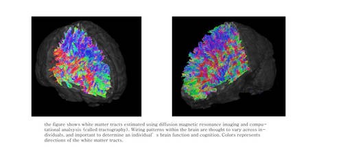
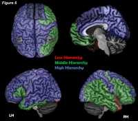
Response: The Krakencoder is a tool that allows us to compactly represent brain networks, or the connections between different parts of the brain. This compact representation helps us to take a step toward achieving the goal of understanding how complex human behavior, like thinking, social interactions, and emotion, arise from the complex network that is the human brain.
Prof. Kuceyeski[/caption]
MedicalResearch.com Interview with:
Prof. Amy Kuceyeski Ph.D.
Professor of Mathematics in Radiology and Neuroscience
Weill Cornell Medicine
MedicalResearch.com: What is the purpose of the Krankencoder tool?
Response: The Krakencoder is a tool that allows us to compactly represent brain networks, or the connections between different parts of the brain. This compact representation helps us to take a step toward achieving the goal of understanding how complex human behavior, like thinking, social interactions, and emotion, arise from the complex network that is the human brain.

 Dr. Akefe[/caption]
Isaac O Akefe DVM, PhD
Clem Jones Centre for Ageing Dementia Research
Queensland Brain Institute
The University of Queensland St Lucia
Academy for Medical Education, Medical School
Brisbane QLD Australia
MedicalResearch.com: What is the background for this study?
Response: The brain is the body’s fattiest organ, with fatty compounds called lipids making up 60% of its weight. Fatty acids are the building blocks of a class of lipids called phospholipids.
In our study, we first showed that levels of saturated fatty acids increase in the brain during neuronal communication and long-term memory formation, but we didn’t know what was causing these changes.
Dr. Akefe[/caption]
Isaac O Akefe DVM, PhD
Clem Jones Centre for Ageing Dementia Research
Queensland Brain Institute
The University of Queensland St Lucia
Academy for Medical Education, Medical School
Brisbane QLD Australia
MedicalResearch.com: What is the background for this study?
Response: The brain is the body’s fattiest organ, with fatty compounds called lipids making up 60% of its weight. Fatty acids are the building blocks of a class of lipids called phospholipids.
In our study, we first showed that levels of saturated fatty acids increase in the brain during neuronal communication and long-term memory formation, but we didn’t know what was causing these changes.

 Prof. Durazzo[/caption]
Timothy C. Durazzo, PhD
Clinical Neuropsychologist and Research Scientist
Mental Illness Research and Education Clinical Centers
VA Palo Alto Health Care System
Professor, Department of Psychiatry and Behavioral Sciences
Stanford University School of Medicine
MedicalResearch.com: What is the background for this study?
-There are a limited number of studies investigating changes in human brain structure, in individuals with an alcohol use disorder, with longer term abstinence after treatment.
-Our study was the first to assess for change in cortical thickness over approximately 7 months of abstinence in those seeking treatment of alcohol use disorder.
-Cortical thickness in humans is genetically and phenotypically distinct from other brain structural measures such as cortical volume and surface area.
-Therefore, assessment of changes in cortical thickness with longer-term abstinence provides additional information on how human brain structure recovers with sobriety.
Prof. Durazzo[/caption]
Timothy C. Durazzo, PhD
Clinical Neuropsychologist and Research Scientist
Mental Illness Research and Education Clinical Centers
VA Palo Alto Health Care System
Professor, Department of Psychiatry and Behavioral Sciences
Stanford University School of Medicine
MedicalResearch.com: What is the background for this study?
-There are a limited number of studies investigating changes in human brain structure, in individuals with an alcohol use disorder, with longer term abstinence after treatment.
-Our study was the first to assess for change in cortical thickness over approximately 7 months of abstinence in those seeking treatment of alcohol use disorder.
-Cortical thickness in humans is genetically and phenotypically distinct from other brain structural measures such as cortical volume and surface area.
-Therefore, assessment of changes in cortical thickness with longer-term abstinence provides additional information on how human brain structure recovers with sobriety.

 Valentina Paz[/caption]
Valentina Paz, M.Sc
Valentina Paz[/caption]
Valentina Paz, M.Sc 

 Dr, Ferguson[/caption]
Michael Ferguson, PhD
Instructor in Neurology | Harvard Medical School
Lecturer on Neurospirituality | Harvard Divinity School
Center for Brain Circuit Therapeutics
Brigham and Women’s Hospital
MedicalResearch.com: What is the background for this study?
Response: Over 80% of the global population consider themselves religious with even more identifying as spiritual, but the neural substrates of spirituality and religiosity remain unresolved.
MedicalResearch.com: What are the main findings? Where is this circuit located in the brain? What other effects does this circuit control or influence?
Response: We found that brain lesions associated with self-reported spirituality map to a human brain circuit centered on the periaqueductal grey.
Dr, Ferguson[/caption]
Michael Ferguson, PhD
Instructor in Neurology | Harvard Medical School
Lecturer on Neurospirituality | Harvard Divinity School
Center for Brain Circuit Therapeutics
Brigham and Women’s Hospital
MedicalResearch.com: What is the background for this study?
Response: Over 80% of the global population consider themselves religious with even more identifying as spiritual, but the neural substrates of spirituality and religiosity remain unresolved.
MedicalResearch.com: What are the main findings? Where is this circuit located in the brain? What other effects does this circuit control or influence?
Response: We found that brain lesions associated with self-reported spirituality map to a human brain circuit centered on the periaqueductal grey.


 Dr. Vazza


 Dr. Qing Chen[/caption]
Qing Chen, M.D., Ph.D.
Assistant Professor, Immunology, Microenvironment & Metastasis Program
Scientific Director, Imaging Facility
The Wistar Institute
MedicalResearch.com: What is the background for this study?
Response: We are focusing on how a specific type of brain cells, astrocytes, helps the cancer cells from melanoma and breast cancer to form metastatic lesions.
Dr. Qing Chen[/caption]
Qing Chen, M.D., Ph.D.
Assistant Professor, Immunology, Microenvironment & Metastasis Program
Scientific Director, Imaging Facility
The Wistar Institute
MedicalResearch.com: What is the background for this study?
Response: We are focusing on how a specific type of brain cells, astrocytes, helps the cancer cells from melanoma and breast cancer to form metastatic lesions.