MedicalResearch.com Interview with:
[caption id="attachment_23730" align="alignleft" width="114"]

Dr. Zhimin Lu[/caption]
Zhimin (James) Lu, M. D., Ph. D
Ruby E. Rutherford Distinguished Professor
Department of Neuro-Oncology
MD Anderson Cancer Center
Houston, TX 77030
MedicalResearch.com: What is the background for this study?
Dr. Lu: Among primary liver cancers, hepatocellular carcinoma (HCC) is the most common histological subtype, accounting for 70-85% of all cases. HCC incidence is increasing in many parts of the world, including developing countries and developed countries such as the United States. HCC has a very poor prognosis, and the overall 3-year survival rate for patients with HCC is approximately 5%. The potentially curative treatments of HCC are resection and liver transplantation. However, most patients with hepatocellular carcinoma present with advanced disease and underlying liver dysfunction and are not suitable candidates for these treatments. Thus, they generally have a poor prognosis, with a median survival time of less than 1 year. The increasing incidence and mortality rates of hepatocellular carcinoma, along with a lack of effective curative treatment options for advanced HCC, have rendered this disease a major health problem worldwide. Thus, a better understanding of HCC tumorigenesis and the development of better diagnostic and therapeutic approaches based on an understanding of the molecular mechanisms that drive hepatocellular carcinoma progression are greatly needed.
The liver, as a major metabolic organ, catalyzes dietary sugar. Dietary sugar encompasses several carbohydrates, including starch, sucrose, and high-fructose corn syrup, each of which is composed of glucose with or without fructose. Starch, which is found in bread and rice, is a glucose polymer. Sucrose is a disaccharide made up of 50% glucose and 50% fructose. High-fructose corn syrup, a common constituent of soft drinks, is a mixture of approximately 40% glucose and 60% fructose. Dietary fructose is also derived from fruits and vegetables. A molecule of glucose has the same caloric value as a molecule of fructose. However, the human body treats these carbohydrates quite differently. Glucose is used directly by tissues such as the muscles and brain as an energy source. Excess glucose is stored in the liver as glycogen. In contrast, dietary fructose, which is epidemiologically linked with obesity and metabolic syndrome, is almost exclusively metabolized by the liver.
Hepatocellular carcinoma cells enhance glucose uptake and lactate production regardless of the oxygen supply, a phenomenon known as the Warburg effect. However, whether fructose metabolism is differentially regulated in hepatocellular carcinoma and normal liver tissue and, if so, the extent to which this altered carbohydrate metabolism contributes to hepatocellular carcinoma development is unknown.

 Jayant S Vaidya MBBS MS DNB FRCS PhD
Professor of Surgery and Oncology,
Scientific Director, Clinical Trials Group,
Division of Surgery and Interventional Science,
University College London
Whittington Health - Clinical Lead for Breast Cancer
Royal Free Hospital
University College London Hospital
MedicalResearch.com: What is the background for this study? What are the main findings?
Prof. Vaidya: TARGIT-A randomised clinical trial (ISRCTN34086741) compared giving TARGIT IORT during lumpectomy vs. traditional EBRT given over several weeks after lumpectomy for breast cancer; local-recurrence-free-survival was similar in the two arms of the trial, particularly when TARGIT was given simultaneously with lumpectomy. Also, there were significantly fewer deaths from other causes with TARGIT IORT.
This study calculated journeys made by patients with breast cancer to receive their radiotherapy, using the geographic and treatment data from a large randomised trial.
The study then assessed the same outcomes (travel distances, travel time and CO2emissions) in two semi-rural breast cancers—the results of this assessment confirm and reinforce the original results: the benefit of the use of TARGIT for patients from two semi=rural breast centres was even larger (753 miles (1212 km), 30 h, 215 kg CO2 per patient).
Jayant S Vaidya MBBS MS DNB FRCS PhD
Professor of Surgery and Oncology,
Scientific Director, Clinical Trials Group,
Division of Surgery and Interventional Science,
University College London
Whittington Health - Clinical Lead for Breast Cancer
Royal Free Hospital
University College London Hospital
MedicalResearch.com: What is the background for this study? What are the main findings?
Prof. Vaidya: TARGIT-A randomised clinical trial (ISRCTN34086741) compared giving TARGIT IORT during lumpectomy vs. traditional EBRT given over several weeks after lumpectomy for breast cancer; local-recurrence-free-survival was similar in the two arms of the trial, particularly when TARGIT was given simultaneously with lumpectomy. Also, there were significantly fewer deaths from other causes with TARGIT IORT.
This study calculated journeys made by patients with breast cancer to receive their radiotherapy, using the geographic and treatment data from a large randomised trial.
The study then assessed the same outcomes (travel distances, travel time and CO2emissions) in two semi-rural breast cancers—the results of this assessment confirm and reinforce the original results: the benefit of the use of TARGIT for patients from two semi=rural breast centres was even larger (753 miles (1212 km), 30 h, 215 kg CO2 per patient).
 Dr. Corrine Leach[/caption]
Corinne Leach, MPH, MS, PHD
Strategic Director, Cancer and Aging Research
American Cancer Society, Inc.
Atlanta, GA 30303
MedicalResearch.com: What is the background for this study? What are the main findings?
Dr. Leach: Using linked data from cancer registries and the Medicare Health Outcomes Survey, we prospectively examined the short-term impact of cancer on the functioning, development of and worsening of age-related health conditions among 921 older adults who developed cancer compared to 4,605 propensity score matched controls. We found that cancer groups demonstrated greater declines in activities of daily living and physical functioning compared to controls with the greatest change for lung cancer patients. Having a cancer diagnosis increased risk for depression but did not increase the odds of developing arthritis in the hand/hip, incontinence (except for prostate cancer), or vision/hearing problems. Having a cancer diagnosis also did not exacerbate the severity of arthritis or foot neuropathy.
Dr. Corrine Leach[/caption]
Corinne Leach, MPH, MS, PHD
Strategic Director, Cancer and Aging Research
American Cancer Society, Inc.
Atlanta, GA 30303
MedicalResearch.com: What is the background for this study? What are the main findings?
Dr. Leach: Using linked data from cancer registries and the Medicare Health Outcomes Survey, we prospectively examined the short-term impact of cancer on the functioning, development of and worsening of age-related health conditions among 921 older adults who developed cancer compared to 4,605 propensity score matched controls. We found that cancer groups demonstrated greater declines in activities of daily living and physical functioning compared to controls with the greatest change for lung cancer patients. Having a cancer diagnosis increased risk for depression but did not increase the odds of developing arthritis in the hand/hip, incontinence (except for prostate cancer), or vision/hearing problems. Having a cancer diagnosis also did not exacerbate the severity of arthritis or foot neuropathy.
 Dr. Josefin Segelman[/caption]
Josefin Segelman MD, PhD
Senior consultant colorectal surgeon
Department of Molecular Medicine and Surgery
Karolinska Institutet
Ersta Hospital
Stockholm Sweden
MedicalResearch.com: What is the background for this study?
Dr. Segelman: Hormonal factors influence the development of colorectal cancer. Observational studies and clinical trials have reported a protective effect of hormone replacement therapy and oral contraceptives. Oophorectomy alters endogenous levels of sex hormones, but the effect on colorectal cancer risk is unclear. Removal of the ovaries alters levels of sex hormones in both pre- and postmenopausal women. In premenopausal women, bilateral oophorectomy is followed by surgical menopause as the endogenous estrogen levels drop. Both before and after natural menopause, bilateral oophorectomy promptly decreases endogenous androgen levels by half as the ovaries and adrenals are equally important for androgen production.
MedicalResearch.com: What are the main findings?
Dr. Segelman: The present nationwide cohort study explored the association between removal of the ovaries for benign indications and subsequent risk of colorectal cancer. Among 195 973 women who underwent the procedure from 1965 – 2011, there was a 30% increased risk of colorectal cancer compared with the general population. After adjustment for various factors, women who underwent bilateral oophorectomy had a higher risk of rectal cancer than those who had unilateral oophorectomy (HR 2.28, 95% CI 1.33-3.91).
Dr. Josefin Segelman[/caption]
Josefin Segelman MD, PhD
Senior consultant colorectal surgeon
Department of Molecular Medicine and Surgery
Karolinska Institutet
Ersta Hospital
Stockholm Sweden
MedicalResearch.com: What is the background for this study?
Dr. Segelman: Hormonal factors influence the development of colorectal cancer. Observational studies and clinical trials have reported a protective effect of hormone replacement therapy and oral contraceptives. Oophorectomy alters endogenous levels of sex hormones, but the effect on colorectal cancer risk is unclear. Removal of the ovaries alters levels of sex hormones in both pre- and postmenopausal women. In premenopausal women, bilateral oophorectomy is followed by surgical menopause as the endogenous estrogen levels drop. Both before and after natural menopause, bilateral oophorectomy promptly decreases endogenous androgen levels by half as the ovaries and adrenals are equally important for androgen production.
MedicalResearch.com: What are the main findings?
Dr. Segelman: The present nationwide cohort study explored the association between removal of the ovaries for benign indications and subsequent risk of colorectal cancer. Among 195 973 women who underwent the procedure from 1965 – 2011, there was a 30% increased risk of colorectal cancer compared with the general population. After adjustment for various factors, women who underwent bilateral oophorectomy had a higher risk of rectal cancer than those who had unilateral oophorectomy (HR 2.28, 95% CI 1.33-3.91).
 Dr. Reina Haque[/caption]
Reina Haque, PhD MPH
Research scientist
Kaiser Permanente Southern California Department of Research & Evaluation
MedicalResearch.com: What is the background for this study? What are the main findings?
Dr. Haque: The study fills an important knowledge gap about the long-term association of aromatase inhibitors on cardiovascular disease risk in breast cancer survivors.
This was a retrospective cohort study that included a cohort of 13,273 postmenopausal breast cancer survivors who were diagnosed with breast cancer, either estrogen or progesterone receptor positive, from 1991 to 2010. The patients were followed through 2011, or a maximum of 21 years. The study participants were divided into four groups based on the drugs they received: 31.7 percent were treated only with tamoxifen; 28.6 percent only with aromatase inhibitors; 20.2 percent used both; and 19.4 percent did not use any of these drugs. These oral drugs are used to combat breast cancer recurrence, but may have long-term side effects on other organs.
The study determined that the risk of cardiac ischemia (which can lead to a heart attack) and stroke were not elevated in patients who only took aromatase inhibitors compared to those who only took tamoxifen. These results provide reassurance that aromatase inhibitors may not increase risk of the potentially fatal cardiovascular outcomes compared to tamoxifen.
Dr. Reina Haque[/caption]
Reina Haque, PhD MPH
Research scientist
Kaiser Permanente Southern California Department of Research & Evaluation
MedicalResearch.com: What is the background for this study? What are the main findings?
Dr. Haque: The study fills an important knowledge gap about the long-term association of aromatase inhibitors on cardiovascular disease risk in breast cancer survivors.
This was a retrospective cohort study that included a cohort of 13,273 postmenopausal breast cancer survivors who were diagnosed with breast cancer, either estrogen or progesterone receptor positive, from 1991 to 2010. The patients were followed through 2011, or a maximum of 21 years. The study participants were divided into four groups based on the drugs they received: 31.7 percent were treated only with tamoxifen; 28.6 percent only with aromatase inhibitors; 20.2 percent used both; and 19.4 percent did not use any of these drugs. These oral drugs are used to combat breast cancer recurrence, but may have long-term side effects on other organs.
The study determined that the risk of cardiac ischemia (which can lead to a heart attack) and stroke were not elevated in patients who only took aromatase inhibitors compared to those who only took tamoxifen. These results provide reassurance that aromatase inhibitors may not increase risk of the potentially fatal cardiovascular outcomes compared to tamoxifen.
 Dr. Orit Markowitz[/caption]
Orit Markowitz, MD, FAAD
Director of Pigmented Lesions and Skin Cancer
Assistant Professor of Dermatology
Mount Sinai Medical Center, NY, NY
Director of Pigmented lesions clinic
Brooklyn VA, Brooklyn, NY
Adjunct Professor, Dermatology
SUNY Downstate Medical Center, Brooklyn, NY
Chief of Dermatology
Queens General Hospital, Jamaica, NY
MedicalResearch.com: How common is skin cancer? Is the incidence rising in US adults? Who is most at risk?
Dr. Markowitz: The annual incidence of skin cancer is more than breast, colon, lung, and prostate cancer combined. Of the 7 most common skin cancers in the US melanoma is the only one whose incidence is increasing. The highest risk group for skin cancer are fair skin, adults with a history of sun exposure.
Dr. Orit Markowitz[/caption]
Orit Markowitz, MD, FAAD
Director of Pigmented Lesions and Skin Cancer
Assistant Professor of Dermatology
Mount Sinai Medical Center, NY, NY
Director of Pigmented lesions clinic
Brooklyn VA, Brooklyn, NY
Adjunct Professor, Dermatology
SUNY Downstate Medical Center, Brooklyn, NY
Chief of Dermatology
Queens General Hospital, Jamaica, NY
MedicalResearch.com: How common is skin cancer? Is the incidence rising in US adults? Who is most at risk?
Dr. Markowitz: The annual incidence of skin cancer is more than breast, colon, lung, and prostate cancer combined. Of the 7 most common skin cancers in the US melanoma is the only one whose incidence is increasing. The highest risk group for skin cancer are fair skin, adults with a history of sun exposure.

 Dr. Asal Mohamadi Johnson[/caption]
Asal Mohamadi Johnson, PhD, MPH
Assistant Professor of Epidemiology, Integrative Health Science
Stetson University
DeLand, FL 32723
MedicalResearch.com: What is the background for this study?
Dr. Johnson: Public health research is primarily focused on neighborhood poverty and racial disparities by illustrating differences between white and black individuals or communities. For example, it has been established that African Americans have higher cancer mortality rates and are less likely to receive appropriate treatment that whites. What we wanted to know in this study was the impact of living in segregated areas apart from other area level characteristics such as poverty or education. Instead of solely looking at health disparities between whites and black patients, our study focused on differences in survival among black patients with early stage Non-Small Cell Lung Cancer (NSCLC) living in different levels of neighborhood segregation.
Dr. Asal Mohamadi Johnson[/caption]
Asal Mohamadi Johnson, PhD, MPH
Assistant Professor of Epidemiology, Integrative Health Science
Stetson University
DeLand, FL 32723
MedicalResearch.com: What is the background for this study?
Dr. Johnson: Public health research is primarily focused on neighborhood poverty and racial disparities by illustrating differences between white and black individuals or communities. For example, it has been established that African Americans have higher cancer mortality rates and are less likely to receive appropriate treatment that whites. What we wanted to know in this study was the impact of living in segregated areas apart from other area level characteristics such as poverty or education. Instead of solely looking at health disparities between whites and black patients, our study focused on differences in survival among black patients with early stage Non-Small Cell Lung Cancer (NSCLC) living in different levels of neighborhood segregation.
 Dr. Paul Nghiem[/caption]
Paul Nghiem, MD, PhD
Professor & Head, University of Washington Dermatology
George F. Odland Endowed Chair
Affiliate Investigator, Fred Hutchinson Cancer Research Center
Professor, Adjunct, of Pathology and Oral Health Sciences
Clinical Director, Skin Oncology, Seattle Cancer Care Alliance
UW Medical Center at Lake Union
Seattle WA 98109
MedicalResearch.com: What is the background for this study? What are the main findings?
Dr. Nghiem: Merkel cell carcinoma (MCC) is about 30 times less common than malignant melanoma, but about 3 times more likely to kill a patient than a melanoma. There is no FDA-approved therapy for this cancer & chemotherapy typically only provides about 90 days prior to the cancer progressing. Because of the strong links between MCC and the immune system, including the fact that most MCCs are caused by a virus, there was interest in trying to use immune checkpoint therapy to treat advanced Merkel cell carcinoma. The response to immune stimulation with anti-PD1 therapy was about as frequent as to chemotherapy (56% of patients responded) but importantly, among the responders, 86% remained in ongoing responses at a median of 7.6 months. While still early, this appears to be strikingly more durable than responses to chemotherapy.
Dr. Paul Nghiem[/caption]
Paul Nghiem, MD, PhD
Professor & Head, University of Washington Dermatology
George F. Odland Endowed Chair
Affiliate Investigator, Fred Hutchinson Cancer Research Center
Professor, Adjunct, of Pathology and Oral Health Sciences
Clinical Director, Skin Oncology, Seattle Cancer Care Alliance
UW Medical Center at Lake Union
Seattle WA 98109
MedicalResearch.com: What is the background for this study? What are the main findings?
Dr. Nghiem: Merkel cell carcinoma (MCC) is about 30 times less common than malignant melanoma, but about 3 times more likely to kill a patient than a melanoma. There is no FDA-approved therapy for this cancer & chemotherapy typically only provides about 90 days prior to the cancer progressing. Because of the strong links between MCC and the immune system, including the fact that most MCCs are caused by a virus, there was interest in trying to use immune checkpoint therapy to treat advanced Merkel cell carcinoma. The response to immune stimulation with anti-PD1 therapy was about as frequent as to chemotherapy (56% of patients responded) but importantly, among the responders, 86% remained in ongoing responses at a median of 7.6 months. While still early, this appears to be strikingly more durable than responses to chemotherapy.
 Dr. Stephen Freedland[/caption]
Stephen J. Freedland, MD
Associate Director, Faculty Development Samuel Oschin Comprehensive Cancer Institute
Co-Director, Cancer Genetics and Prevention Program
Director, Center for Integrated Research in Cancer and Lifestyle
Professor, Surgery
Warschaw Robertson Law Families Chair in Prostate Cancer
Cedars-Sinai, Los Angeles
MedicalResearch.com: What is the background for this study? What are the main findings?
Dr. Freedland: PSA is a marker of prostate pathology. While often used to screen for prostate cancer, it is not prostate specific and can be elevated due to inflammation or enlarged prostate or other reasons. Whether it predicts the development of urinary symptoms is not clear. Among men with minimal to no urinary symptoms, we found that the higher the PSA, the greater the risk of future development of urinary symptoms.
MedicalResearch.com: What should clinicians and patients take away from your report?
Dr. Freedland: The readers should know that if a man has an elevated PSA and a negative prostate biopsy, the higher the PSA, the greater the risk of future urinary symptoms. These are men who may need closer follow-up.
Dr. Stephen Freedland[/caption]
Stephen J. Freedland, MD
Associate Director, Faculty Development Samuel Oschin Comprehensive Cancer Institute
Co-Director, Cancer Genetics and Prevention Program
Director, Center for Integrated Research in Cancer and Lifestyle
Professor, Surgery
Warschaw Robertson Law Families Chair in Prostate Cancer
Cedars-Sinai, Los Angeles
MedicalResearch.com: What is the background for this study? What are the main findings?
Dr. Freedland: PSA is a marker of prostate pathology. While often used to screen for prostate cancer, it is not prostate specific and can be elevated due to inflammation or enlarged prostate or other reasons. Whether it predicts the development of urinary symptoms is not clear. Among men with minimal to no urinary symptoms, we found that the higher the PSA, the greater the risk of future development of urinary symptoms.
MedicalResearch.com: What should clinicians and patients take away from your report?
Dr. Freedland: The readers should know that if a man has an elevated PSA and a negative prostate biopsy, the higher the PSA, the greater the risk of future urinary symptoms. These are men who may need closer follow-up.  Dr. J. William Harbour[/caption]
 Dr. Stacie Dusetzina[/caption]
Stacie B. Dusetzina, PhD
Assistant Professor
Division of Pharmaceutical Outcomes and Policy
Eshelman School of Pharmacy
University of North Carolina at Chapel Hill
Chapel Hill, NC
MedicalResearch.com: What is the background for this study? What are the main findings?
Dr. Dusetzina: Drug prices are of significant policy interest, particularly the prices for so-called “specialty” medications which are used to treat rare and/or complex conditions like cancer. In this study I estimated monthly price for orally-administered cancer treatments that were approved between 2000 and 2014. First I looked at the price of the drug during the year of initial FDA approval and then I looked at annual changes in the price after the year of approval. The main findings are that, even after inflation adjustment, the monthly price paid for orally-administered cancer treatments is increasing rapidly both at the time of approval and in subsequent years.
As an example, if you compare average monthly prices during the first year post-approval for treatments approved between 2000-2010 to those approved after 2010 there was a major increase in launch prices from $5,529 per month to $9,013 per month. Year-to-year changes in price after launch varied a lot by drug ranging from decreases in price of -2.7% per year to increases of 11.4% per year. However, nearly all of the products studied increased in price over time.
Dr. Stacie Dusetzina[/caption]
Stacie B. Dusetzina, PhD
Assistant Professor
Division of Pharmaceutical Outcomes and Policy
Eshelman School of Pharmacy
University of North Carolina at Chapel Hill
Chapel Hill, NC
MedicalResearch.com: What is the background for this study? What are the main findings?
Dr. Dusetzina: Drug prices are of significant policy interest, particularly the prices for so-called “specialty” medications which are used to treat rare and/or complex conditions like cancer. In this study I estimated monthly price for orally-administered cancer treatments that were approved between 2000 and 2014. First I looked at the price of the drug during the year of initial FDA approval and then I looked at annual changes in the price after the year of approval. The main findings are that, even after inflation adjustment, the monthly price paid for orally-administered cancer treatments is increasing rapidly both at the time of approval and in subsequent years.
As an example, if you compare average monthly prices during the first year post-approval for treatments approved between 2000-2010 to those approved after 2010 there was a major increase in launch prices from $5,529 per month to $9,013 per month. Year-to-year changes in price after launch varied a lot by drug ranging from decreases in price of -2.7% per year to increases of 11.4% per year. However, nearly all of the products studied increased in price over time.
 Donghao Lu[/caption]
Donghao Lu MD, PhD candidate
Department of Medical Epidemiology & Biostatistics,
Karolinska Institutet
Stockholm
MedicalResearch.com: What is the background for this study? What are the main findings?
Dr. Lu: Psychiatric comorbidities are common among cancer patients. However, whether or not there is already increased risk of psychiatric disorders during the diagnostic workup leading to a cancer diagnosis was largely unknown.
We found that, among cancer patients, the risks for several common and potentially stress-related mental disorders, including depression, anxiety, substance abuse, somatoform/conversion disorder and stress reaction/adjustment disorder started to increase from ten months before cancer diagnosis, peaked during the first week after diagnosis, compared to cancer-free individuals in Sweden.
Donghao Lu[/caption]
Donghao Lu MD, PhD candidate
Department of Medical Epidemiology & Biostatistics,
Karolinska Institutet
Stockholm
MedicalResearch.com: What is the background for this study? What are the main findings?
Dr. Lu: Psychiatric comorbidities are common among cancer patients. However, whether or not there is already increased risk of psychiatric disorders during the diagnostic workup leading to a cancer diagnosis was largely unknown.
We found that, among cancer patients, the risks for several common and potentially stress-related mental disorders, including depression, anxiety, substance abuse, somatoform/conversion disorder and stress reaction/adjustment disorder started to increase from ten months before cancer diagnosis, peaked during the first week after diagnosis, compared to cancer-free individuals in Sweden.
 Dr. Stamatia Destounis[/caption]
Stamatia Destounis, MD, FSBI, FACR
Elizabeth Wende Breast Care, LLC,
Clinical Professor of Imaging Sciences
University of Rochester
School of Medicine and Dentistry
Rochester NY 14620
MedicalResearch.com: What is the background for this study? What are the main findings?
Dr. Destounis: Identification of women who have an increased risk of breast cancer is important, as they are often eligible for additional screening methods, such as breast MRI. One criterion for eligibility for screening breast MRI is >20% lifetime risk of breast cancer, as determined by risk assessment models through genetic counseling.
At my facility, we have incorporated a genetics program. Through the program we are flagging and identifying a large volume of patients who are potentially eligible for additional services. This study was conducted to determine the value of screening MRI in the patient subgroup who have undergone genetic counseling at my facility. In this group we found 50% of patients who were referred for counseling were also recommended to have screening MRI. However, only 21.3% of those recommended actually pursued the exam. Of those patients who did have a screening MRI, 4 were diagnosed with breast cancer, all of which were invasive and node negative. We ultimately had a 10% biopsy rate and 50% cancer detection rate in this subgroup.
Dr. Stamatia Destounis[/caption]
Stamatia Destounis, MD, FSBI, FACR
Elizabeth Wende Breast Care, LLC,
Clinical Professor of Imaging Sciences
University of Rochester
School of Medicine and Dentistry
Rochester NY 14620
MedicalResearch.com: What is the background for this study? What are the main findings?
Dr. Destounis: Identification of women who have an increased risk of breast cancer is important, as they are often eligible for additional screening methods, such as breast MRI. One criterion for eligibility for screening breast MRI is >20% lifetime risk of breast cancer, as determined by risk assessment models through genetic counseling.
At my facility, we have incorporated a genetics program. Through the program we are flagging and identifying a large volume of patients who are potentially eligible for additional services. This study was conducted to determine the value of screening MRI in the patient subgroup who have undergone genetic counseling at my facility. In this group we found 50% of patients who were referred for counseling were also recommended to have screening MRI. However, only 21.3% of those recommended actually pursued the exam. Of those patients who did have a screening MRI, 4 were diagnosed with breast cancer, all of which were invasive and node negative. We ultimately had a 10% biopsy rate and 50% cancer detection rate in this subgroup.
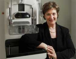 Dr. Elizabeth Rafferty[/caption]
Elizabeth A. Rafferty, MD
Department of Radiology,
Massachusetts General Hospital, Boston
Now with L&M Radiology, West Acton,
Massachusetts
MedicalResearch.com: What is the background for this study? What are the main findings?
Dr. Rafferty: Breast tomosynthesis has been approved for mammographic screening in the United States for just over 5 years, and many single center studies have demonstrated its improved performance for screening outcomes over digital mammography alone. Our previously published multi-center analysis, (JAMA 2014;311(24), the largest study on this topic to date, demonstrated significantly improved cancer detection and reduced recall rates for women undergoing tomosynthesis compared with digital mammography alone. In the current issue of JAMA we evaluate the differential screening performance after implementation of breast tomosynthesis as a function of breast density.
While tomosynthesis continues to be increasingly available, questions remained about which women should be imaged with this technique. In particular, does this technology offer additional benefit for all women, or only for women with dense breasts. The size of the database compiled by the centers participating in this study allowed us to evaluate this important question.
The most critical finding of our study was that the use of tomosynthesis for breast cancer screening significantly improved invasive cancer detection rates while simultaneously significantly reducing recall rates both for women with dense and non-dense breast tissue. Having said that, the magnitude of the benefit was largest for women with heterogeneously dense breast tissue; for this population, tomosynthesis increased the detection of invasive cancers by 50% while simultaneously reducing the recall rate by 14%.
Dr. Elizabeth Rafferty[/caption]
Elizabeth A. Rafferty, MD
Department of Radiology,
Massachusetts General Hospital, Boston
Now with L&M Radiology, West Acton,
Massachusetts
MedicalResearch.com: What is the background for this study? What are the main findings?
Dr. Rafferty: Breast tomosynthesis has been approved for mammographic screening in the United States for just over 5 years, and many single center studies have demonstrated its improved performance for screening outcomes over digital mammography alone. Our previously published multi-center analysis, (JAMA 2014;311(24), the largest study on this topic to date, demonstrated significantly improved cancer detection and reduced recall rates for women undergoing tomosynthesis compared with digital mammography alone. In the current issue of JAMA we evaluate the differential screening performance after implementation of breast tomosynthesis as a function of breast density.
While tomosynthesis continues to be increasingly available, questions remained about which women should be imaged with this technique. In particular, does this technology offer additional benefit for all women, or only for women with dense breasts. The size of the database compiled by the centers participating in this study allowed us to evaluate this important question.
The most critical finding of our study was that the use of tomosynthesis for breast cancer screening significantly improved invasive cancer detection rates while simultaneously significantly reducing recall rates both for women with dense and non-dense breast tissue. Having said that, the magnitude of the benefit was largest for women with heterogeneously dense breast tissue; for this population, tomosynthesis increased the detection of invasive cancers by 50% while simultaneously reducing the recall rate by 14%.
 Dr. Reddy[/caption]
E. Premkumar Reddy, Ph.D.
Professor, Department of Oncological Sciences
and Department of Structural and Chemical Biology
Director, Experimental Cancer Therapeutics
Mount Sinai School of Medicine
New York, NY 10029
MedicalResearch.com: What is the background for this study? What are the main findings?
Dr. Reddy: It is now well established that cancer cells harbor mutations in their genome which are responsible for uncontrolled proliferation. Nearly 30 years ago, we as well as two other groups discovered that RAS genes are often mutated in human cancers. Later studies showed that nearly 25-30% of human cancers contain this mutation and these mutations can be caused by chemical carcinogens such as tobacco smoke. Since then there has been an intense effort to understand the biological functions of RAS and to develop drugs that block the activity of these mutant RAS genes. Although molecular oncologists have made significant headway in understanding these mutations and their impact on cellular signaling, little progress has been made towards developing drugs that systematically target the RAS oncogenes. This lack of progress has led many in the field to label RAS as “undruggable”. However, basic research conducted by scientists in this field has revealed that RAS proteins function as ON-OFF switches to signal cells to grow or not to grow because of their ability to bind to a large number of cellular proteins and transmit this signal to their binding partners. These findings also revealed that mutations in RAS genes leaves them in a permanent “ON” position which continues to transmit growth signals permanently. Since most efforts to develop drugs that bind to RAS proteins and reverse their activity failed, we took a different approach to block these signals. Since transmission of growth signals by RAS genes requires their interaction with cellular proteins, all of which contain a domain called “RAS-Binding Domain (RBD)” we created a drug named “Rigosertib” that binds to these RBDs and block their binding to RAS, thereby interrupting RAS signals. When Rigosertib was tested in animals, it could readily inhibit the growth of human tumors that contain RAS mutations. Our studies also show that Rigosertib is a very safe drug with minimal side-effects.
Dr. Reddy[/caption]
E. Premkumar Reddy, Ph.D.
Professor, Department of Oncological Sciences
and Department of Structural and Chemical Biology
Director, Experimental Cancer Therapeutics
Mount Sinai School of Medicine
New York, NY 10029
MedicalResearch.com: What is the background for this study? What are the main findings?
Dr. Reddy: It is now well established that cancer cells harbor mutations in their genome which are responsible for uncontrolled proliferation. Nearly 30 years ago, we as well as two other groups discovered that RAS genes are often mutated in human cancers. Later studies showed that nearly 25-30% of human cancers contain this mutation and these mutations can be caused by chemical carcinogens such as tobacco smoke. Since then there has been an intense effort to understand the biological functions of RAS and to develop drugs that block the activity of these mutant RAS genes. Although molecular oncologists have made significant headway in understanding these mutations and their impact on cellular signaling, little progress has been made towards developing drugs that systematically target the RAS oncogenes. This lack of progress has led many in the field to label RAS as “undruggable”. However, basic research conducted by scientists in this field has revealed that RAS proteins function as ON-OFF switches to signal cells to grow or not to grow because of their ability to bind to a large number of cellular proteins and transmit this signal to their binding partners. These findings also revealed that mutations in RAS genes leaves them in a permanent “ON” position which continues to transmit growth signals permanently. Since most efforts to develop drugs that bind to RAS proteins and reverse their activity failed, we took a different approach to block these signals. Since transmission of growth signals by RAS genes requires their interaction with cellular proteins, all of which contain a domain called “RAS-Binding Domain (RBD)” we created a drug named “Rigosertib” that binds to these RBDs and block their binding to RAS, thereby interrupting RAS signals. When Rigosertib was tested in animals, it could readily inhibit the growth of human tumors that contain RAS mutations. Our studies also show that Rigosertib is a very safe drug with minimal side-effects.
 Dr. Nelly Tan[/caption]
Dr. Nelly Tan MD
David Geffen School of Medicine
Department of Radiology
UCLA
MedicalResearch.com: What is the background for this study? What are the main findings?
Dr. Tan: Standard of care for prostate cancer diagnosis has been to perform ultrasound guided random (non-targeted) prostate biopsy (TRUS) which is neither sensitive or specific. The main limitation had been our inability to detect and localize prostate cancer through imaging.
Over the past 10 years, MRI has taken center stage for detection and localization of prostate cancer and has shown to improve prostate cancer diagnosis, risk stratification, and staging of the disease. Over the past few years, MRI guided biopsy techniques (in the form of Ultrasound-MRI (US-MRI) fusion and in-bore direct MRI guided biopsy) have been reported. We reported our performance of direct in-bore MRI guided biopsy at UCLA. Our study showed a prostate cancer diagnosis of 59% in all patients and 80% of patients with prostate cancer had clinically significant cancer.
Dr. Nelly Tan[/caption]
Dr. Nelly Tan MD
David Geffen School of Medicine
Department of Radiology
UCLA
MedicalResearch.com: What is the background for this study? What are the main findings?
Dr. Tan: Standard of care for prostate cancer diagnosis has been to perform ultrasound guided random (non-targeted) prostate biopsy (TRUS) which is neither sensitive or specific. The main limitation had been our inability to detect and localize prostate cancer through imaging.
Over the past 10 years, MRI has taken center stage for detection and localization of prostate cancer and has shown to improve prostate cancer diagnosis, risk stratification, and staging of the disease. Over the past few years, MRI guided biopsy techniques (in the form of Ultrasound-MRI (US-MRI) fusion and in-bore direct MRI guided biopsy) have been reported. We reported our performance of direct in-bore MRI guided biopsy at UCLA. Our study showed a prostate cancer diagnosis of 59% in all patients and 80% of patients with prostate cancer had clinically significant cancer.