More Cancer Research on MedicalResearch.com
[caption id="attachment_20709" align="alignleft" width="156"]

Dr. Erin Hahn[/caption]
MedicalResearch.com Interview with:
Erin E. Hahn, PhD, MPH
Research Scientist
Southern California Permanente Medical Group
Kaiser Permanente Research
Department of Research & Evaluation
Pasadena, CA 91101
Medical Research: What is the background for this study?
Dr. Hahn: Adolescent and young adults, or AYAs, who are diagnosed and treated for Hodgkin lymphoma have very high overall survival rates. However, these patients are at high risk for short and long term health issues related to their cancer treatment, including cancer recurrence, cardiac and pulmonary problems, and developing new primary cancers. Some of these issues arise during treatment and persist over time, called long-term effects, and some develop years later, called late effects.
Evidence and consensus based guidelines are available from organizations like the National Comprehensive Cancer Network and the Children’s Oncology Group to help manage the post treatment care of Adolescent and young adults Hodgkin lymphoma survivors. Examining adherence to guidelines is an important part of high quality care, and can help us find and address gaps in care.
Guideline recommended care for these patients includes: oncology visits, imaging and labs, preventive care, counseling and education, risk based screening for late effects. Risk-based screening is based on a patient’s treatment. The type of health screening a patient needs is determined by the treatment exposure they had, such as certain types of chemotherapy or high-dose radiation that have known late effects
Medical Research: What are the main findings?
Dr. Hahn: For this pilot study, I was interested to see if post-treatment Adolescent and young adults
Hodgkin lymphoma patients in an integrated health care system received recommended short and long term care. The study setting is Kaiser Permanente Southern California (KPSC). KPSC provides care for almost 4 million members with 14 medical centers, and they have a long-standing electronic medical record.
For our population, we included AYA patients diagnosed with classical Hodgkin lymphoma between 15 and 39 years of age, diagnosed between 2000 and 2010. We wanted to find patients who were diagnosed, treated, and followed for at least 2 years within this single system. We have a sample of 354 patients, which is great. It has been traditionally difficult to find and follow these patients/obtain accurate medical information that isn’t only self-report data. We were able to extract chemotherapy, radiation, and other care details from the electronic medical record.
We first looked at receipt of short term recommended care, within the first year after treatment had ended. We looked specifically at oncology visits, use of recommended CT scan and lab tests, and preventive care, such as the flu vaccine. The great majority of patients had the recommended oncology visits, CT scan, and lab tests. However, receipt of the flu vaccine was lower, at 20%. When we looked at a composite measure of all 4 recommended services, only about half of the patients received all four recommended services within the first year after treatment.
We also looked at use of a longer term recommended service for cardiac issues. Cardiac screening is recommended for patients who are 10 years out from their treatment and who received high-dose anthracyclines, plus radiation to the chest. This is the highest risk group for cardiac damage. Almost everyone received annual blood pressure screening, but only about 30% received screening with an electrocardiogram, echocardiogram, or MUGA scan.

 Dr. Nina Bhardwaj[/caption]
Nina Bhardwaj, MD, PhD and
Director of Immunotherapy and professor of Hematology and Medical Oncology
[caption id="attachment_21044" align="alignleft" width="130"]
Dr. Nina Bhardwaj[/caption]
Nina Bhardwaj, MD, PhD and
Director of Immunotherapy and professor of Hematology and Medical Oncology
[caption id="attachment_21044" align="alignleft" width="130"] Dr. Benjamin Greenbaum[/caption]
Benjamin Greenbaum, PhD
Assistant Professor
The Tisch Cancer Institute at the Icahn School of Medicine at Mount Sinai
Medical Research: How did the discovery of the group of non-coding RNA molecules in cancer cells that sets off an immune response come about?
Dr. Greenbaum: Our work is a collaboration between my lab, which is computational, and the Bhardwaj lab, focused on cancer immunology. I had previously made the observation that certain RNA viruses were avoiding certain motifs, such as CpG dinucleotide containing motifs, and the Bhardwaj lab tested whether those motifs could set off an immune response. Recent work had shown that tumors transcribe unusual RNA with immunological consequences, so we investigated whether the same sort of approaches we had used for viral RNA worked here.
Dr. Bhardwaj: It has recently become clear that, due to epigenetic alterations, tumors transcribe non-coding RNAs that are typically silenced. Often such RNA emanates from the “dark matter” genome. Many of these regions consist of repetitive elements and endogenous retroelements that are rarely transcribed in normal tissue. At the same time, due to immunotherapy, understanding the role of the immune system and immune activation in tumors has become critically important. The activation of specific elements of the innate immune system in a tumor may have either beneficial or detrimental effects for patients. Moreover, recent work has suggested that endogenous element activation can lead to improved immunotherapy outcomes. Therefore, it is critically important to understand the nature of innate immune activation in tumors and what triggers are responsible for these responses.
We have been developing methods to detect abnormal patterns in viral RNA that may indicate activation of the innate immune system. We have found that patterns of motif usage avoided in the evolution of viruses, such as influenza, indicate RNA features that provoke an innate immune response. The innate immune system is capable of sensing motifs in viruses. We tested directly whether these avoided patterns are immunostimulatory.
Medical Research: What are the main findings?
Dr. Bhardwaj: We used a novel quantitative approach, derived from methods in statistical physics, to characterize all of the non-coding RNA transcribed by normal tissue and compared them to the non-coding RNA found in tumors. We found that while the non-coding RNA transcribed in normal tissue displays patterns of motif usage consisting with that of coding RNA, the RNA transcribed in tumors, yet rarely found in normal tissue, can have motif usage more typically associated with viral and bacterial genomes. We predicted a handful of such RNA are immunostimulatory and validated this prediction in antigen presenting cells. We then showed that this sensing may come from a subset of the innate immune system associated with pathogen RNA sensing. We called these RNA “i-ncRNA”, for immunostimulatory non-coding RNA.
Dr. Benjamin Greenbaum[/caption]
Benjamin Greenbaum, PhD
Assistant Professor
The Tisch Cancer Institute at the Icahn School of Medicine at Mount Sinai
Medical Research: How did the discovery of the group of non-coding RNA molecules in cancer cells that sets off an immune response come about?
Dr. Greenbaum: Our work is a collaboration between my lab, which is computational, and the Bhardwaj lab, focused on cancer immunology. I had previously made the observation that certain RNA viruses were avoiding certain motifs, such as CpG dinucleotide containing motifs, and the Bhardwaj lab tested whether those motifs could set off an immune response. Recent work had shown that tumors transcribe unusual RNA with immunological consequences, so we investigated whether the same sort of approaches we had used for viral RNA worked here.
Dr. Bhardwaj: It has recently become clear that, due to epigenetic alterations, tumors transcribe non-coding RNAs that are typically silenced. Often such RNA emanates from the “dark matter” genome. Many of these regions consist of repetitive elements and endogenous retroelements that are rarely transcribed in normal tissue. At the same time, due to immunotherapy, understanding the role of the immune system and immune activation in tumors has become critically important. The activation of specific elements of the innate immune system in a tumor may have either beneficial or detrimental effects for patients. Moreover, recent work has suggested that endogenous element activation can lead to improved immunotherapy outcomes. Therefore, it is critically important to understand the nature of innate immune activation in tumors and what triggers are responsible for these responses.
We have been developing methods to detect abnormal patterns in viral RNA that may indicate activation of the innate immune system. We have found that patterns of motif usage avoided in the evolution of viruses, such as influenza, indicate RNA features that provoke an innate immune response. The innate immune system is capable of sensing motifs in viruses. We tested directly whether these avoided patterns are immunostimulatory.
Medical Research: What are the main findings?
Dr. Bhardwaj: We used a novel quantitative approach, derived from methods in statistical physics, to characterize all of the non-coding RNA transcribed by normal tissue and compared them to the non-coding RNA found in tumors. We found that while the non-coding RNA transcribed in normal tissue displays patterns of motif usage consisting with that of coding RNA, the RNA transcribed in tumors, yet rarely found in normal tissue, can have motif usage more typically associated with viral and bacterial genomes. We predicted a handful of such RNA are immunostimulatory and validated this prediction in antigen presenting cells. We then showed that this sensing may come from a subset of the innate immune system associated with pathogen RNA sensing. We called these RNA “i-ncRNA”, for immunostimulatory non-coding RNA.

 Dr. Lazovich[/caption]
MedicalResearch.com Interview with:
DeAnn Lazovich, Ph.D.
Associate Professor
Division of Epidemiology and Community Health
University of Minnesota
Minneapolis, MN 55454
Medical Research: What is the background for this study? What are the main findings?
Dr. Lazovich: In Minnesota, as well as nationally, melanoma rates have been increasing more steeply in women than men younger than age 50 years since about the mid-1990s. Some have speculated that this could be due to women's indoor tanning use, as women use indoor tanning much more than men do. We had data on indoor
Dr. Lazovich[/caption]
MedicalResearch.com Interview with:
DeAnn Lazovich, Ph.D.
Associate Professor
Division of Epidemiology and Community Health
University of Minnesota
Minneapolis, MN 55454
Medical Research: What is the background for this study? What are the main findings?
Dr. Lazovich: In Minnesota, as well as nationally, melanoma rates have been increasing more steeply in women than men younger than age 50 years since about the mid-1990s. Some have speculated that this could be due to women's indoor tanning use, as women use indoor tanning much more than men do. We had data on indoor 
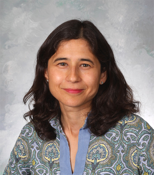

 Dr. Thomas Kirchhoff[/caption]
MedicalResearch.com Interview with:
Tomas Kirchhoff, PhD
Assistant Professor, Departments of Population Health and Environmental Medicine
NYU Langone Medical Center
Member, Laura and Isaac Perlmutter Cancer Center
NYU Langone
Medical Research: What is the background for this study?
Dr. Kirchhoff: Melanoma is the deadliest form of skin cancer, and the cause of approximately 80% of all skin cancer patients annually. One factor that can help reverse this negative trend is efficient prediction of which patients at early melanoma stage will likely progress to more advanced metastatic disease. Current clinical predictors of patient survival, based on tumor characteristics, are important, but are relatively non-specific to inform melanoma prognosis to an individual patient level. It is critical to identify other factors that can serve as more personalized markers of predicting the course of melanoma.
Medical Research: What are the main findings?
Dr. Kirchhoff: In our study, we found that inherited genetic markers that impact activity of certain immune genes correlate with
Dr. Thomas Kirchhoff[/caption]
MedicalResearch.com Interview with:
Tomas Kirchhoff, PhD
Assistant Professor, Departments of Population Health and Environmental Medicine
NYU Langone Medical Center
Member, Laura and Isaac Perlmutter Cancer Center
NYU Langone
Medical Research: What is the background for this study?
Dr. Kirchhoff: Melanoma is the deadliest form of skin cancer, and the cause of approximately 80% of all skin cancer patients annually. One factor that can help reverse this negative trend is efficient prediction of which patients at early melanoma stage will likely progress to more advanced metastatic disease. Current clinical predictors of patient survival, based on tumor characteristics, are important, but are relatively non-specific to inform melanoma prognosis to an individual patient level. It is critical to identify other factors that can serve as more personalized markers of predicting the course of melanoma.
Medical Research: What are the main findings?
Dr. Kirchhoff: In our study, we found that inherited genetic markers that impact activity of certain immune genes correlate with  Prof. Ian Wong[/caption]
MedicalResearch.com Interview with:
Professor Ian C K Wong
Fellow of Royal Pharmaceutical Society
Fellow of Royal College of Paediatrics and Child Health (Honorary)
Fellow of the Higher Education Academy
Chair in Pharmacy Practice
Head of Research Department of Practice and Policy
UCL School of Pharmacy
London
Medical Research: What is the background for this study? What are the main findings?
Dr. Wong: Previous studies had showed an increased cardiovascular risk associated with clarithromycin (a widely used antibiotic) but the duration of effect remained unclear. Therefore, we conducted this study to investigate the duration of cardiovascular adverse effect provided that the risk exists after patients receiving clarithromycin in Hong Kong. We used three study designs to examine the association (temporal relationship) between clarithromycin and cardiovascular adverse outcomes such as myocardial infarction, arrhythmia, stroke, cardiac mortality at different time points.
Prof. Ian Wong[/caption]
MedicalResearch.com Interview with:
Professor Ian C K Wong
Fellow of Royal Pharmaceutical Society
Fellow of Royal College of Paediatrics and Child Health (Honorary)
Fellow of the Higher Education Academy
Chair in Pharmacy Practice
Head of Research Department of Practice and Policy
UCL School of Pharmacy
London
Medical Research: What is the background for this study? What are the main findings?
Dr. Wong: Previous studies had showed an increased cardiovascular risk associated with clarithromycin (a widely used antibiotic) but the duration of effect remained unclear. Therefore, we conducted this study to investigate the duration of cardiovascular adverse effect provided that the risk exists after patients receiving clarithromycin in Hong Kong. We used three study designs to examine the association (temporal relationship) between clarithromycin and cardiovascular adverse outcomes such as myocardial infarction, arrhythmia, stroke, cardiac mortality at different time points.
 Dr. Chirag Patil[/caption]
MedicalResearch.com Interview with:
Dr. Chirag Patil, MD
American Board Certified Neurosurgeon
Brain & Spine Tumor Program
Lead Investigator, Precision Medicine Initiative Against Brain Cancer
Program Director, Neurosurgical Residence training program
Director, Center for Neurosurgical Outcomes Research Cedars-Sinai Medical Center, Los Angeles, California
MedicalResearch.com Editor’s note: Dr. Patil’s research is focused on developing a method of personalized cancer treatment through the harnessing of genome wide mutational analysis of a specific patient’s cancer.
MedicalResearch.com: Would you tell us a little about yourself and your research interests?
Dr. Patil: I am a Stanford-trained, Board Certified Neurosurgeon and cancer researcher at Cedars-Sinai Medical Center in Los Angeles, California. I primarily focus on the care of patients with malignant brain tumors, particularly glioblastomas. I received my undergraduate degree from Cornell, followed by a medical degree from the University of California, San Francisco (UCSF), where I was a Regent’s scholar. I completed a residency in neurosurgery and a fellowship in stereotactic radiology at Stanford University. I also have a master’s degree in epidemiology with a focus on clinical trial design and mathematical modeling from Stanford.
MedicalResearch.com: Can you tell us about some of your research interests?
Dr. Patil: I am keenly interested in and focused on developing precision science-powered novel brain tumor therapies, immuno-therapies, and patient-centered “big data” outcomes research. I lead the recently-funded Cedars-Sinai Precision Medicine Initiative Against Brain Cancer, which utilizes tumor genomics to build a mathematical computer model, i.e., a virtual cancer cell of each patient’s unique tumor. The White House and several other stakeholders have taken keep interest in this research initiative as an example of a leading precision medicine program.
Dr. Chirag Patil[/caption]
MedicalResearch.com Interview with:
Dr. Chirag Patil, MD
American Board Certified Neurosurgeon
Brain & Spine Tumor Program
Lead Investigator, Precision Medicine Initiative Against Brain Cancer
Program Director, Neurosurgical Residence training program
Director, Center for Neurosurgical Outcomes Research Cedars-Sinai Medical Center, Los Angeles, California
MedicalResearch.com Editor’s note: Dr. Patil’s research is focused on developing a method of personalized cancer treatment through the harnessing of genome wide mutational analysis of a specific patient’s cancer.
MedicalResearch.com: Would you tell us a little about yourself and your research interests?
Dr. Patil: I am a Stanford-trained, Board Certified Neurosurgeon and cancer researcher at Cedars-Sinai Medical Center in Los Angeles, California. I primarily focus on the care of patients with malignant brain tumors, particularly glioblastomas. I received my undergraduate degree from Cornell, followed by a medical degree from the University of California, San Francisco (UCSF), where I was a Regent’s scholar. I completed a residency in neurosurgery and a fellowship in stereotactic radiology at Stanford University. I also have a master’s degree in epidemiology with a focus on clinical trial design and mathematical modeling from Stanford.
MedicalResearch.com: Can you tell us about some of your research interests?
Dr. Patil: I am keenly interested in and focused on developing precision science-powered novel brain tumor therapies, immuno-therapies, and patient-centered “big data” outcomes research. I lead the recently-funded Cedars-Sinai Precision Medicine Initiative Against Brain Cancer, which utilizes tumor genomics to build a mathematical computer model, i.e., a virtual cancer cell of each patient’s unique tumor. The White House and several other stakeholders have taken keep interest in this research initiative as an example of a leading precision medicine program.
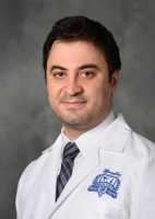 Dr. Firas Abdollah[/caption]
MedicalResearch.com Interview with:
Firas Abdollah, M.D., F.E.B.U.
(Fellow of European Board of Urology) Urology Fellow with the Center for Outcomes Research, Analytics and Evaluation
Vattikuti Urology Institute at Henry Ford Hospital in Detroit
MedicalResearch: What is the background for this study? What are the main findings?
Dr. Abdollah: Cancer screening aims to detect tumors early, before they become symptomatic. Evidence suggests that detection and treatment of early-stage tumors may reduce cancer mortality among screened individuals. Despite this potential benefit, screening programs may also cause harm. Notably, screening may identify low-risk indolent tumors that would never become clinically evident in the absence of screening (overdiagnosis), subjecting patients to the harms of unnecessary treatment. Such considerations are central to screening for prostate and breast cancers, the most prevalent solid tumors in men and women, respectively. These tumors are often slow growing, and guidelines recommend against screening (non-recommended screening) for these tumors in individuals with limited life expectancy, i.e. those with a life expectancy less than 10 years. Unfortunately, our study found that this practice is not uncommon in the US. Using a nationwide representative survey conducted in 2012, we found that among 149,514 individuals 65 years or older, 76,419 (51.1%) received any prostate/breast screening. Among these, 23,532 (30.8%) individuals had a life expectancy of less than 10 years. These numbers imply that among the screened population over 65 years old, almost one in three individuals received a non-recommended screening. This corresponds to an overall rate of non-recommended screening of 15.7% (23,532 of 149,514 individuals).
Another important finding of our study was that there were important variations in the rate of non-recommended screening from state to state; i.e. the chance of an individual older than 65 to receive a non-recommended screening varies based on his/her geographical location in United States.
Finally, on a state-by-state level, there was a correlation (40%) between non-recommended screening for prostate and breast cancer, i.e. states that are more likely to offer non-recommended screening for
Dr. Firas Abdollah[/caption]
MedicalResearch.com Interview with:
Firas Abdollah, M.D., F.E.B.U.
(Fellow of European Board of Urology) Urology Fellow with the Center for Outcomes Research, Analytics and Evaluation
Vattikuti Urology Institute at Henry Ford Hospital in Detroit
MedicalResearch: What is the background for this study? What are the main findings?
Dr. Abdollah: Cancer screening aims to detect tumors early, before they become symptomatic. Evidence suggests that detection and treatment of early-stage tumors may reduce cancer mortality among screened individuals. Despite this potential benefit, screening programs may also cause harm. Notably, screening may identify low-risk indolent tumors that would never become clinically evident in the absence of screening (overdiagnosis), subjecting patients to the harms of unnecessary treatment. Such considerations are central to screening for prostate and breast cancers, the most prevalent solid tumors in men and women, respectively. These tumors are often slow growing, and guidelines recommend against screening (non-recommended screening) for these tumors in individuals with limited life expectancy, i.e. those with a life expectancy less than 10 years. Unfortunately, our study found that this practice is not uncommon in the US. Using a nationwide representative survey conducted in 2012, we found that among 149,514 individuals 65 years or older, 76,419 (51.1%) received any prostate/breast screening. Among these, 23,532 (30.8%) individuals had a life expectancy of less than 10 years. These numbers imply that among the screened population over 65 years old, almost one in three individuals received a non-recommended screening. This corresponds to an overall rate of non-recommended screening of 15.7% (23,532 of 149,514 individuals).
Another important finding of our study was that there were important variations in the rate of non-recommended screening from state to state; i.e. the chance of an individual older than 65 to receive a non-recommended screening varies based on his/her geographical location in United States.
Finally, on a state-by-state level, there was a correlation (40%) between non-recommended screening for prostate and breast cancer, i.e. states that are more likely to offer non-recommended screening for  Dr. Dirk Timmerman[/caption]
 Dr. Jennifer Stein[/caption]
 Dr. Murchison[/caption]
MedicalResearch.com Interview with:
Dr. Elizabeth Murchison
Menzies Institute for Medical Research
University of Tasmania
Save the Tasmanian Devil Program
Tasmanian Department of Primary Industries, Parks, Water and the Environment
Hobart Australia
Department of Veterinary Medicine
University of Cambridge, Cambridge UK
Medical Research: What is the background for this study?
Dr. Murchison: Transmissible cancers are cancers that can be transmitted between individuals by the transfer of living cancer cells. Transmissible cancers emerge only very rarely in nature, and until now only three examples were known. One of the three known naturally occurring transmissible cancers affects Tasmanian devils, the world’s largest carnivorous marsupial. This disease, which causes disfiguring facial tumours, was first observed in the late 1990s, and since then the disease has spread widely through the Tasmanian devil population. This transmissible cancer first emerged as a cancer in a single individual Tasmanian devil that probably lived about 30 years ago; this devil’s cancer cells have continued to survive by transmitting between hosts by biting.
Medical Research: What are the main findings?
Dr. Murchison: In late 2014, routine monitoring of the Tasmanian devil population led to the discovery of a male devil with facial tumours that resembled the known Tasmanian devil transmissible facial cancer. However, genetic analysis of this tumour indicated that the tumour in this devil was derived from a second transmissible cancer that was genetically unrelated to the first transmissible cancer in this species. Indeed, the genetic profile of this second cancer indicated that it had originally emerged from a male animal. This second cancer has subsequently been found in nine additional devils in the same part of Tasmania.
Dr. Murchison[/caption]
MedicalResearch.com Interview with:
Dr. Elizabeth Murchison
Menzies Institute for Medical Research
University of Tasmania
Save the Tasmanian Devil Program
Tasmanian Department of Primary Industries, Parks, Water and the Environment
Hobart Australia
Department of Veterinary Medicine
University of Cambridge, Cambridge UK
Medical Research: What is the background for this study?
Dr. Murchison: Transmissible cancers are cancers that can be transmitted between individuals by the transfer of living cancer cells. Transmissible cancers emerge only very rarely in nature, and until now only three examples were known. One of the three known naturally occurring transmissible cancers affects Tasmanian devils, the world’s largest carnivorous marsupial. This disease, which causes disfiguring facial tumours, was first observed in the late 1990s, and since then the disease has spread widely through the Tasmanian devil population. This transmissible cancer first emerged as a cancer in a single individual Tasmanian devil that probably lived about 30 years ago; this devil’s cancer cells have continued to survive by transmitting between hosts by biting.
Medical Research: What are the main findings?
Dr. Murchison: In late 2014, routine monitoring of the Tasmanian devil population led to the discovery of a male devil with facial tumours that resembled the known Tasmanian devil transmissible facial cancer. However, genetic analysis of this tumour indicated that the tumour in this devil was derived from a second transmissible cancer that was genetically unrelated to the first transmissible cancer in this species. Indeed, the genetic profile of this second cancer indicated that it had originally emerged from a male animal. This second cancer has subsequently been found in nine additional devils in the same part of Tasmania.
 Pritesh Karia[/caption]
MedicalResearch.com Interview with:
Pritesh Karia[/caption]
MedicalResearch.com Interview with: Tanning Bed CDC Image[/caption]
MedicalResearch.com Interview with:
Myra Sendelweck M.Eng
M.D. Candidate 2018 and
Robert Dellavalle, MD, PhD, MSPH
Chief, Dermatology Service Denver VA Medical Center Denver, CO
University of Colorado School of Medicine
MedicalResearch: What is the background for this study? What are the main findings?
Response: Indoor tanning has increasingly been recognized amongst providers as a public health concern. Recent literature suggests an association between indoor tanning and other risky health behaviors in adolescents.
We were intrigued by this association. We analyzed a survey of Colorado high school students and found that those who tanned were also more likely to use various substances, such as steroids, alcohol, marijuana, and illicit drugs. Tanners were over five times as likely to report steroid use!
Tanning Bed CDC Image[/caption]
MedicalResearch.com Interview with:
Myra Sendelweck M.Eng
M.D. Candidate 2018 and
Robert Dellavalle, MD, PhD, MSPH
Chief, Dermatology Service Denver VA Medical Center Denver, CO
University of Colorado School of Medicine
MedicalResearch: What is the background for this study? What are the main findings?
Response: Indoor tanning has increasingly been recognized amongst providers as a public health concern. Recent literature suggests an association between indoor tanning and other risky health behaviors in adolescents.
We were intrigued by this association. We analyzed a survey of Colorado high school students and found that those who tanned were also more likely to use various substances, such as steroids, alcohol, marijuana, and illicit drugs. Tanners were over five times as likely to report steroid use!
 Dr. Benjamin Neel[/caption]
 Dr. Elena Batrakova[/caption]
 Dr. Dai Fukumura[/caption]
MedicalResearch.com Interview with:
Dai Fukumura, M.D., Ph.D.
Joao Incio, M.D.
and Rakesh K. Jain, Ph.D
Edwin L. Steele Laboratory
Department of Radiation Oncology
Massachusetts General Hospital
Harvard Medical School
Medical Research: What is the background for this study? What are the main findings?
Dr. Fukumura: This study focused on pancreatic ductal adenocarcinoma, the most common form of pancreatic cancer, which accounts for almost 40,000 cancer death in the U.S. ever year. Half of those diagnosed with this form of pancreatic cancer are overweight or obese, and up to 80 percent have type 2 diabetes or are insulin resistant. Diabetic patients taking metformin – a commonly used generic medication for type 2 diabetes – are known to have a reduced risk of developing pancreatic cancer; and among patients who develop the tumor, those taking the drug may have a reduced risk of death. But prior to the current study the mechanism of metformin’s action against pancreatic cancer was unclear, and no potential biomarkers of response to metformin had been reported.
We have uncovered a novel mechanism behind the ability of the diabetes drug metformin to inhibit the progression of pancreatic cancer. Metformin decreases the inflammation and fibrosis characteristic of the most common form of pancreatic cancer. We found that metformin alleviates desmoplasia – an accumulation of dense connective tissue and tumor-associated immune cells that is a hallmark of pancreatic cancer – by inhibiting the activation of the pancreatic stellate cells that produce the extracellular matrix and by reprogramming immune cells to reduce inflammation. Our findings in cellular and animal models and in patient tumor samples also indicate that this beneficial effect may be most prevalent in overweight and obese patients, who appear to have tumors with increased fibrosis.
Dr. Dai Fukumura[/caption]
MedicalResearch.com Interview with:
Dai Fukumura, M.D., Ph.D.
Joao Incio, M.D.
and Rakesh K. Jain, Ph.D
Edwin L. Steele Laboratory
Department of Radiation Oncology
Massachusetts General Hospital
Harvard Medical School
Medical Research: What is the background for this study? What are the main findings?
Dr. Fukumura: This study focused on pancreatic ductal adenocarcinoma, the most common form of pancreatic cancer, which accounts for almost 40,000 cancer death in the U.S. ever year. Half of those diagnosed with this form of pancreatic cancer are overweight or obese, and up to 80 percent have type 2 diabetes or are insulin resistant. Diabetic patients taking metformin – a commonly used generic medication for type 2 diabetes – are known to have a reduced risk of developing pancreatic cancer; and among patients who develop the tumor, those taking the drug may have a reduced risk of death. But prior to the current study the mechanism of metformin’s action against pancreatic cancer was unclear, and no potential biomarkers of response to metformin had been reported.
We have uncovered a novel mechanism behind the ability of the diabetes drug metformin to inhibit the progression of pancreatic cancer. Metformin decreases the inflammation and fibrosis characteristic of the most common form of pancreatic cancer. We found that metformin alleviates desmoplasia – an accumulation of dense connective tissue and tumor-associated immune cells that is a hallmark of pancreatic cancer – by inhibiting the activation of the pancreatic stellate cells that produce the extracellular matrix and by reprogramming immune cells to reduce inflammation. Our findings in cellular and animal models and in patient tumor samples also indicate that this beneficial effect may be most prevalent in overweight and obese patients, who appear to have tumors with increased fibrosis.
 Dr. NaNa Keum[/caption]
 Dr. R. Jeffrey Karnes[/caption]
MedicalResearch.com Interview with:
R. Jeffrey Karnes MD
Department of Urology, Mayo Clinic,
Rochester, MN 55905
MedicalResearch: What is the background for this study? What are the main findings?
Dr. Karnes: Cancer recurrence following radical prostatectomy is a concern for men undergoing definitive surgical treatment for prostate cancer. Approximately 20-35% of patients develop a rising prostate specific antigen following radical prostatectomy for clinically localized prostate cancer. PSA monitoring is an important tool for cancer surveillance; however, a standard PSA cutpoint to indicate biochemical recurrence has yet to be established. Over 60 different definitions have been described in literature. This variation creates confusion for the patients and clinicians. By studying a large group of patients who underwent radical prostatectomy at Mayo Clinic, we found that a PSA cutpoint of 0.4 ng/mL is the optimal definition for biochemical recurrence.
Dr. R. Jeffrey Karnes[/caption]
MedicalResearch.com Interview with:
R. Jeffrey Karnes MD
Department of Urology, Mayo Clinic,
Rochester, MN 55905
MedicalResearch: What is the background for this study? What are the main findings?
Dr. Karnes: Cancer recurrence following radical prostatectomy is a concern for men undergoing definitive surgical treatment for prostate cancer. Approximately 20-35% of patients develop a rising prostate specific antigen following radical prostatectomy for clinically localized prostate cancer. PSA monitoring is an important tool for cancer surveillance; however, a standard PSA cutpoint to indicate biochemical recurrence has yet to be established. Over 60 different definitions have been described in literature. This variation creates confusion for the patients and clinicians. By studying a large group of patients who underwent radical prostatectomy at Mayo Clinic, we found that a PSA cutpoint of 0.4 ng/mL is the optimal definition for biochemical recurrence.
 Dr. Boolbol[/caption]
MedicalResearch.com Interview with:
Susan K. Boolbol, MD, FACS
Chief, Division of Breast Surgery
Chief, Appel-Venet Comprehensive Breast Service
Co-Director, Breast Surgery Fellowship
Mount Sinai Beth Israel
Associate Professor of Surgery
Icahn School of Medicine at Mount Sinai
New York, NY 10003
Medical Research: What is the background for these new recommendations?
Dr. Boolbol: To make this final recommendation, the Task Force conducted a comprehensive review of the science since its 2009 recommendation and considered the public comments it received on its 2015 draft recommendation statement. Based on all of this, the task force issued their recommendations.
Medical Research: What are the main changes from current guidelines?
Dr. Boolbol: Presently, there are several different guidelines and recommendations regarding screening mammography. Depending on the group issuing the guidelines, the recommendations vary from
Dr. Boolbol[/caption]
MedicalResearch.com Interview with:
Susan K. Boolbol, MD, FACS
Chief, Division of Breast Surgery
Chief, Appel-Venet Comprehensive Breast Service
Co-Director, Breast Surgery Fellowship
Mount Sinai Beth Israel
Associate Professor of Surgery
Icahn School of Medicine at Mount Sinai
New York, NY 10003
Medical Research: What is the background for these new recommendations?
Dr. Boolbol: To make this final recommendation, the Task Force conducted a comprehensive review of the science since its 2009 recommendation and considered the public comments it received on its 2015 draft recommendation statement. Based on all of this, the task force issued their recommendations.
Medical Research: What are the main changes from current guidelines?
Dr. Boolbol: Presently, there are several different guidelines and recommendations regarding screening mammography. Depending on the group issuing the guidelines, the recommendations vary from  Dr. Sergio Acuna[/caption]
MedicalResearch.com Interview with:
Sergio A. Acuna, MD
Graduate Student at St. Michael's Hospital and IHPME
University of Toronto
Medical Research: What is the background for this study?
Dr. Acuna: Solid organ transplant recipients are known to be at greater risk of developing cancers compared to the general population; however, because they are also at high increased risk of mortality from non-cancer causes, the risk of cancer morality in this population is unclear. As previous studies on this topic have reported disparate findings, the cancer mortality risk in this population remained uncertain.
Medical Research: What are the main findings?
Dr. Acuna: Our study provides conclusive evidence that solid organ transplant recipients are at increased risk of cancer mortality. Our findings demonstrate that solid organ transplant recipients are at increased risk of cancer death compared to the general population regardless of age, transplanted organ, and year of transplantation, and indicate cancer is a substantial cause of death in this population.
Dr. Sergio Acuna[/caption]
MedicalResearch.com Interview with:
Sergio A. Acuna, MD
Graduate Student at St. Michael's Hospital and IHPME
University of Toronto
Medical Research: What is the background for this study?
Dr. Acuna: Solid organ transplant recipients are known to be at greater risk of developing cancers compared to the general population; however, because they are also at high increased risk of mortality from non-cancer causes, the risk of cancer morality in this population is unclear. As previous studies on this topic have reported disparate findings, the cancer mortality risk in this population remained uncertain.
Medical Research: What are the main findings?
Dr. Acuna: Our study provides conclusive evidence that solid organ transplant recipients are at increased risk of cancer mortality. Our findings demonstrate that solid organ transplant recipients are at increased risk of cancer death compared to the general population regardless of age, transplanted organ, and year of transplantation, and indicate cancer is a substantial cause of death in this population.
 Prof. Hennekens[/caption]
MedicalResearch.com Interview with:
Professor Charles Hennekens MD Dr.P.H
Sir Richard Doll Professor
Senior Academic Advisor to the Dean
Charles E. Schmidt College of Medicine
Florida Atlantic University
777 Glades Road
Boca Raton, FL 33431
Medical Research: What is the background for this study? What are the main findings?
Prof. Hennekens: Randomized evidence indicates clear benefits of mammography in middle age and, at present, most guidelines recommend regular mammography for women up to age 74. In collaboration with colleagues at Baylor Medical College and Meharry Medical School we were able to link the Surveillance, Epidemiology, and End Results (SEER) data to the Medicare administrative claims data. We found that, up to 84 years, screening was more common among whites than blacks and women receiving regular annual screening mammography had lower risks of mortality from breast cancer.
Prof. Hennekens[/caption]
MedicalResearch.com Interview with:
Professor Charles Hennekens MD Dr.P.H
Sir Richard Doll Professor
Senior Academic Advisor to the Dean
Charles E. Schmidt College of Medicine
Florida Atlantic University
777 Glades Road
Boca Raton, FL 33431
Medical Research: What is the background for this study? What are the main findings?
Prof. Hennekens: Randomized evidence indicates clear benefits of mammography in middle age and, at present, most guidelines recommend regular mammography for women up to age 74. In collaboration with colleagues at Baylor Medical College and Meharry Medical School we were able to link the Surveillance, Epidemiology, and End Results (SEER) data to the Medicare administrative claims data. We found that, up to 84 years, screening was more common among whites than blacks and women receiving regular annual screening mammography had lower risks of mortality from breast cancer.
 Dr. Daniel Mulrooney[/caption]
MedicalResearch.com Interview with:
Daniel A. Mulrooney, MD, MS
Cancer Survivorship
Jude Children's Research Hospital
TN 38105-3678
Medical Research: What is the background for this study? What are the main findings?
Dr. Mulrooney: This is a cross-sectional analysis performed in the St. Jude Lifetime Cohort Study (SJLIFE), an ongoing study designed to facilitate longitudinal evaluation of health outcomes among adults previously treated for childhood cancer. Following patients over the life spectrum can be challenging making it difficult to understand the long-term health effects of childhood cancer therapy. Previous studies have relied on self-report, registry, or death certificate data. Our study is novel because we clinically evaluated cancer survivors on the St. Jude campus and identified substantial, asymptomatic cardiac disease (cardiomyopathy, coronary artery disease, valvular disease, and conduction/rhythm disorders).
Dr. Daniel Mulrooney[/caption]
MedicalResearch.com Interview with:
Daniel A. Mulrooney, MD, MS
Cancer Survivorship
Jude Children's Research Hospital
TN 38105-3678
Medical Research: What is the background for this study? What are the main findings?
Dr. Mulrooney: This is a cross-sectional analysis performed in the St. Jude Lifetime Cohort Study (SJLIFE), an ongoing study designed to facilitate longitudinal evaluation of health outcomes among adults previously treated for childhood cancer. Following patients over the life spectrum can be challenging making it difficult to understand the long-term health effects of childhood cancer therapy. Previous studies have relied on self-report, registry, or death certificate data. Our study is novel because we clinically evaluated cancer survivors on the St. Jude campus and identified substantial, asymptomatic cardiac disease (cardiomyopathy, coronary artery disease, valvular disease, and conduction/rhythm disorders).
 Dr. Bissan Al-Lazikini[/caption]
MedicalResearch.com Interview with:
Dr Bissan Al-Lazikani
Team leader in computational biology
The Institute of Cancer Research
London
Medical Research: What is the background for the canSAR database? What are the main uses for the tool?
Dr. Al-Lazikani: Drug discovery is a difficult, time consuming and expensive venture that frequently ends in late stage drug failures - especially in oncology.
As with any complex venture, decisions throughout the drug discovery pipeline can be empowered by having access to the right information at the right time. But for drug discovery this means bringing together billions of experimental data from very diverse areas of science spanning genomics, proteomics, chemistry and more.
We developed canSAR to help guide our own drug discovery efforts by integrating these huge, diverse data and by analysing the data and deriving hidden links and knowledge from them. This means that we can answer questions in minutes that would have taken weeks using previously available public resources. But, more importantly, canSAR analyses and links these data in a way that allows us to derive knowledge that was hidden before. For example, one of the main ways canSAR is used is to help select the best druggable targets for drug discovery. Using canSAR we were able to uncover many druggable cancer proteins that were previously overlooked, and we are delighted to see that several of these proteins are now the subjects of drug discovery and development projects both by us and by others.
We took the decision to make canSAR publicly and freely available because we believe that cancer drug discovery is a vast challenge that requires openness and data sharing worldwide. It has been embraced by the community is being used by tens of thousands of cancer scientists worldwide, both in academia and industry, to generate hypotheses for experiments and select targets for drug discovery.
Dr. Bissan Al-Lazikini[/caption]
MedicalResearch.com Interview with:
Dr Bissan Al-Lazikani
Team leader in computational biology
The Institute of Cancer Research
London
Medical Research: What is the background for the canSAR database? What are the main uses for the tool?
Dr. Al-Lazikani: Drug discovery is a difficult, time consuming and expensive venture that frequently ends in late stage drug failures - especially in oncology.
As with any complex venture, decisions throughout the drug discovery pipeline can be empowered by having access to the right information at the right time. But for drug discovery this means bringing together billions of experimental data from very diverse areas of science spanning genomics, proteomics, chemistry and more.
We developed canSAR to help guide our own drug discovery efforts by integrating these huge, diverse data and by analysing the data and deriving hidden links and knowledge from them. This means that we can answer questions in minutes that would have taken weeks using previously available public resources. But, more importantly, canSAR analyses and links these data in a way that allows us to derive knowledge that was hidden before. For example, one of the main ways canSAR is used is to help select the best druggable targets for drug discovery. Using canSAR we were able to uncover many druggable cancer proteins that were previously overlooked, and we are delighted to see that several of these proteins are now the subjects of drug discovery and development projects both by us and by others.
We took the decision to make canSAR publicly and freely available because we believe that cancer drug discovery is a vast challenge that requires openness and data sharing worldwide. It has been embraced by the community is being used by tens of thousands of cancer scientists worldwide, both in academia and industry, to generate hypotheses for experiments and select targets for drug discovery.