
10 Apr Advanced Heart Failure Patients Should Have Option of LVAD Device
Posted at 15:40h
in Author Interviews
MedicalResearch.com Interview with:
Amrut V. Ambardekar, MD
Medical Director Cardiac Transplant Program
Division of Cardiology, Section of Advanced Heart Failure-Transplant Cardiology
University of Colorado
MedicalResearch.com: What is the background for this study? What are the main findings?
Response: As left ventricular assist device (LVAD) technology has improved, the appropriate timing for implant of these devices (essential a form of an artificial heart pump) in patients with advanced heart failure is unknown.
The goal of the MedaMACS study was to describe the prognosis of a group of patients with advanced heart failure who currently do not require intravenous therapies, and determine how they compare to a similar group of patients who received a LVAD.
The main finding from this study is that the “sickest” group of patients with advanced heart failure on oral medical therapy (known as INTERMACS profile 4 patients) have very poor outcomes with a strong trend for improvement in survival with LVAD therapy.
The other take home message is that among all of the patients enrolled in the study on medical therapy, only approximately half were alive after an average of 12 months of follow up without needing a heart transplant or LVAD placement.


 Dr. Orit Markowitz[/caption]
Orit Markowitz, MD
Director of Pigmented Lesions and Skin Cancer
The Mount Sinai Hospital and
Assistant Professor of Dermatology
Icahn School of Medicine at Mount Sinai
Director of Pigmented lesions clinic
Brooklyn VA,
Adjunct Professor, Dermatology
SUNY Downstate Medical Center, Brooklyn, NY
Chief of Dermatology
Queens General Hospital, Jamaica, NY
MedicalResearch.com Editors’ Note: As part of an ongoing series of occasional article on cancer prevention, Dr. Markowitz from The Mount Sinai Hospital discusses skin cancer and the use Optical Coherence Tomography in skin cancer diagnosis and treatment.
MedicalResearch.com: How common is the problem of non-melanoma skin cancer? Are they difficult to detect and treat?
Dr. Markowitz: Skin cancer is the most commonly diagnosed cancer in the United States. Non melanoma skin cancers, including basal cell carcinomas and squamous cell carcinomas, are the most common malignancies of the skin, constituting around 80 percent of all skin cancers. The annual cost of treating skin cancers in the U.S. is estimated at $8.1 billion, with $3.3 billion for melanoma.
Dr. Orit Markowitz[/caption]
Orit Markowitz, MD
Director of Pigmented Lesions and Skin Cancer
The Mount Sinai Hospital and
Assistant Professor of Dermatology
Icahn School of Medicine at Mount Sinai
Director of Pigmented lesions clinic
Brooklyn VA,
Adjunct Professor, Dermatology
SUNY Downstate Medical Center, Brooklyn, NY
Chief of Dermatology
Queens General Hospital, Jamaica, NY
MedicalResearch.com Editors’ Note: As part of an ongoing series of occasional article on cancer prevention, Dr. Markowitz from The Mount Sinai Hospital discusses skin cancer and the use Optical Coherence Tomography in skin cancer diagnosis and treatment.
MedicalResearch.com: How common is the problem of non-melanoma skin cancer? Are they difficult to detect and treat?
Dr. Markowitz: Skin cancer is the most commonly diagnosed cancer in the United States. Non melanoma skin cancers, including basal cell carcinomas and squamous cell carcinomas, are the most common malignancies of the skin, constituting around 80 percent of all skin cancers. The annual cost of treating skin cancers in the U.S. is estimated at $8.1 billion, with $3.3 billion for melanoma.
 Dr. James Freeman[/caption]
Dr. James V. Freeman MD
Assistant professor of cardiology and
Assistant Clinical Professor of Nursing
Internal Medicine
Yale School of Medicine
MedicalResearch.com: What is the background for this study? What are the main findings?
Dr. Freeman: Randomized trials of left atrial appendage (LAA) closure with the Watchman device have shown varying results, and its cost-effectiveness compared to anticoagulation has not been evaluated using all available contemporary trial data.
We used a Markov decision model to estimate lifetime quality-adjusted survival, costs, and cost-effectiveness of LAA closure with Watchman, compared directly with warfarin and indirectly with dabigatran, using data from the long-term (mean 3.8 year) follow-up of PROTECT AF and PREVAIL randomized trials. Using data from PROTECT AF, the incremental cost-effectiveness ratios (ICER) compared to warfarin and dabigatran were $20,486 and $23,422 per quality adjusted life year (QALY), respectively. Using data from PREVAIL, LAA closure was dominated by warfarin and dabigatran, meaning that it was less effective (8.44, 8.54, and 8.59 QALYs, respectively) and more costly.
Dr. James Freeman[/caption]
Dr. James V. Freeman MD
Assistant professor of cardiology and
Assistant Clinical Professor of Nursing
Internal Medicine
Yale School of Medicine
MedicalResearch.com: What is the background for this study? What are the main findings?
Dr. Freeman: Randomized trials of left atrial appendage (LAA) closure with the Watchman device have shown varying results, and its cost-effectiveness compared to anticoagulation has not been evaluated using all available contemporary trial data.
We used a Markov decision model to estimate lifetime quality-adjusted survival, costs, and cost-effectiveness of LAA closure with Watchman, compared directly with warfarin and indirectly with dabigatran, using data from the long-term (mean 3.8 year) follow-up of PROTECT AF and PREVAIL randomized trials. Using data from PROTECT AF, the incremental cost-effectiveness ratios (ICER) compared to warfarin and dabigatran were $20,486 and $23,422 per quality adjusted life year (QALY), respectively. Using data from PREVAIL, LAA closure was dominated by warfarin and dabigatran, meaning that it was less effective (8.44, 8.54, and 8.59 QALYs, respectively) and more costly.
 Dr. Donald Burke[/caption]
Donald S. Burke, M.D.
Dean of the University of Pittsburgh Graduate School of Public Health
Director of the University of Pittsburgh Center for Vaccine Research
MedicalResearch.com: What is the background for this study? What are the main findings?
Dr. Burke: At the University of Pittsburgh we developed a unique method for detecting antibodies in the blood of patients in a proof-of-principle study that opens the door to development of simple diagnostic tests for diseases for which no microbial cause is known, including auto-immune diseases, cancers and other conditions.
We used a technique pioneered by co-author Thomas Kodadek, Ph.D., of the Scripps Research Institute, that synthesizes random molecular shapes called “peptoids” hooked onto microscopic plastic beads. The technique can produce millions of molecular shapes. The peptoids are not organic, but if they match to the corresponding shape on an antibody, that antibody will connect to them, allowing the scientist to pull out that bead and examine that peptoid and its corresponding antibody.
My team chemically generated a huge library of random molecular shapes. Then, using blood from HIV-infected patients and from non-infected people, we screened a million of these random molecular shapes to find the ones that bound only to antibodies present in the blood of HIV-infected patients, but not the healthy controls. No HIV proteins or structures were used to construct or select the peptoids, but the approach, nonetheless, successfully led to selection of the best molecular shapes to use in screening for HIV antibodies.
We then resynthesized that HIV-antibody-targeting peptoid in mass and tested it by screening hundreds of samples from the Multicenter AIDS Cohort Study (MACS), a confidential research study of the natural history of treated and untreated HIV/AIDS in men who have sex with men (supported by the National Institutes of Health). Study co-author Charles Rinaldo, Ph.D., chair of Pitt Public Health’s Department of Infectious Diseases and Microbiology and director of the Pittsburgh arm of the MACS, selected the samples, but blinded the testers to which samples were HIV-positive or -negative. The test distinguished between the samples of HIV-positive blood and HIV-negative blood with a high degree of accuracy.
Dr. Donald Burke[/caption]
Donald S. Burke, M.D.
Dean of the University of Pittsburgh Graduate School of Public Health
Director of the University of Pittsburgh Center for Vaccine Research
MedicalResearch.com: What is the background for this study? What are the main findings?
Dr. Burke: At the University of Pittsburgh we developed a unique method for detecting antibodies in the blood of patients in a proof-of-principle study that opens the door to development of simple diagnostic tests for diseases for which no microbial cause is known, including auto-immune diseases, cancers and other conditions.
We used a technique pioneered by co-author Thomas Kodadek, Ph.D., of the Scripps Research Institute, that synthesizes random molecular shapes called “peptoids” hooked onto microscopic plastic beads. The technique can produce millions of molecular shapes. The peptoids are not organic, but if they match to the corresponding shape on an antibody, that antibody will connect to them, allowing the scientist to pull out that bead and examine that peptoid and its corresponding antibody.
My team chemically generated a huge library of random molecular shapes. Then, using blood from HIV-infected patients and from non-infected people, we screened a million of these random molecular shapes to find the ones that bound only to antibodies present in the blood of HIV-infected patients, but not the healthy controls. No HIV proteins or structures were used to construct or select the peptoids, but the approach, nonetheless, successfully led to selection of the best molecular shapes to use in screening for HIV antibodies.
We then resynthesized that HIV-antibody-targeting peptoid in mass and tested it by screening hundreds of samples from the Multicenter AIDS Cohort Study (MACS), a confidential research study of the natural history of treated and untreated HIV/AIDS in men who have sex with men (supported by the National Institutes of Health). Study co-author Charles Rinaldo, Ph.D., chair of Pitt Public Health’s Department of Infectious Diseases and Microbiology and director of the Pittsburgh arm of the MACS, selected the samples, but blinded the testers to which samples were HIV-positive or -negative. The test distinguished between the samples of HIV-positive blood and HIV-negative blood with a high degree of accuracy.
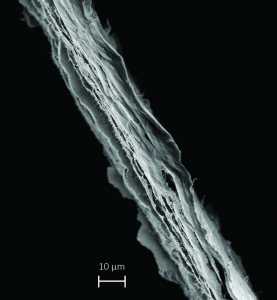 Mille Feuille Paper Filter[/caption]
MedicalResearch.com: What is the background for this study? What are the main findings?
Dr. Mihranyan: We describe for the first time a paper filter that can remove even the worst-case viruses from water with high efficiency and at industrially relevant rates. The filter is produced from 100% naturally derived cellulose and is formed into paper sheets using very simple processing, which is essentially the same as that for making paper on a large scale. Filter paper is used ubiquitously in every day life from coffee filters to chemistry classrooms but these filters have normally too large pores to retain microbes, let alone viruses.
We show for the first time that we can remove viruses as small as 20 nm! How is it possible? We use cellulose nanofibers from green algae and we possess know-how to control the distribution of the pores inside the paper to be able to remove such small particles. One important aspect, which we discuss in detail in the article, is the special internal layered structure of the filter, which is remarkably similar to French pastry mille-feuille- hence, the name mille-feuille filter.
Mille Feuille Paper Filter[/caption]
MedicalResearch.com: What is the background for this study? What are the main findings?
Dr. Mihranyan: We describe for the first time a paper filter that can remove even the worst-case viruses from water with high efficiency and at industrially relevant rates. The filter is produced from 100% naturally derived cellulose and is formed into paper sheets using very simple processing, which is essentially the same as that for making paper on a large scale. Filter paper is used ubiquitously in every day life from coffee filters to chemistry classrooms but these filters have normally too large pores to retain microbes, let alone viruses.
We show for the first time that we can remove viruses as small as 20 nm! How is it possible? We use cellulose nanofibers from green algae and we possess know-how to control the distribution of the pores inside the paper to be able to remove such small particles. One important aspect, which we discuss in detail in the article, is the special internal layered structure of the filter, which is remarkably similar to French pastry mille-feuille- hence, the name mille-feuille filter.
 Dr. Sushanta Mitra[/caption]
Sushanta K. Mitra, PhD, PEng
Associate Vice-President Research
Kaneff Professor in Micro & Nanotechnology for Social Innovation
FCSME, FASME, FEIC, FRSC, FCAE, FAAAS Y
York University Toronto
MedicalResearch.com: What is the background for this study? What are the main findings?
Dr. Mitra: As a mechanical engineer I got interested in the water problem when I had discussions with Tata Consultancy Services (TCS), India and the tertiary public health centre doctors near Mumbai, where the doctors had to deal with large number of patients with water-borne diseases. This was hugely a challenge from resource point of view as the doctors would much preferred to have their attention focused on more pressing diseases. They approached me about developing tools for rapid detection of water-borne pathogen in drinking water. Hence, my journey started on water quality monitoring.
MedicalResearch.com: What is the background for this study? What are the main findings?
Dr. Mitra: Here, we have developed a low-cost compact E. coli and total coliform detection system, which uses commercially available plunger-tube assembly. We incorporate a hydrogel (porous matrix) inside the tube so that the plunger-tube assembly act as a concentrator and a detector at the same time. Specially formulated enzymatic substrates are caged inside the hydrogel so that an E. coli cell trapped within the hydrogel will be lysed and react with the enzymatic substrates to produce a red color.
Dr. Sushanta Mitra[/caption]
Sushanta K. Mitra, PhD, PEng
Associate Vice-President Research
Kaneff Professor in Micro & Nanotechnology for Social Innovation
FCSME, FASME, FEIC, FRSC, FCAE, FAAAS Y
York University Toronto
MedicalResearch.com: What is the background for this study? What are the main findings?
Dr. Mitra: As a mechanical engineer I got interested in the water problem when I had discussions with Tata Consultancy Services (TCS), India and the tertiary public health centre doctors near Mumbai, where the doctors had to deal with large number of patients with water-borne diseases. This was hugely a challenge from resource point of view as the doctors would much preferred to have their attention focused on more pressing diseases. They approached me about developing tools for rapid detection of water-borne pathogen in drinking water. Hence, my journey started on water quality monitoring.
MedicalResearch.com: What is the background for this study? What are the main findings?
Dr. Mitra: Here, we have developed a low-cost compact E. coli and total coliform detection system, which uses commercially available plunger-tube assembly. We incorporate a hydrogel (porous matrix) inside the tube so that the plunger-tube assembly act as a concentrator and a detector at the same time. Specially formulated enzymatic substrates are caged inside the hydrogel so that an E. coli cell trapped within the hydrogel will be lysed and react with the enzymatic substrates to produce a red color.
 Sheldon j. .J.Kwok[/caption]
Sheldon J.J. Kwok
MD/PhD Candidate
Harvard-MIT Health Sciences and Technology | Harvard Medical School
Yun Bio-Optics Lab
Wellman Center for Photomedicine
MGH
MedicalResearch.com: What is the background for this study?
Response: Corneal collagen crosslinking (CXL) using UV light and riboflavin has become a popular and effective technique for treating corneal ectatic disorders, such as keratoconus, by mechanically strengthening the corneal stroma. We were interested in enhancing the capabilities of CXL using the principle of two-photon excitation, which uses a femtosecond laser to confine crosslinking to only where the laser is focused. By scanning the laser, this allows us to crosslink any arbitrary three-dimensional region deep inside tissue.
With two-photon collagen crosslinking (2P-CXL), treatment of thin corneas is possible without affecting the underlying endothelium. Irradiation can also be patterned to improve keratocyte viability. Furthermore, selective crosslinking in three dimensions offers the possibility of modulating corneal curvature for refractive error correction.
Sheldon j. .J.Kwok[/caption]
Sheldon J.J. Kwok
MD/PhD Candidate
Harvard-MIT Health Sciences and Technology | Harvard Medical School
Yun Bio-Optics Lab
Wellman Center for Photomedicine
MGH
MedicalResearch.com: What is the background for this study?
Response: Corneal collagen crosslinking (CXL) using UV light and riboflavin has become a popular and effective technique for treating corneal ectatic disorders, such as keratoconus, by mechanically strengthening the corneal stroma. We were interested in enhancing the capabilities of CXL using the principle of two-photon excitation, which uses a femtosecond laser to confine crosslinking to only where the laser is focused. By scanning the laser, this allows us to crosslink any arbitrary three-dimensional region deep inside tissue.
With two-photon collagen crosslinking (2P-CXL), treatment of thin corneas is possible without affecting the underlying endothelium. Irradiation can also be patterned to improve keratocyte viability. Furthermore, selective crosslinking in three dimensions offers the possibility of modulating corneal curvature for refractive error correction.
 Dr. Laura E. Niklason[/caption]
Dr Laura E Niklason, MD PhD
Department of Anesthesia & Biomedical Engineering
Yale University, New Haven, CT
MedicalResearch.com: What is the background for this study? What are the main findings?
Dr. Niklason: For end stage renal disease patients who are not candidates for fistula, dialysis access grafts are the best option for chronic hemodialysis. However, polytetrafluoroethylene arteriovenous grafts suffer from high rates of thrombosis, infection and intimal hyperplasia at the venous anastomosis.
We are conducting two, single arm Phase II trials where a novel bioengineered human acellular vessel (HAV) was implanted into the arms of patients for hemodialysis access. Primary endpoints were safety (freedom from immune response/infection, aneurysm, or mechanical failure, and incidence of adverse events), and efficacy as assessed by primary, primary assisted and secondary patencies at 6 months. Secondary endpoints included patency and intervention rates at 12, 18 and 24 months, and changes in panel reactive antibodies following implantation. All patients were followed for at least one year, or had a censoring event.
Human acellular vessels were implanted into 60 patients at 6 centers in the US and Poland. The average duration of follow-up was 16 months (range 12 to 30); all patients have completed at least 12 months of follow-up (or been censored).
Dr. Laura E. Niklason[/caption]
Dr Laura E Niklason, MD PhD
Department of Anesthesia & Biomedical Engineering
Yale University, New Haven, CT
MedicalResearch.com: What is the background for this study? What are the main findings?
Dr. Niklason: For end stage renal disease patients who are not candidates for fistula, dialysis access grafts are the best option for chronic hemodialysis. However, polytetrafluoroethylene arteriovenous grafts suffer from high rates of thrombosis, infection and intimal hyperplasia at the venous anastomosis.
We are conducting two, single arm Phase II trials where a novel bioengineered human acellular vessel (HAV) was implanted into the arms of patients for hemodialysis access. Primary endpoints were safety (freedom from immune response/infection, aneurysm, or mechanical failure, and incidence of adverse events), and efficacy as assessed by primary, primary assisted and secondary patencies at 6 months. Secondary endpoints included patency and intervention rates at 12, 18 and 24 months, and changes in panel reactive antibodies following implantation. All patients were followed for at least one year, or had a censoring event.
Human acellular vessels were implanted into 60 patients at 6 centers in the US and Poland. The average duration of follow-up was 16 months (range 12 to 30); all patients have completed at least 12 months of follow-up (or been censored).