
Technology
01 Jan New Microfluidic Technology Creates Microscale 3D Livers in a Droplet
Posted at 19:25h
in Author Interviews, Brigham & Women's - Harvard, Hepatitis - Liver Disease, Technology
MedicalResearch.com Interview with:
[caption id="attachment_30880" align="alignleft" width="133"]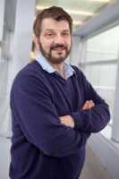 Prof. David A. Weitz[/caption]
Prof. David A. Weitz
Mallinckrodt Professor of Physics and Applied Physics
School of Engineering and Applied Sciences
Harvard University
MedicalResearch.com: What is the background for this technology study? What are the main findings?
Response: Currently, it is very time-consuming and expensive to develop new drugs. One reason is that many drugs fail in clinical trials after animal studies, simply because animals are very different from humans. One promising means of solving this problem is to replace animal experiments with artificial human tissues that can be used to directly screen a drug. However, it is a challenge to construct artificial human tissues, as almost all human tissues are composed of multiple types of cells and extracellular matrices in 3D structures.
In our studies, we have successfully developed a droplet-based microfluidic technique to fabricate large numbers of monodisperse, portable microtissues. We spatially assemble different types of cells in a 3D core-shell structure and construct an artificial human microtissue in each individual drop. The specific structures we create in the microdoplets are designed to mimic the behavior of the liver, and hence we call these structures a ‘liver in a drop.’
Prof. David A. Weitz[/caption]
Prof. David A. Weitz
Mallinckrodt Professor of Physics and Applied Physics
School of Engineering and Applied Sciences
Harvard University
MedicalResearch.com: What is the background for this technology study? What are the main findings?
Response: Currently, it is very time-consuming and expensive to develop new drugs. One reason is that many drugs fail in clinical trials after animal studies, simply because animals are very different from humans. One promising means of solving this problem is to replace animal experiments with artificial human tissues that can be used to directly screen a drug. However, it is a challenge to construct artificial human tissues, as almost all human tissues are composed of multiple types of cells and extracellular matrices in 3D structures.
In our studies, we have successfully developed a droplet-based microfluidic technique to fabricate large numbers of monodisperse, portable microtissues. We spatially assemble different types of cells in a 3D core-shell structure and construct an artificial human microtissue in each individual drop. The specific structures we create in the microdoplets are designed to mimic the behavior of the liver, and hence we call these structures a ‘liver in a drop.’
27 Dec Vaccine Nanodiscs Can Trigger More Cancer Fighting Immune Cells
Posted at 18:48h
in Author Interviews, Cancer Research, Immunotherapy, Nature, Technology, University of Michigan
MedicalResearch.com Interview with:
[caption id="attachment_30791" align="alignleft" width="168"] Dr. James Moon[/caption]
James Moon, PhD
John Gideon Searle Assistant Professor
University of Michigan
Dept. of Pharmaceutical Sciences and Biomedical Engineering
Biointerfaces Institute
Ann Arbor, MI, 48109
MedicalResearch.com: What is the background for this study?
Response: The field of cancer immunotherapy has recently made a breakthrough with the clinical success of immune checkpoint inhibitors, which work by removing the brakes on immunosuppressed T-cells. However, these approaches generally work by augmenting pre-existing T-cell immunity and benefit only a subset of patients. In addition, because the majority of somatic mutations in cancer cells are unique to each patient, cancer immunotherapy may benefit from a personalized approach.
Dr. James Moon[/caption]
James Moon, PhD
John Gideon Searle Assistant Professor
University of Michigan
Dept. of Pharmaceutical Sciences and Biomedical Engineering
Biointerfaces Institute
Ann Arbor, MI, 48109
MedicalResearch.com: What is the background for this study?
Response: The field of cancer immunotherapy has recently made a breakthrough with the clinical success of immune checkpoint inhibitors, which work by removing the brakes on immunosuppressed T-cells. However, these approaches generally work by augmenting pre-existing T-cell immunity and benefit only a subset of patients. In addition, because the majority of somatic mutations in cancer cells are unique to each patient, cancer immunotherapy may benefit from a personalized approach.
16 Dec Non-Invasive Interface Allows Subjects To Control Objects With Just Thoughts
MedicalResearch.com Interview with: [caption id="attachment_30535" align="alignleft" width="118"] Dr. Bin He[/caption]
Bin He, Ph.D.
Director, Institute for Engineering in Medicine
Director, Center for Neuroengineering
Distinguished McKnight University Professor of Biomedical Engineering
Medtronic-Bakken Endowed Chair for Engineering in Medicine
University of Minnesota, Minneapolis, MN 55455
MedicalResearch.com: What is the background for this study? What are the main findings?
Response: This work is aimed at developing a noninvasive brains-computer interface to allow disabled patients to control their environment by just thinking about it.
We found 8 human subjects were able to accomplish 3D reach and grasp tasks without using any muscle activities but just thinking about it.
Dr. Bin He[/caption]
Bin He, Ph.D.
Director, Institute for Engineering in Medicine
Director, Center for Neuroengineering
Distinguished McKnight University Professor of Biomedical Engineering
Medtronic-Bakken Endowed Chair for Engineering in Medicine
University of Minnesota, Minneapolis, MN 55455
MedicalResearch.com: What is the background for this study? What are the main findings?
Response: This work is aimed at developing a noninvasive brains-computer interface to allow disabled patients to control their environment by just thinking about it.
We found 8 human subjects were able to accomplish 3D reach and grasp tasks without using any muscle activities but just thinking about it.
09 Dec Computerized Working Memory Training in Pediatric Sickle Cell Disease
MedicalResearch.com Interview with: [caption id="attachment_30413" align="alignleft" width="200"] Dr. Steven J. Hardy[/caption]
Steven J. Hardy, Phd
Licensed Clinical Psychologist
Divisions of Hematology and Oncology
Children’s National Health System
Assistant Professor of Pediatrics and Psychiatry & Behavioral Sciences
George Washington School of Medicine and Health Sciences Washington, DC
MedicalResearch.com: What is the background for this study? What are the main findings?
Response: Children with sickle cell disease exhibit neurocognitive deficits as a consequence of either silent or overt cerebral infarction or disease-related non-infarct central nervous system effects (likely resulting from chronic anemia and hypoxic events). These complications often lead to impairment in executive functioning (e.g., working memory, attention, inhibition, cognitive flexibility), which can make it difficult to focus in class, plan for long-term school projects, remember and carry out multi-step tasks or assignments, and stay organized. The literature on interventions to reduce neurocognitive sequelae of sickle cell disease is extremely limited.
Our research team investigated a promising home-based, computerized cognitive training program (Cogmed) involving repeated practice on performance-adapted exercises targeting working memory with a sample of youth (ages 7 – 16) with sickle cell disease. Of the participants who have enrolled in the study (n = 70), 49% exhibited working memory deficits (<25% in the general population have a working memory deficit) and were randomized to an eight-week waitlist or to begin Cogmed immediately. Participants who used Cogmed demonstrated significant improvements on multiple measures of working memory, while those randomized to the waitlist group only exhibited such improvements after receiving Cogmed. Approximately 25% of participants completed the recommended number of Cogmed sessions (20 – 25 sessions). However, analyses revealed that participants who completed at least 10 sessions (about 50% of the participants) showed comparable levels of working memory improvement.
Dr. Steven J. Hardy[/caption]
Steven J. Hardy, Phd
Licensed Clinical Psychologist
Divisions of Hematology and Oncology
Children’s National Health System
Assistant Professor of Pediatrics and Psychiatry & Behavioral Sciences
George Washington School of Medicine and Health Sciences Washington, DC
MedicalResearch.com: What is the background for this study? What are the main findings?
Response: Children with sickle cell disease exhibit neurocognitive deficits as a consequence of either silent or overt cerebral infarction or disease-related non-infarct central nervous system effects (likely resulting from chronic anemia and hypoxic events). These complications often lead to impairment in executive functioning (e.g., working memory, attention, inhibition, cognitive flexibility), which can make it difficult to focus in class, plan for long-term school projects, remember and carry out multi-step tasks or assignments, and stay organized. The literature on interventions to reduce neurocognitive sequelae of sickle cell disease is extremely limited.
Our research team investigated a promising home-based, computerized cognitive training program (Cogmed) involving repeated practice on performance-adapted exercises targeting working memory with a sample of youth (ages 7 – 16) with sickle cell disease. Of the participants who have enrolled in the study (n = 70), 49% exhibited working memory deficits (<25% in the general population have a working memory deficit) and were randomized to an eight-week waitlist or to begin Cogmed immediately. Participants who used Cogmed demonstrated significant improvements on multiple measures of working memory, while those randomized to the waitlist group only exhibited such improvements after receiving Cogmed. Approximately 25% of participants completed the recommended number of Cogmed sessions (20 – 25 sessions). However, analyses revealed that participants who completed at least 10 sessions (about 50% of the participants) showed comparable levels of working memory improvement.
05 Dec Key Barriers To Development of Artificial Red Blood Cells Overcome
Posted at 20:26h
in Accidents & Violence, Anemia, Author Interviews, Hematology, Surgical Research, Technology
MedicalResearch.com Interview with:
[caption id="attachment_30198" align="alignleft" width="133"] Dr. Allan Doctor[/caption]
Allan Doctor, MD
Pediatric Critical Care Medicine
Professor of Pediatrics and (Associate) Biochemistry
Washington University School of Medicine &
Saint Louis Children’s Hospital
St. Louis, Missouri
MedicalResearch.com: What is the background for this study? What are the main findings?
Response: Our research team has developed the first nanoscale artificial cells designed to emulate vital functions of natural red blood cells. If ultimately confirmed safe for use in humans, this nanotechnology-based product, called ‘ErythroMer’, could represent a new and innovative alternative to blood transfusions that would be especially valuable in situations where stored blood is needed, but difficult to obtain or use, such as in pre-hospital or battlefield settings. The artificial cells are designed to be freeze-dried, stored for extended periods at ambient temperatures, and simply reconstituted with water for immediate use.
This year, the National Academy of Sciences estimated that 30,000 civilian trauma deaths/year are preventable and of these, two-thirds arise from hemorrhage in the pre-hospital phase of care. One key goal for our team is to advance treatment for trauma victims or soldiers in austere environments by initiating resuscitation in the field, particularly when transport is prolonged. ErythroMer could be a blood substitute that medics carry in their pack and literally take it out, add water, and inject. There are currently no simple, practical means to bring transfusion to most trauma victims outside of hospitals. Delays in resuscitation significantly impact outcomes; it is our goal to push timely, effective care to field settings.
Dr. Allan Doctor[/caption]
Allan Doctor, MD
Pediatric Critical Care Medicine
Professor of Pediatrics and (Associate) Biochemistry
Washington University School of Medicine &
Saint Louis Children’s Hospital
St. Louis, Missouri
MedicalResearch.com: What is the background for this study? What are the main findings?
Response: Our research team has developed the first nanoscale artificial cells designed to emulate vital functions of natural red blood cells. If ultimately confirmed safe for use in humans, this nanotechnology-based product, called ‘ErythroMer’, could represent a new and innovative alternative to blood transfusions that would be especially valuable in situations where stored blood is needed, but difficult to obtain or use, such as in pre-hospital or battlefield settings. The artificial cells are designed to be freeze-dried, stored for extended periods at ambient temperatures, and simply reconstituted with water for immediate use.
This year, the National Academy of Sciences estimated that 30,000 civilian trauma deaths/year are preventable and of these, two-thirds arise from hemorrhage in the pre-hospital phase of care. One key goal for our team is to advance treatment for trauma victims or soldiers in austere environments by initiating resuscitation in the field, particularly when transport is prolonged. ErythroMer could be a blood substitute that medics carry in their pack and literally take it out, add water, and inject. There are currently no simple, practical means to bring transfusion to most trauma victims outside of hospitals. Delays in resuscitation significantly impact outcomes; it is our goal to push timely, effective care to field settings.
01 Dec Humanoid Robot Deepens Understanding of How Toddlers Learn What Words Mean
MedicalResearch.com Interview with: [caption id="attachment_30093" align="alignleft" width="350"]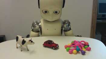 Humanoid Robot Helps Understand How Toddlers Link Words to Objects[/caption]
Dr Katie (Katherine) Twomey
ESRC Future Research Leaders Fellow, Lancaster University
Senior Research Associate, ESRC International Centre for Language and Communicative Development (LuCiD)
MedicalResearch.com: What is the background for this study? What are the main findings?
Response: Although we know that toddlers can quickly work out what new words mean, it's not yet clear exactly how they do it. For example, when they see a new object alongside their favorite toy truck and hear a new word "block", we know that they will link "block" to the new object. They could do this by thinking in detail about what they already know, for example "if my toy is called "truck", then "block" must be the name of the new object". Equally, however, they could quickly link the new word to the new object without thinking about it in-depth.
We tested this second possibility using iCub, a humanoid robot which learns by making quick associations between what it sees and what it hears, without the ability to think in detail about what it already knows. We replicated two studies of toddlers' early word learning with iCub and found that even though it can only learn through making simple links between words and objects, it behaved exactly as children did in the original experiments.
Humanoid Robot Helps Understand How Toddlers Link Words to Objects[/caption]
Dr Katie (Katherine) Twomey
ESRC Future Research Leaders Fellow, Lancaster University
Senior Research Associate, ESRC International Centre for Language and Communicative Development (LuCiD)
MedicalResearch.com: What is the background for this study? What are the main findings?
Response: Although we know that toddlers can quickly work out what new words mean, it's not yet clear exactly how they do it. For example, when they see a new object alongside their favorite toy truck and hear a new word "block", we know that they will link "block" to the new object. They could do this by thinking in detail about what they already know, for example "if my toy is called "truck", then "block" must be the name of the new object". Equally, however, they could quickly link the new word to the new object without thinking about it in-depth.
We tested this second possibility using iCub, a humanoid robot which learns by making quick associations between what it sees and what it hears, without the ability to think in detail about what it already knows. We replicated two studies of toddlers' early word learning with iCub and found that even though it can only learn through making simple links between words and objects, it behaved exactly as children did in the original experiments.
28 Nov Virtual Reality Systems Can Generate Immersive 3D Images of Fetuses
[caption id="attachment_30002" align="alignleft" width="200"] Close-up of fetus at 26 weeks[/caption]
MedicalResearch.com Interview with:
Dr. Heron Werner Junior
Clínica de Diagnóstico por Imagem – CDPI
Rio de Janeiro - Brazil
MedicalResearch.com: What is the background for this study?
Response: A growing number of technological advancements in obtaining and viewing images through noninvasive techniques have brought major breakthroughs in fetal medicine.
In general, two main technologies are used to obtain images within the uterus during pregnancy i.e. ultrasound (US) and magnetic resonance imaging (MRI).
Close-up of fetus at 26 weeks[/caption]
MedicalResearch.com Interview with:
Dr. Heron Werner Junior
Clínica de Diagnóstico por Imagem – CDPI
Rio de Janeiro - Brazil
MedicalResearch.com: What is the background for this study?
Response: A growing number of technological advancements in obtaining and viewing images through noninvasive techniques have brought major breakthroughs in fetal medicine.
In general, two main technologies are used to obtain images within the uterus during pregnancy i.e. ultrasound (US) and magnetic resonance imaging (MRI).
21 Nov Tiny Skin-Mounted Sensor Can Monitor Heart, GI and Voice Sounds
MedicalResearch.com Interview with: [caption id="attachment_29851" align="alignleft" width="300"]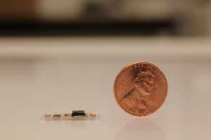 Tiny Skin Sensor Can Monitor Body Sounds[/caption]
YuHao Liu (Howard)
Materials Research Laboratory
University of Illinois
Urbana Champaign
MedicalResearch.com: What is the background for this study? What are the main findings?
Response: Our research group has been working on developing the next-generation wearable technology - integrating electronic tattoos on human skin for healthcare monitoring. This novel platform allows us to explore new sensors and actuators that are better than the commercially available ones.
We have developed sensors that can measure temperature, pressure, hydration, electrophysiology..etc. However, no one ever think about capturing sound from the body until our team developed this new technology. In fact, body sounds can be important indicator in healthcare monitoring or even disease diagnostic.
Tiny Skin Sensor Can Monitor Body Sounds[/caption]
YuHao Liu (Howard)
Materials Research Laboratory
University of Illinois
Urbana Champaign
MedicalResearch.com: What is the background for this study? What are the main findings?
Response: Our research group has been working on developing the next-generation wearable technology - integrating electronic tattoos on human skin for healthcare monitoring. This novel platform allows us to explore new sensors and actuators that are better than the commercially available ones.
We have developed sensors that can measure temperature, pressure, hydration, electrophysiology..etc. However, no one ever think about capturing sound from the body until our team developed this new technology. In fact, body sounds can be important indicator in healthcare monitoring or even disease diagnostic.
20 Nov Rolex Winner PEEK Vision Allows Smartphone-Based Eye Exams In Resource Poor Settings
MedicalResearch.com Interview with: Dr. Andrew Bastawrous, Ophthalmologist Rolex Awards for Enterprise United Kingdom Dr. Bastawrous’ smartphone-based portable eye examination system, Peek Vision, allows patients in low resource setting, to be screened for vision problems and eye diseases, enabling accurate diagnosis and treatment. MedicalResearch.com: What is the background for Peek Vision? What are the main findings of your work with this system? Response: Mama Paul has been blind for almost 20 years, most of her time is spent within the safety of her tiny home. It is basic, but in her mind’s eye she can still picture where the door is, her son - Paul, lives next door and is always willing to help. Losing sight is the sense most people fear losing most. I am severely visually impaired, I am also fortunate to have perfect vision when wearing corrective glasses or contact lenses and privileged to be in the profession of ophthalmology where centuries of research and practice have brought us to a time when so much of blindness is now curable or preventable. There is no feeling like it: when the eye patch comes off someone who hasn’t seen for years, the sheer wonder as they take in their surroundings and their anticipation to see faces that have become voices and places that have become memories. Back in 2011, as I pondered and planned for the challenges that lay ahead of us in Kenya, I had the continual thought that there must be an easier way to reach people, a way that is less expensive, less resource hungry and therefore could be used on a much wider scale. In Kenya, and much of Africa, more people have access to a mobile phone than they do clean running water. It had to be possible to harness this connectivity.19 Nov Brain-Computer Interface Allows Locked-in ALS Patient To Communicate
MedicalResearch.com Interview with: Mariska Van Steensel PhD Nick F. Ramsey, Ph.D. Department of Neurology and Neurosurgery Brain Center Rudolf Magnus University Medical Center Utrecht Utrecht, the Netherlands MedicalResearch.com: What is the background for this study? What are the main findings? Response: Patients who are severely paralyzed due to for example ALS or brain stem stroke are often unable to speak (also called ‘ Locked-in State'), and therefore need assistive devices, such as an eye tracker, for their communication. When these devices fail (e.g. due to environmental lighting or eye movement problems), people may indicate yes or no with eye blinks in response to closed questions. This leaves patients in a highly dependent position, since questions asked may or may not represent their actual wish or comment. In the current study, we used a technology called brain-computer interfacing (BCI), to allow a patient with late-stage amyotrophic lateral sclerosis (ALS) to control a communication device using her brain signals. The patient was implanted with subdural electrodes that covered the brain area that is normally responsible for hand movement. The electrodes were connected with wires, subcutaneously, to an amplifier/transmitter device that was placed subcutaneously under the clavicle. The patient was able to generate a signal equivalent to a mouse-click with this brain-computer interface by attempting to move her hand, and used it to make selections of letters or words on her communication device, with high accuracy and a speed of 2 letters per minute. She used the brain-computer interface system to communicate whenever she was outside, as her eye-tracker device does not function well in that situation.18 Nov New Capsule Can Reside in Stomach and Deliver Drugs for Several Weeks After Single Ingestion
Posted at 02:39h
in Author Interviews, Brigham & Women's - Harvard, Compliance, Gastrointestinal Disease, Pharmacology, Technology
MedicalResearch.com Interview with:
[caption id="attachment_29758" align="alignleft" width="143"] Dr. Traverso[/caption]
C. Giovanni Traverso, MB, BChir, PhD
Gastroenterologist and biomedical engineer
Division of Gastroenterology at BWH
Instructor of medicine at Harvard Medical School
MedicalResearch.com: What is the background for this study? What are the main findings?
Response: We developed a drug delivery system capable of safely residing in the stomach for 2 weeks. Furthermore we demonstrated the capacity of the novel dosage form, in the shape of a star, to protect the drug from the acidic stomach environment and also slowly release drug over the course of 14 days.
We applied this new technology towards efforts targeting the elimination of malaria. Specifically, we focused on a drug called ivermectin that has been used to treat parasites but also has the benefit of being toxic to malaria-carrying mosquitos when they bite someone who has ivermectin in their system.
Dr. Traverso[/caption]
C. Giovanni Traverso, MB, BChir, PhD
Gastroenterologist and biomedical engineer
Division of Gastroenterology at BWH
Instructor of medicine at Harvard Medical School
MedicalResearch.com: What is the background for this study? What are the main findings?
Response: We developed a drug delivery system capable of safely residing in the stomach for 2 weeks. Furthermore we demonstrated the capacity of the novel dosage form, in the shape of a star, to protect the drug from the acidic stomach environment and also slowly release drug over the course of 14 days.
We applied this new technology towards efforts targeting the elimination of malaria. Specifically, we focused on a drug called ivermectin that has been used to treat parasites but also has the benefit of being toxic to malaria-carrying mosquitos when they bite someone who has ivermectin in their system.
16 Nov Supermagnetized Crystals May Deliver Precision Drugs Directly To Medical Targets
MedicalResearch.com Interview with: Dr. Kezheng Chen Lab of Functional and Biomedical Nanomaterials College of Materials Science and Engineering Qingdao University of Science and Technology Qingdao China MedicalResearch.com: What is the background for this study? What are the main findings? Response: From a view point of practical applications, superparamagnetism is usually highly desirable, because it can prevent the magnetic particles from irreversible aggregation and ensure an excellent dispersity once the applied magnetic field is removed. Up to now, the largest size of reported superparamagnetic clusters is around several hundreds of nanometers, which are composed of numerous nanocrystals, and hence exhibiting polycrystalline nature. In this sense, how to realize well pronounced superparamagnetism in large-size single crystals is of great interest to the general public.11 Nov Action Video Games Improved Amblyopia In Two Weeks
MedicalResearch.com Interview with: Krista Kelly, PhD Postdoctoral Fellow Crystal Charity Ball Pediatric Vision Evaluation Center Retina Foundation of the Southwest Dallas, TX 75231 MedicalResearch.com: What is the background for this study? Response: Amblyopia is one of the most common causes of monocular impairment in children, affecting 1 or 2 children in every US classroom. Patching of the fellow eye has been used for decades to improve visual acuity in the amblyopic eye. But patching does not always restore normal vision and does not teach the two eyes to work together. A novel technique originally designed by Drs Robert Hess and Ben Thompson at McGill University that works to reduce interocular suppression by rebalancing the contrast between the eyes has shown promising results in amblyopic adults. Dr Eileen Birch at the Retina Foundation of the Southwest worked with Dr Hess to adapt this contrast re-balancing approach to an iPad game platform suitable for children. Her research showed that the games were successful in improving visual acuity in amblyopic children as well. However, these initial games were rudimentary and resulted in low compliance.01 Nov Study Recommends Children Avoid Mobile Screens Before Bedtime
MedicalResearch.com Interview with: [caption id="attachment_29335" align="alignleft" width="200"] Dr. Ben Carter[/caption]
Dr Ben Carter PhD
Senior Lecturer in Medical Statistics
Statistics Editor for the Cochrane Skin Group
(Honorary Associate Professor, Nottingham University)
Institute of Primary Care and Public Health
Cardiff University
MedicalResearch.com: What is the background for this study? What are the main findings?
Response: This study leads from the growing use of mobile and media device use in children. We report the impact of devices leads to poorer sleep outcomes.
MedicalResearch.com: What should readers take away from your report?
Response: Using or even merely access to your mobile and media device should be restricted 90 minutes prior to bedtime.
Dr. Ben Carter[/caption]
Dr Ben Carter PhD
Senior Lecturer in Medical Statistics
Statistics Editor for the Cochrane Skin Group
(Honorary Associate Professor, Nottingham University)
Institute of Primary Care and Public Health
Cardiff University
MedicalResearch.com: What is the background for this study? What are the main findings?
Response: This study leads from the growing use of mobile and media device use in children. We report the impact of devices leads to poorer sleep outcomes.
MedicalResearch.com: What should readers take away from your report?
Response: Using or even merely access to your mobile and media device should be restricted 90 minutes prior to bedtime.
26 Oct Low-Intensity Ultrasound Not Effective In Accelerating Healing of Tibial Fractures
MedicalResearch.com Interview with: [caption id="attachment_29117" align="alignleft" width="150"]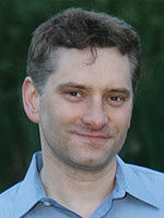 Jason Busse[/caption]
Jason Busse PhD
Associate Professor, Department of Anesthesia
McMaster University
Hamilton, ON
MedicalResearch.com: What is the background for this study? What are the main findings?
Response: Our group previously reviewed the evidence regarding the effectiveness of low-intensity pulsed ultrasound (LIPUS) for fracture healing. We found moderate to very low quality evidence for LIPUS in accelerating functional recovery among patients with fracture, and that most trials only explored effects on surrogate outcomes (e.g. radiographic healing); only five of 13 trials directly assessed functional end points - of these, one was positive. We concluded that large trials of high methodological quality, focusing on patient important outcomes such as quality of life and return to function, were needed to establish the role of LIPUS in fracture healing.
We have now completed such a study. Our large, international trial of LIPUS for surgically managed tibial fractures found the addition of LIPUS does not improve functional recovery or accelerate radiographic healing.
Jason Busse[/caption]
Jason Busse PhD
Associate Professor, Department of Anesthesia
McMaster University
Hamilton, ON
MedicalResearch.com: What is the background for this study? What are the main findings?
Response: Our group previously reviewed the evidence regarding the effectiveness of low-intensity pulsed ultrasound (LIPUS) for fracture healing. We found moderate to very low quality evidence for LIPUS in accelerating functional recovery among patients with fracture, and that most trials only explored effects on surrogate outcomes (e.g. radiographic healing); only five of 13 trials directly assessed functional end points - of these, one was positive. We concluded that large trials of high methodological quality, focusing on patient important outcomes such as quality of life and return to function, were needed to establish the role of LIPUS in fracture healing.
We have now completed such a study. Our large, international trial of LIPUS for surgically managed tibial fractures found the addition of LIPUS does not improve functional recovery or accelerate radiographic healing.
19 Oct New Technology Can Quickly Detect TB and Evaluate Therapeutic Efficacy
MedicalResearch.com Interview with: [caption id="attachment_29007" align="alignleft" width="200"]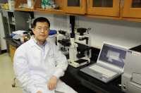 Dr. Ying Kong[/caption]
Ying Kong Ph.D.
Assistant Professor
University of Tennessee Health Science Center
Department of Microbiology, Immunology, and Biochemistry
Memphis, TN 38163
MedicalResearch.com: What is the background for this study?
Response: Tuberculosis (TB) is a public health concern worldwide, with high morbidity and mortality. The causative agent of TB, M. tuberculosis, grows very slowly in culture. For research of TB, we need to quantitate bacterial numbers in order to evaluate drug and vaccine efficacy or to identify bacterial genes that are critical for survival in hosts or causing disease.
M. tuberculosis divides every ~20 hours, which is much slower than other bacteria such as E. coli and Salmonella typhimurium, which divide every 20 minutes. Conventionally, quantitation of M. tuberculosis needs to spread M. tuberculosis on agar plates and wait for four weeks to obtain visible colonies, and then to count colony forming units. For the fast-growing bacteria, it takes only 18 hours to obtain visible colonies on agar plates. We and other groups have developed fluorescent protein labeled M. tuberculosis strains in order to quantitate M. tuberculosis in real time by measuring fluorescence. In this way, we are able to estimate bacterial number right after fluorescence measurement, which only takes a few minutes. However, this technology is not a diagnostic tool for clinical use, because the M. tuberculosis strains that we used were recombinant strains transformed with fluorescent protein genes. Another imaging technology that we have developed, REF, is for diagnosis purpose, which has been described in details in our other papers (Xie H, et al. Rapid point-of-care detection of the tuberculosis pathogen using a BlaC-specific fluorogenic probe. Nat Chem. 2012 Oct;4(10):802-9. Cheng Y, et al. Fluorogenic probes with substitutions at the 2 and 7 positions of cephalosporin are highly BlaC-specific for rapid Mycobacterium tuberculosis detection. Angew Chem Int Ed Engl. 2014 Aug 25;53(35):9360-4.).
Dr. Ying Kong[/caption]
Ying Kong Ph.D.
Assistant Professor
University of Tennessee Health Science Center
Department of Microbiology, Immunology, and Biochemistry
Memphis, TN 38163
MedicalResearch.com: What is the background for this study?
Response: Tuberculosis (TB) is a public health concern worldwide, with high morbidity and mortality. The causative agent of TB, M. tuberculosis, grows very slowly in culture. For research of TB, we need to quantitate bacterial numbers in order to evaluate drug and vaccine efficacy or to identify bacterial genes that are critical for survival in hosts or causing disease.
M. tuberculosis divides every ~20 hours, which is much slower than other bacteria such as E. coli and Salmonella typhimurium, which divide every 20 minutes. Conventionally, quantitation of M. tuberculosis needs to spread M. tuberculosis on agar plates and wait for four weeks to obtain visible colonies, and then to count colony forming units. For the fast-growing bacteria, it takes only 18 hours to obtain visible colonies on agar plates. We and other groups have developed fluorescent protein labeled M. tuberculosis strains in order to quantitate M. tuberculosis in real time by measuring fluorescence. In this way, we are able to estimate bacterial number right after fluorescence measurement, which only takes a few minutes. However, this technology is not a diagnostic tool for clinical use, because the M. tuberculosis strains that we used were recombinant strains transformed with fluorescent protein genes. Another imaging technology that we have developed, REF, is for diagnosis purpose, which has been described in details in our other papers (Xie H, et al. Rapid point-of-care detection of the tuberculosis pathogen using a BlaC-specific fluorogenic probe. Nat Chem. 2012 Oct;4(10):802-9. Cheng Y, et al. Fluorogenic probes with substitutions at the 2 and 7 positions of cephalosporin are highly BlaC-specific for rapid Mycobacterium tuberculosis detection. Angew Chem Int Ed Engl. 2014 Aug 25;53(35):9360-4.).
15 Oct Smartphone App Can Screen For Atrial Fibrillation
MedicalResearch.com Interview with: [caption id="attachment_28905" align="alignleft" width="178"]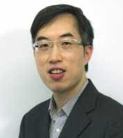 Dr. Ngai-yin Chan[/caption]
Dr. Ngai-yin Chan
Princess Margaret Hospital
Lai Chi Kok, Hong Kong
MedicalResearch.com: What is the background for this study?
Response: Atrial fibrillation (AF) is the most common sustained heart rhythm disorder which can cause stroke, heart failure and an increased risk of death. The risk of stroke can be reduced substantially with drug treatment. However, a quarter of patients with AF causing stroke have silent and asymptomatic AF before stroke. The current guidelines recommend opportunistic screening for AF. Whether systematic community screening for AF with a convenient smartphone ECG can reduce the burden of AF remains unknown.
Dr. Ngai-yin Chan[/caption]
Dr. Ngai-yin Chan
Princess Margaret Hospital
Lai Chi Kok, Hong Kong
MedicalResearch.com: What is the background for this study?
Response: Atrial fibrillation (AF) is the most common sustained heart rhythm disorder which can cause stroke, heart failure and an increased risk of death. The risk of stroke can be reduced substantially with drug treatment. However, a quarter of patients with AF causing stroke have silent and asymptomatic AF before stroke. The current guidelines recommend opportunistic screening for AF. Whether systematic community screening for AF with a convenient smartphone ECG can reduce the burden of AF remains unknown.
12 Oct Technology May Help Physicians Improve the Art of Medical Diagnosis
MedicalResearch.com Interview with: [caption id="attachment_28621" align="alignleft" width="200"]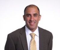 Dr. Ateev Mehrotra[/caption]
Ateev Mehrotra, M.D.
Associate Professor
Department of Health Care Policy
Harvard Medical School
MedicalResearch.com: What is the background for this study? What are the main findings?
Response: Prior research has highlighted that physicians make diagnostic errors roughly 10 to 15 percent of the time. Over the last two decades, computer-based checklists and other “fail-safe” digital apps have been increasingly used to reduce medication errors or streamline infection-prevention protocols. Lately, experts have wondered whether computers might also help reduce diagnostic errors.
In the study, 234 internal medicine physicians were asked to evaluate 45 clinical cases, involving both common and uncommon conditions with varying degrees of severity. For each case, physicians had to identify the most likely diagnosis along with two additional possible diagnoses. Each clinical vignette was solved by at least 20 physicians. The same cases were also evaluated using 19 symptom checkers, websites or apps that use computers that help patients determine potential diagnoses for what is wrong based on their symptoms.
The physicians vastly outperformed the symptom-checker apps, listing the correct diagnosis 72 percent of the time, compared with 34 percent of the time for the digital platforms. Eighty-four percent of clinicians listed the correct diagnosis in the top three possibilities, compared with 51 percent for the digital symptom-checkers.
Dr. Ateev Mehrotra[/caption]
Ateev Mehrotra, M.D.
Associate Professor
Department of Health Care Policy
Harvard Medical School
MedicalResearch.com: What is the background for this study? What are the main findings?
Response: Prior research has highlighted that physicians make diagnostic errors roughly 10 to 15 percent of the time. Over the last two decades, computer-based checklists and other “fail-safe” digital apps have been increasingly used to reduce medication errors or streamline infection-prevention protocols. Lately, experts have wondered whether computers might also help reduce diagnostic errors.
In the study, 234 internal medicine physicians were asked to evaluate 45 clinical cases, involving both common and uncommon conditions with varying degrees of severity. For each case, physicians had to identify the most likely diagnosis along with two additional possible diagnoses. Each clinical vignette was solved by at least 20 physicians. The same cases were also evaluated using 19 symptom checkers, websites or apps that use computers that help patients determine potential diagnoses for what is wrong based on their symptoms.
The physicians vastly outperformed the symptom-checker apps, listing the correct diagnosis 72 percent of the time, compared with 34 percent of the time for the digital platforms. Eighty-four percent of clinicians listed the correct diagnosis in the top three possibilities, compared with 51 percent for the digital symptom-checkers.
07 Oct Machine Learning and Free-Text Analysis of Notes Improves Patient Identification
MedicalResearch.com Interview with: [caption id="attachment_28567" align="alignleft" width="144"] Dr. Saul Blecker,[/caption]
Saul Blecker, MD, MHS
Department of Population Health
New York University Langone School of Medicine,
New York, NY 10016
Saul.Blecker@nyumc.org
MedicalResearch.com: What is the background for this study? What are the main findings?
Response: The identification of conditions or diseases in the electronic health record (EHR) is critical in clinical practice, for quality improvement, and for clinical interventions. Today, a disease such as heart failure is typically identified in real-time using a “problem list”, i.e., a list of conditions for each patient that is maintained by his or her providers, or using simple rules drawn from structured data. In this study, we examined the comparative benefit of using more sophisticated approaches for identifying hospitalized patients with heart failure.
Dr. Saul Blecker,[/caption]
Saul Blecker, MD, MHS
Department of Population Health
New York University Langone School of Medicine,
New York, NY 10016
Saul.Blecker@nyumc.org
MedicalResearch.com: What is the background for this study? What are the main findings?
Response: The identification of conditions or diseases in the electronic health record (EHR) is critical in clinical practice, for quality improvement, and for clinical interventions. Today, a disease such as heart failure is typically identified in real-time using a “problem list”, i.e., a list of conditions for each patient that is maintained by his or her providers, or using simple rules drawn from structured data. In this study, we examined the comparative benefit of using more sophisticated approaches for identifying hospitalized patients with heart failure.
06 Oct Digital Mobile Technology Gives Some Arm Function To Stroke Patients
MedicalResearch.com Interview with: [caption id="attachment_28589" align="alignleft" width="149"]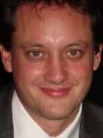 Dr Paul Bentley[/caption]
Dr Paul Bentley MA MRCP PhD
Clinical Senior Lecturer in Clinical Neuroscience
Honorary Consultant Neurologist
Neurology Dept
Imperial College NHS Healthcare Trust
Charing Cross Hospital
London
MedicalResearch.com: What is the background for this study? What are the main findings?
Response: gripAble was designed to help people with arm disability practise physiotherapy when a physiotherapist is not available, or in between physiotherapy sessions. We know that the amount of physiotherapy provision in the UK, after stroke or arm injury, is typically below that which is recommended by professional bodies. Furthermore, increasing research suggests that higher-intensity training can boost functional outcomes. The innovation was designed to help people with a range of disabilities including severe paralysis engage with computer games with their weak arm. At the same time its designed to be portable for use at home or in bed, and low-cost.
gripAble also enables remote measurement and monitoring of arm function, by setting users a series of calibrated tasks played out on the tablet screen. This way doctors and physiotherapists can assess the needs of a patient, and gain an idea of how well a patient is responding to home physiotherapy.
Dr Paul Bentley[/caption]
Dr Paul Bentley MA MRCP PhD
Clinical Senior Lecturer in Clinical Neuroscience
Honorary Consultant Neurologist
Neurology Dept
Imperial College NHS Healthcare Trust
Charing Cross Hospital
London
MedicalResearch.com: What is the background for this study? What are the main findings?
Response: gripAble was designed to help people with arm disability practise physiotherapy when a physiotherapist is not available, or in between physiotherapy sessions. We know that the amount of physiotherapy provision in the UK, after stroke or arm injury, is typically below that which is recommended by professional bodies. Furthermore, increasing research suggests that higher-intensity training can boost functional outcomes. The innovation was designed to help people with a range of disabilities including severe paralysis engage with computer games with their weak arm. At the same time its designed to be portable for use at home or in bed, and low-cost.
gripAble also enables remote measurement and monitoring of arm function, by setting users a series of calibrated tasks played out on the tablet screen. This way doctors and physiotherapists can assess the needs of a patient, and gain an idea of how well a patient is responding to home physiotherapy.
05 Oct UV Sensitive ‘Band-aid’ Makes Monitoring Sun Exposure Easy
MedicalResearch.com Interview with: [caption id="attachment_28574" align="alignleft" width="164"]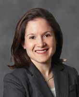 Dr. Andrea M Armani[/caption]
Dr. Andrea M Armani PhD
Fluor Early Career Chair and Associate Professor
Mork Family Department of Chemical Engineering and Materials Science
University of Southern California, Los Angeles, California
MedicalResearch.com: What is the background for this study?
Response: The “Internet of Things” (IoT) has seen an explosion in online sensor technologies, including UV sensors and monitors; for example, those from Apple and Samsung. However, they require connectivity and power, and they are integrated into delicate electronic systems that are not compatible with outdoor, athletic activities such as swimming, which is precisely when you should monitor UV exposure. Therefore, somewhat ironically, the technologies developed to meet the demands of the IoT are not ideal for cumulative UV exposure detection.
Our goal was to develop a single use patch – like a smart “band-aid” – for the beach to alert users when they had been in the sun for an hour and needed to re-apply sunscreen or get out of the sun altogether. This application required a rugged system that was waterproof, bendable, and compatible with sunscreen. Additionally, the sensor readout needed to be easy to interpret. These requirements influenced our design and material selection.
Dr. Andrea M Armani[/caption]
Dr. Andrea M Armani PhD
Fluor Early Career Chair and Associate Professor
Mork Family Department of Chemical Engineering and Materials Science
University of Southern California, Los Angeles, California
MedicalResearch.com: What is the background for this study?
Response: The “Internet of Things” (IoT) has seen an explosion in online sensor technologies, including UV sensors and monitors; for example, those from Apple and Samsung. However, they require connectivity and power, and they are integrated into delicate electronic systems that are not compatible with outdoor, athletic activities such as swimming, which is precisely when you should monitor UV exposure. Therefore, somewhat ironically, the technologies developed to meet the demands of the IoT are not ideal for cumulative UV exposure detection.
Our goal was to develop a single use patch – like a smart “band-aid” – for the beach to alert users when they had been in the sun for an hour and needed to re-apply sunscreen or get out of the sun altogether. This application required a rugged system that was waterproof, bendable, and compatible with sunscreen. Additionally, the sensor readout needed to be easy to interpret. These requirements influenced our design and material selection.
03 Oct Police Body-Worn Cameras Drastically Reduce Complaints Against Police
MedicalResearch.com Interview with: Barak Ariel, PhD Jerry Lee Fellow in Experimental Criminology and Lecturer in Experimental Criminology University of Cambridge Alex Sutherland, D.Phil. RAND Europe MedicalResearch.com: What is the background for this study? Response: The very first randomised-controlled experiment on cameras in Rialto (California) was originally intended to ‘cut red tape’, but Rialto Police Department decided instead to look at whether wearing the cameras could help reduce complaints and police use-of-force. That study found that cameras did cut use-of-force and complaints, with the latter almost to zero in the 12 months of the experiment. Given that each complaint costs, millions of dollars in the US, there was a lot of interest following that study, and that research was seen as crucial in the widespread adoption of Body-Worn Cameras in the US and other countries. Although Rialto is typical of a lot of mid-sized police forces in the US, it is just one location at one time – so the roll out that was taking place had a very weak evidence base. The same results might not be found in other locations and given that many police forces started to roll out cameras without evidence, there was a strong justification for replicating the study. This paper (and two others already published) report results from x10 RCTs that took place in the UK and US, with more than 2 million residents, over a period of a year.23 Sep Bioflux Device Monitors Heart Rhythm and Sends in Real Time To 24 Hour Monitoring Lab
MedicalResearch.com Interview with: Waqaas Al-Siddiq, Founder and CEO of Biotricity
MedicalResearch.com: Would you tell us a little about yourself? How did you get interested in combining healthcare with technology?
[caption id="attachment_28381" align="alignleft" width="200"]
Waqaas Al-Siddiq, Founder and CEO of Biotricity
MedicalResearch.com: Would you tell us a little about yourself? How did you get interested in combining healthcare with technology?
[caption id="attachment_28381" align="alignleft" width="200"] Waqaas Al-Siddiq[/caption]
Response: I am the president and CEO of biotricity which is a healthcare technology company dedicated to providing diagnostic and post-diagnostic solutions for both the physician and consumer for long-term chronic care management. I got interested in combining healthcare and technology while I was doing research for monitoring remote environments in critical scenarios. I thought that it would be very interesting to apply that to healthcare because it’s a problem that no one has figured out how to solve yet. And it’s a problem that is driving healthcare costs out of control.
Waqaas Al-Siddiq[/caption]
Response: I am the president and CEO of biotricity which is a healthcare technology company dedicated to providing diagnostic and post-diagnostic solutions for both the physician and consumer for long-term chronic care management. I got interested in combining healthcare and technology while I was doing research for monitoring remote environments in critical scenarios. I thought that it would be very interesting to apply that to healthcare because it’s a problem that no one has figured out how to solve yet. And it’s a problem that is driving healthcare costs out of control.
23 Sep Scaffold Can Potentially Deliver Therapeutic miRNA To Decrease Breast Cancer Metastases
Posted at 21:41h
in Author Interviews, Breast Cancer, Brigham & Women's - Harvard, Nature, Technology
MedicalResearch.com Interview with:
[caption id="attachment_28309" align="alignleft" width="157"] Dr. Natalie Artzi[/caption]
Natalie Artzi PhD
principal research scientist
MIT's Institute for Medical Engineering and Science and
Assistant professor of medicine
Brigham and Women's Hospital
With co-authors:
Avital Gilam, João Conde, Daphna Weissglas-Volkov, Nuria Oliva, Eitan Friedman, Noam Shomron
MedicalResearch.com: What is the background for this study? What are the main findings?
Response: Metastases are the primary cause for mortality in breast cancer, the most common cancer in women regardless of ethnicity. Recent studies show that germline sequence variants, such as single-nucleotide polymorphisms (SNPs) in miRNA-binding sites, can disrupt the downregulation by miRNAs, with a profound effect on gene expression levels and consequentially on the phenotype, including increased risk for cancer.
In the current study, we aimed to determine the potential effect of SNPs within miRNA-binding sites on metastatic breast cancer progression and their potential use as suppression targets to prevent metastasis.
Our collaborators at Tel-Aviv Universityin a research led by Dr. Noam Shomron found that the SNP, rs1071738, located in a target site for miR-96 and miR-182 on the 3’-UTR of the PALLD gene, encodes the Palladin actin-associated protein, which is a documented player in breast cancer motility. In vitro experiments revealed a functional downregulation of Palladin levels by miR-96 and miR-182, which subsequently reduces migration and invasion abilities of breast cancer cells.
My lab then showed in an in vivo experiment that the use of nanoparticles embedded in a hydrogel scaffold as a miRNA delivery vehicle enables an efficient and specific delivery of miR-96/miR-182 directly to breast tumours, which results in marked reduction of breast cancer metastasis. We then proceeded to study the effect of combination therapy in which we will use a chemotherapy drug to shrink the primary tumor and the miRNAs to prevent metastasis. The intercalation of a chemotherapy drug, cisplatin, to the miR-conjugated nanoparticles further improved the effect, leading to significant reduction in both primary tumour growth and metastasis.
Our study highlights the therapeutic potential of miRNAs, and specifically miR-96 and miR-182, and support the importance of Palladin regulation in breast cancer metastasis.
Dr. Natalie Artzi[/caption]
Natalie Artzi PhD
principal research scientist
MIT's Institute for Medical Engineering and Science and
Assistant professor of medicine
Brigham and Women's Hospital
With co-authors:
Avital Gilam, João Conde, Daphna Weissglas-Volkov, Nuria Oliva, Eitan Friedman, Noam Shomron
MedicalResearch.com: What is the background for this study? What are the main findings?
Response: Metastases are the primary cause for mortality in breast cancer, the most common cancer in women regardless of ethnicity. Recent studies show that germline sequence variants, such as single-nucleotide polymorphisms (SNPs) in miRNA-binding sites, can disrupt the downregulation by miRNAs, with a profound effect on gene expression levels and consequentially on the phenotype, including increased risk for cancer.
In the current study, we aimed to determine the potential effect of SNPs within miRNA-binding sites on metastatic breast cancer progression and their potential use as suppression targets to prevent metastasis.
Our collaborators at Tel-Aviv Universityin a research led by Dr. Noam Shomron found that the SNP, rs1071738, located in a target site for miR-96 and miR-182 on the 3’-UTR of the PALLD gene, encodes the Palladin actin-associated protein, which is a documented player in breast cancer motility. In vitro experiments revealed a functional downregulation of Palladin levels by miR-96 and miR-182, which subsequently reduces migration and invasion abilities of breast cancer cells.
My lab then showed in an in vivo experiment that the use of nanoparticles embedded in a hydrogel scaffold as a miRNA delivery vehicle enables an efficient and specific delivery of miR-96/miR-182 directly to breast tumours, which results in marked reduction of breast cancer metastasis. We then proceeded to study the effect of combination therapy in which we will use a chemotherapy drug to shrink the primary tumor and the miRNAs to prevent metastasis. The intercalation of a chemotherapy drug, cisplatin, to the miR-conjugated nanoparticles further improved the effect, leading to significant reduction in both primary tumour growth and metastasis.
Our study highlights the therapeutic potential of miRNAs, and specifically miR-96 and miR-182, and support the importance of Palladin regulation in breast cancer metastasis.
23 Sep Designing an Implantable Scaffold To Capture Cancer Cells and Personalize Treatment
MedicalResearch.com Interview with: [caption id="attachment_28279" align="alignleft" width="200"] Grace Bushnell[/caption]
Grace G. Bushnell PhD candidate
Department of Biomedical Engineering
University of Michigan
Ann Arbor, Michigan
MedicalResearch.com: What is the background for this study? What are the main findings?
Response: This study builds off of previous work from the Shea and Jeruss laboratories at the University of Michigan, which reported that implanted scaffolds can capture early metastatic cells in vivo prior to the colonization of other organs and serve as a platform for early detection of metastatic disease. Furthermore, the presence of a scaffold reduced the tumor burden in other metastatic locations.
In this work, the major finding was that early detection using a scaffold combined with a therapeutic intervention led to a survival advantage relative to mice that did not receive a scaffold. The scaffolds had been designed to persist in vivo for longer periods of time than in the original study. Additionally, the scaffold was implanted prior to the inoculation of metastatic cancer cells in the mouse. The role of the immune system in the process was further refined, as the immune cell composition at the scaffold changed significantly after disease initiation. These studies demonstrate that early intervention in a metastatic setting can lead to enhanced survival. This principle of early intervention is well established for the primary tumor, and these studies suggest that this principle may be extended to metastatic disease.
Grace Bushnell[/caption]
Grace G. Bushnell PhD candidate
Department of Biomedical Engineering
University of Michigan
Ann Arbor, Michigan
MedicalResearch.com: What is the background for this study? What are the main findings?
Response: This study builds off of previous work from the Shea and Jeruss laboratories at the University of Michigan, which reported that implanted scaffolds can capture early metastatic cells in vivo prior to the colonization of other organs and serve as a platform for early detection of metastatic disease. Furthermore, the presence of a scaffold reduced the tumor burden in other metastatic locations.
In this work, the major finding was that early detection using a scaffold combined with a therapeutic intervention led to a survival advantage relative to mice that did not receive a scaffold. The scaffolds had been designed to persist in vivo for longer periods of time than in the original study. Additionally, the scaffold was implanted prior to the inoculation of metastatic cancer cells in the mouse. The role of the immune system in the process was further refined, as the immune cell composition at the scaffold changed significantly after disease initiation. These studies demonstrate that early intervention in a metastatic setting can lead to enhanced survival. This principle of early intervention is well established for the primary tumor, and these studies suggest that this principle may be extended to metastatic disease.
22 Sep 3D Models and Immersive Virtual Reality Simulation for Complex Skull Base Surgery
MedicalResearch.com Interview with: [caption id="attachment_28265" align="alignleft" width="160"] Dr. Alfred Marc Calo Iloreta[/caption]
Alfred Marc Calo Iloreta, MD
Assistant Professor
Skull Base Surgery and Rhinology
Department of Otolaryngology -
Head and Neck Surgery
Icahn School of Medicine at Mount Sinai
New York, New York
MedicalResearch.com: Would you tell us a little about yourself? How did you become interested in ENT and your subspecialty in particular?
Response: I am a ENT/Head and Neck Surgeon practicing in Manhattan at the Mount Sinai Hospital. I trained here in New York City for residency and also completed a fellowship in Rhinology and Skull Base Surgery.
I chose this field and sub-specialty because of the intricate and complex anatomy of the head and neck. In addition rhinology and skull base surgery utilizes multiple advanced technologies from high definition optics, to neuronavigation to allow us to work with this complex anatomy.
Dr. Alfred Marc Calo Iloreta[/caption]
Alfred Marc Calo Iloreta, MD
Assistant Professor
Skull Base Surgery and Rhinology
Department of Otolaryngology -
Head and Neck Surgery
Icahn School of Medicine at Mount Sinai
New York, New York
MedicalResearch.com: Would you tell us a little about yourself? How did you become interested in ENT and your subspecialty in particular?
Response: I am a ENT/Head and Neck Surgeon practicing in Manhattan at the Mount Sinai Hospital. I trained here in New York City for residency and also completed a fellowship in Rhinology and Skull Base Surgery.
I chose this field and sub-specialty because of the intricate and complex anatomy of the head and neck. In addition rhinology and skull base surgery utilizes multiple advanced technologies from high definition optics, to neuronavigation to allow us to work with this complex anatomy.
19 Sep Computer Bests Neuroradiologists in Distinguishing Tumor Recurrence From Radiation Necrosis
Posted at 21:39h
in Author Interviews, Brain Cancer - Brain Tumors, Case Western, MRI, Radiology, Technology
MedicalResearch.com Interview with:
[caption id="attachment_28107" align="alignleft" width="200"] Dr. Pallavi Tiwari[/caption]
Dr. Pallavi Tiwari PhD
Assistant Professor biomedical engineering
Case Western Reserve University
MedicalResearch.com: What is the background for this study? What are the main findings?
Response: One of the biggest challenges in neuro-oncology currently is distinguishing radionecrosis, a side-effect of aggressive radiation, from tumor recurrence on imaging. Surgical intervention is the only means of definitive diagnosis, but suffers from considerable morbidity and mortality. The treatments for radionecrosis and cancer recurrence are very different. Early identification of the two conditions can help speed prognosis, therapy, and improve patient outcomes.
The purpose of this feasibility study was to evaluate the role of machine learning algorithms along with computer extracted texture features, also known as radiomic features, in distinguishing radionecrosis and tumor recurrence on routine MRI scans (T1w, T2w, FLAIR). The radiomic algorithms were trained on 43 studies from our local collaborating institution - University Hospitals Case Medical Center, and tested on 15 studies at a collaborating institution, University of Texas Southwest Medical Center. We further compared the performance of the radiomic techniques with two expert readers.
Our results demonstrated that radiomic features can identify subtle differences in quantitative measurements of tumor heterogeneity on routine MRIs, that are not visually appreciable to human readers. Of the 15 test studies, the radiomics algorithm could identify 12 of 15 correctly, while expert 1 could identify 7 of 15, and expert 2, 8 of 15.
Dr. Pallavi Tiwari[/caption]
Dr. Pallavi Tiwari PhD
Assistant Professor biomedical engineering
Case Western Reserve University
MedicalResearch.com: What is the background for this study? What are the main findings?
Response: One of the biggest challenges in neuro-oncology currently is distinguishing radionecrosis, a side-effect of aggressive radiation, from tumor recurrence on imaging. Surgical intervention is the only means of definitive diagnosis, but suffers from considerable morbidity and mortality. The treatments for radionecrosis and cancer recurrence are very different. Early identification of the two conditions can help speed prognosis, therapy, and improve patient outcomes.
The purpose of this feasibility study was to evaluate the role of machine learning algorithms along with computer extracted texture features, also known as radiomic features, in distinguishing radionecrosis and tumor recurrence on routine MRI scans (T1w, T2w, FLAIR). The radiomic algorithms were trained on 43 studies from our local collaborating institution - University Hospitals Case Medical Center, and tested on 15 studies at a collaborating institution, University of Texas Southwest Medical Center. We further compared the performance of the radiomic techniques with two expert readers.
Our results demonstrated that radiomic features can identify subtle differences in quantitative measurements of tumor heterogeneity on routine MRIs, that are not visually appreciable to human readers. Of the 15 test studies, the radiomics algorithm could identify 12 of 15 correctly, while expert 1 could identify 7 of 15, and expert 2, 8 of 15.
19 Sep Addition of Virtual Reality To Treadmill Training Reduced Falls in Older Adults
MedicalResearch.com Interview with: [caption id="attachment_28141" align="alignleft" width="200"]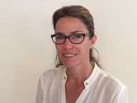 Dr. Anat Mirelman[/caption]
Anat Mirelman, PhD
Director- Laboratory of Early Markers of Neurodegeneration (LEMON)
Center for the study of Movement , Cognition and Mobility (CMCM)
Department of Neurology
Tel Aviv Sourasky Medical Center
Sackler School of Medicine, Tel Aviv University
Tel Aviv 64239, Israel
MedicalResearch.com: What is the background for this study? What are the main findings?
Response: The main aim of this research was to evaluate the efficacy of using a motor cognitive training using virtual reality in reducing fall frequency and fall risk in older adults.
Falls are a leading cause of morbidity and mortality in older adults. The prevalence of falls is huge, with one out of every 3 adults aged 65 years or older will fall at least once per year, with approximately half of these fallers suffering multiple falls in this period . These statistics are even higher in neurodegenerative conditions such as in Parkinson’s disease and in people with cognitive impairments. For example, studies have shown that as much as 80% of people with Parkinson’s disease fall each year. So many older adults are falling.
The consequences of falls are huge.
The most dramatic result is hip fracture. But this is relatively rare. However, even in the absence of a fracture or other injury, falls often lead to fear of falling, social isolation, and depression, which in turn often leads to inactivity, muscle weakness, impaired balance and gait, more falls, more social isolation. In other words, falls often start a vicious cycle, which has many important negative health consequences. Falls are associated with morbidity and mortality and they also have a huge economic impact. In many western countries, 1-2% of healthcare dollars are spent on falls.
For many years, age-associated changes in muscle strength, balance and gait were viewed as the key factors that contribute to the risk of falls. However, more recently, we and others showed that certain aspects of cognition are also critical to safe ambulation. For example people with AD often fall, almost to the same amount as people with PD, highlighting the cognitive component of falls. This makes sense intuitively if we Imagine the cognitive skills we need just to cross a busy intersection. These tasks require executive function, specially, planning, the ability to avoid obstacles, and the ability to perform two or more tasks at the same time.
Dr. Anat Mirelman[/caption]
Anat Mirelman, PhD
Director- Laboratory of Early Markers of Neurodegeneration (LEMON)
Center for the study of Movement , Cognition and Mobility (CMCM)
Department of Neurology
Tel Aviv Sourasky Medical Center
Sackler School of Medicine, Tel Aviv University
Tel Aviv 64239, Israel
MedicalResearch.com: What is the background for this study? What are the main findings?
Response: The main aim of this research was to evaluate the efficacy of using a motor cognitive training using virtual reality in reducing fall frequency and fall risk in older adults.
Falls are a leading cause of morbidity and mortality in older adults. The prevalence of falls is huge, with one out of every 3 adults aged 65 years or older will fall at least once per year, with approximately half of these fallers suffering multiple falls in this period . These statistics are even higher in neurodegenerative conditions such as in Parkinson’s disease and in people with cognitive impairments. For example, studies have shown that as much as 80% of people with Parkinson’s disease fall each year. So many older adults are falling.
The consequences of falls are huge.
The most dramatic result is hip fracture. But this is relatively rare. However, even in the absence of a fracture or other injury, falls often lead to fear of falling, social isolation, and depression, which in turn often leads to inactivity, muscle weakness, impaired balance and gait, more falls, more social isolation. In other words, falls often start a vicious cycle, which has many important negative health consequences. Falls are associated with morbidity and mortality and they also have a huge economic impact. In many western countries, 1-2% of healthcare dollars are spent on falls.
For many years, age-associated changes in muscle strength, balance and gait were viewed as the key factors that contribute to the risk of falls. However, more recently, we and others showed that certain aspects of cognition are also critical to safe ambulation. For example people with AD often fall, almost to the same amount as people with PD, highlighting the cognitive component of falls. This makes sense intuitively if we Imagine the cognitive skills we need just to cross a busy intersection. These tasks require executive function, specially, planning, the ability to avoid obstacles, and the ability to perform two or more tasks at the same time.
15 Sep Taking Happy Photos Can Improve Mood and Reduce Stress
MedicalResearch.com Interview with: [caption id="attachment_27968" align="alignleft" width="200"] Dr. Yu Chen[/caption]
Yu Chen, Ph.D.
Post-doc researcher
Department of Informatics
University of California, Irvine
MedicalResearch.com: What is the background for this study? What are the main findings?
Response: College students are facing increasing amount of stress these days. We are interested in leveraging information technology to help them become happier. We week to implement happiness-boosting exercises in positive psychology using technology in a lightweight way. Since college students frequently take photos using their smartphones, we started to investigate how to use smartphone photography to help students conduct the happiness-boosting exercises.
Participants were divided into three groups and instructed to take a photo per day in one of the following three conditions:
1) a smiling selfie;
2) a photo of something that makes himself/herself happy;
3) a photo of something that makes another person happy, which is then sent to that person.
We found that participants have become more positive after purposefully taking the assigned type of photo for three weeks. Participants who took photos that make others happy also became calmer. Some participants who took smiling selfies reported becoming more confident and comfortable with their smiles. Those who took photos to make themselves happy reported becoming more reflective and appreciative. Participants who took photos to make others happy found connecting with strong ties help them reduce stress.
Dr. Yu Chen[/caption]
Yu Chen, Ph.D.
Post-doc researcher
Department of Informatics
University of California, Irvine
MedicalResearch.com: What is the background for this study? What are the main findings?
Response: College students are facing increasing amount of stress these days. We are interested in leveraging information technology to help them become happier. We week to implement happiness-boosting exercises in positive psychology using technology in a lightweight way. Since college students frequently take photos using their smartphones, we started to investigate how to use smartphone photography to help students conduct the happiness-boosting exercises.
Participants were divided into three groups and instructed to take a photo per day in one of the following three conditions:
1) a smiling selfie;
2) a photo of something that makes himself/herself happy;
3) a photo of something that makes another person happy, which is then sent to that person.
We found that participants have become more positive after purposefully taking the assigned type of photo for three weeks. Participants who took photos that make others happy also became calmer. Some participants who took smiling selfies reported becoming more confident and comfortable with their smiles. Those who took photos to make themselves happy reported becoming more reflective and appreciative. Participants who took photos to make others happy found connecting with strong ties help them reduce stress.