Author Interviews, COVID -19 Coronavirus, Medical Imaging / 27.08.2020

MedicalResearch.com Interview with: Bruce G. Haffty, MD FACR FASTRO FASCO Associate Vice Chancellor Cancer Programs Rutgers Biomedical and Health Sciences Professor and Chair, Dept. Radiation Oncology Rutgers Robert Wood Johnson and New Jersey Medical Schools Rutgers Cancer Institute of New Jersey MedicalResearch.com: What is the background for this study? : What are the main findings? Response: That at peak times of COVID in NJ, in a tertiary care hospital with lots of COVID patients, where cancer patients still need to get treated, there was no evidence of surface COVID contamination, which should be reassuring to patients requiring radiation treatment in a busy hospital with a high in patient population of COVID patients. It should be noted that patients and staff were routinely mask wearing, observing social distancing and routinely hand washing as well as screening patients as they came in to the department with temperature checks and questions regarding symptoms. (more…)
Author Interviews, Medical Imaging, Technology / 11.12.2019

MedicalResearch.com Interview with: Dr. David Steiner, MD PhD Google Health, USA MedicalResearch.com: What is the background for this study? Response: Advances in artificial intelligence raise promising opportunities for improved interpretation of chest X-rays and many other types of medical images. However, even before researchers begin to address the critical question of clinical validation, there is important work to be done establishing strategies for evaluating and comparing different artificial intelligence algorithms. One challenge is defining and collecting the correct clinical interpretation or “label” for the large number of chest X-rays needed to train and evaluate these algorithms. Another important challenge is evaluating the algorithm on a dataset that actually represents the diversity of the cases encountered in clinical practice. For example, it might be relatively easy to make an algorithm that performs perfectly on a few hundred or so “easy” cases, but this of course might not be particularly useful in practice. (more…)
Author Interviews, Cost of Health Care, JAMA / 22.05.2019

MedicalResearch.com Interview with: Dan Ly, MD, MPP Ph.D. Program in Health Policy Harvard MedicalResearch.com: What is the background for this study? 
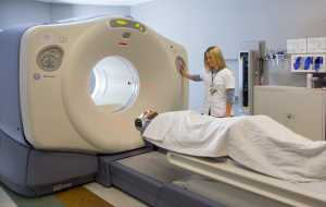
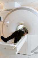
Response: There is some mixed evidence regarding whether state level tort reform reduces defensive medicine, or the practicing of medicine in such a way to reduce medical liability. This includes “positive” defensive medicine, or performing certain tests and procedures to reduce such liability. Other research finds that the perception of malpractice risk drives such defensive medicine, including the use of diagnostic imaging, such as CT scans and MRIs. I was interested in exploring what influenced the perception of this risk, hypothesizing that, for a physician, a report of an injury against one’s colleague might increase the perception of this risk and lead to an increase the use of diagnostic imaging. (more…)
Author Interviews, MRI, Prostate Cancer, Technology / 12.02.2019

MedicalResearch.com Interview with: Gaurav Pandey, Ph.D. Assistant Professor Department of Genetics and Genomic Sciences Icahn Institute of Data Science and Genomic Technology Icahn School of Medicine at Mount Sinai, New York  MedicalResearch.com: What is the background for this study?  Response: Multiparametric magnetic resonance imaging (mpMRI) has become increasingly important for the clinical assessment of prostate cancer (PCa), most routinely through PI-RADS v2, but its interpretation is generally variable due to its relatively subjective nature. Radiomics, a methodology that can analyze a large number of features of images that are difficult to study solely by visual assessment, combined with machine learning methods have shown potential for improving the accuracy and objectivity of mpMRI-based prostate cancer assessment. However, previous studies in this direction are generally limited to a small number of classification methods, evaluation using the AUC score only, and a non-rigorous assessment of all possible combinations of radiomics and machine learning methods. (more…)
Author Interviews, FDA, JAMA, Medical Imaging / 04.12.2018

MedicalResearch.com Interview with: Aldo Badano, Ph.D. Deputy Director, Division of Imaging, Diagnostics, and Software Reliability Office of Science and Engineering Laboratories Center for Devices and Radiological Health Silver Spring, MD 20993 Aldo Badano, Ph.D. Deputy Director, Division of Imaging, Diagnostics, and Software Reliability Office of Science and Engineering Laboratories Center for Devices and Radiological Health Silver Spring, MD 20993  MedicalResearch.com: What is the background for this study? What are the main findings?  Response: Expensive and lengthy clinical trials can delay regulatory evaluation of innovative technologies, affecting patient access to high-quality medical products. Although computational modeling is increasingly being used in product development, it is rarely at the center of regulatory applications. Within this context, the VICTRE project attempted to replicate a previously conducted imaging clinical trial using only computational models. The VICTRE trial involved no human subjects and no clinicians. All trial steps were conducted in silico. The fundamental question the article addresses is whether in silico imaging trials are at a mature development stage to play a significant role in the regulatory evaluation of new medical imaging systems. The VICTRE trial consisted of in silico imaging of 2986 virtual patients comparing digital mammography (DM) and digital breast tomosynthesis (DBT) systems. The improved lesion detection performance favoring DBT for all breast sizes and lesion types was consistent with results from a comparative trial using human patients and radiologists.  (more…)
Alzheimer's - Dementia, Author Interviews, MRI / 29.11.2018

MedicalResearch.com Interview with: Cyrus A. Raji, MD PhD Asst Prof of Radiology, Mallinckrodt Institute of Radiology Neuroradiology Faculty and the Neuoimaging Laboratories Washington University School of Medicine St. Louis MedicalResearch.com: What is the background for this study? What are the main findings?  Response: Alzheimer's disease is the most common cause of dementia and every patient suspected of having this disorder receives an MRI scan of the brain. MRI scans of the brain in dementia are currently limited to evaluating for structural lesions that could be leading to memory loss such as stroke or tumor. What this study sought to accomplish was to determine if a newer type of MRI scan called diffusion tensor imaging (DTI) can predict who will experience cognitive decline and dementia. We found that DTI can predict persons who will demented 2.6 years before the earliest onset of symptoms. This study was done in 61 individuals, 30 converters and 31 non-converters, from the Alzheimer's Disease Neuroimaging Initiative and we found that DTI metrics could predict dementia 2.6 years later with 89-95% accuracy. (more…)
Author Interviews, Breast Cancer, Cost of Health Care, Mammograms, Medical Imaging / 29.07.2018

MedicalResearch.com Interview with: Michal Horný PhD Assistant Professor Emory University School of Medicine, Department of Radiology and Imaging Sciences Emory University Rollins School of Public Health Department of Health Policy and Management Atlanta, GA 30322 MedicalResearch.com: What is the background for this study? Response: Increased breast tissue density is a common finding at screening mammography. Approximately 30-50% of women have so-called “dense breasts” but many of them are not aware of it. The problem is that the increased tissue density can potentially mask early cancers. In other words, if there is cancer hiding in dense breast tissue, it could be difficult to spot it. To improve the awareness of breast tissue density, a patient group called Are You Dense Advocacy, Inc., started lobbying state and federal policymakers to pass laws mandating health care providers to notify women about their breast density assessments. As a result, 31 states have already enacted some form of legislation regarding dense breast tissue. (more…)
Author Interviews, Neurological Disorders, Neurology, Personalized Medicine, Radiology, Surgical Research / 13.07.2018

MedicalResearch.com Interview with: Yasser Iturria-Medina, PhD Primary Investigator, Ludmer Centre for Neuroinformatics & Mental Health Assistant Professor, Department of Neurology and Neurosurgery Faculty of Medicine McGill University MedicalResearch.com: What is the background for this study? What are the main findings? Response: There are millions of patients following therapeutic interventions that will not benefit them. In this study, we aimed to illustrate that it is possible to identify the most beneficial intervention for each patient, in correspondence with the principles of the personalized medicine (PM). Our results show that using multimodal imaging and computational models it is possible to predict individualized therapeutic needs. The predictions are in correspondence with the individual molecular properties, which validate our findings and the used computational techniques. The results highly also the imprecision of the traditional clinical evaluations and categories for understanding the individual therapeutic needs, evidencing the positive impact that would have to use multimodal data and data-driven techniques in the clinic, in addition to the medical doctor's criterion/evaluations.   (more…)
Annals Internal Medicine, Author Interviews, Cancer Research, Kidney Disease, Radiology, Surgical Research / 30.06.2018

MedicalResearch.com Interview with: Adam Talenfeld, M.D Assistant Professor of Radiology Weill Cornell Medical College Assistant Attending Radiologist New York-Presbyterian Hospital. MedicalResearch.com: What is the background for this study? What are the main findings? Response: We know that renal function decreases as we age, and we know that decreased renal function is independently associated with increased mortality. This is why medical society guidelines recommend partial nephrectomy, which preserves kidney tissue and function, over radical nephrectomy for the treatment of the smallest kidney cancers, stage T1a tumors, which are under 4 cm diameter. Paradoxically, though, we know older patients are more likely than younger patients to receive radical nephrectomy for these smallest tumors, probably because it’s a simpler surgery than partial nephrectomy. Percutaneous ablation, focal tissue destruction using heat or cold emanating from the tip of a needle, is a newer, image-guided, minimally-invasive, tissue-sparing treatment for solid organ tumors. We wanted to test how well percutaneous ablation would compare to partial nephrectomy and radical nephrectomy for these smallest kidney cancers. We found that percutaneous ablation was associated with similar 5-year overall and cancer-specific survival compared to radical nephrectomy. At the same time, ablation was associated with significantly lower rates of new-onset chronic renal insufficiency and one-fifth as many serious non-urological complications than radical nephrectomy within 30 days of treatment. These were complications, such as deep venous thrombosis or pneumonia, that resulted in emergency department visits or new hospital admissions. The outcomes of percutaneous ablation compared with partial nephrectomy were somewhat less clear, though ablation was again associated with fewer perioperative complications. (more…)
Abuse and Neglect, Hematology, Lancet, Medical Imaging, Transplantation / 07.03.2018

MedicalResearch.com Interview with: Kirsten Williams, M.D. Blood and marrow transplant specialist Children’s National Health System  MedicalResearch.com: What is the background for this study? What are the main findings?  Response: This study addressed a life-threatening complication of bone marrow transplantation called bone marrow failure. Bone marrow transplantation has provided a cure for patients with aggressive leukemias or acquired or genetic marrow dysfunction. The process of bone marrow transplantation involves giving chemotherapy and/or radiation, which removes the diseased blood cells from the bone marrow. After this, new bone marrow stem cells are infused from a healthy individual. They travel to the bone marrow and start the slow process of remaking the blood system. Because these new cells start from infancy, it takes upwards of four to five weeks for new mature healthy cells to emerge into the blood, where they can be identified. Historically, there has been no timely way to determine if the new cells have successfully repopulated unless they can be seen in the blood compartment. This condition of bone marrow failure is life-threatening, because patients don't have white blood cells to protect them from infection. Once bone marrow failure is diagnosed, a second new set of stem cells are infused, often after more chemotherapy is given. However, for many individuals this re-transplantation is too late, because severe infections can be fatal while waiting cells to recover. We were the first group to use a new imaging test to understand how the newly infused bone marrow cells develop inside the patient. We have recently published a way to detect the new bone marrow cell growth as early as five days after the cells are given. We used an investigational nuclear medicine test to reveal this early cell growth, which could be detected weeks before the cells appear in the blood. This radiology test is safe, does not cause any problems and is not invasive. It is called FLT (18F-fluorothymidine) and the contrast is taken up by dividing hematopoietic stem cells. The patients could even see the growth of their new cells inside the bone marrow (which they very much enjoyed while waiting to see recovery of the cells in their blood). We could use the brightness of the image (called SUV) to determine approximately how many weeks remained before the cells were visible in the blood. Finally, we actually could see where the new cells went after they were infused, tracking their settling in various organs and bones. Through this, we could see that cells did not travel directly to all of the bones right away as was previously thought, but rather first went to the liver and spleen, then to the mid-spine (thorax), then to the remainder of the spine and breastplate, and finally to the arms and legs. This pattern of bone marrow development is seen in healthy developing fetuses. In this case, it occurs in a similar pattern in adults undergoing bone marrow transplant. (more…)
Author Interviews, Radiology, Technology / 17.02.2018

MedicalResearch.com Interview with: Eric Karl Oermann, MD Instructor Department of Neurosurgery Mount Sinai Health System New York, New York 10029  MedicalResearch.com: What is the background for this study? What are the main findings?  Response: Supervised machine learning requires data consisting of features and labels. In order to do machine learning with medical imaging, we need ways of obtaining labels, and one promising means of doing so is by utilizing natural language processing (NLP) to extract labels from physician's descriptions of the images (typically contained in reports). Our main finding was that (1) the language employed in Radiology reports is simpler than normal day-to-day language, and (2) that we can build NLP models that obtain excellent results at extracting labels when compared to manually extracted labels from physicians.  (more…)
Author Interviews, Biomarkers, CT Scanning, MRI, Prostate Cancer / 07.02.2018

MedicalResearch.com Interview with: Jeremie Calais PhD Ahmanson Translational Imaging Division UCLA Nuclear Medicine Department Los Angeles, CA 90095Jeremie Calais MD Ahmanson Translational Imaging Division UCLA Nuclear Medicine Department Los Angeles, CA 90095  MedicalResearch.com: What is the background for this study? What are the main findings? Response: The only curative treatment for recurrent prostate cancer after radical prostatectomy is salvage radiotherapy. Unfortunately, current standard imaging modalities are too insensitive to visualize the location of the recurrence until it is too late. As a result, salvage radiotherapy is directed to areas only suspected to harbor the recurrence based upon a "best guess" approach according to standard guidelines that define radiotherapy treatment volumes. PSMA PET/CT is a new imaging technique with sensitivity sufficient to detect and localize the recurrent prostate cancer early enough to potentially guide salvage radiotherapy. The first sign of prostate cancer recurrence is a rising PSA. For salvage radiotherapy to be successful, it should be initiated before the PSA rises above 1 ng/mL, and ideally, closer to 0.2 ng/mL or lower. PSMA PET/CT localizes sites of prostate cancer recurrence in up to 70% of patients with low PSA, below < 1.0. In the US it is not yet FDA approved and currently only used for research purposes. In our current study we included 270 patients with early recurrence of prostate cancer after surgery from Germany and UCLA,  we found that 20 % of the patients had at least one lesion detected by  PSMA PET/CT which was NOT covered by the standard radiation fields. Obviously, salvage radiotherapy is only curative if recurrent disease is completely encompassed by the radiotherapy fields and would have failed in these patients. (more…)
Author Interviews, JAMA, Multiple Sclerosis, Radiology / 04.01.2018

MedicalResearch.com Interview with: Netta Levin MD PhD fMRI lab Neurology Department Hadassah Hebrew University Medical Center Jerusalem  MedicalResearch.com: What is the background for this study? Response: Multiple sclerosis (MS) is an autoimmune demyelinating disease of the central nervous system, manifesting with episodes of local inflammatory processes, called relapses. The most useful surrogate laboratory test for MS is magnetic resonance imaging (MRI), in which dissemination of demyelinating lesions in space and time are the hallmark of the disease. However, there is a discrepancy between the lesion load - the number, size, and location of the lesions - and the clinical state of the patients, reflected in their disability. This discrepancy is known as the “clinico-radiological paradox” and suggests that something other than the well-known mechanisms of demyelination, remyelination, and axonal loss may tip the scale of recovery from an acute episode. Global effects of the local damage and compensatory mechanisms were suggested as an explanation to this paradox. In this study, we compared the visual system of patients with clinically isolated syndrome optic neuritis (ON) to patients with clinically isolated episodes in other functional systems, exploring changes, both anatomical and functional, caused to the system following the demyelinating episode. Optic neuritis was deemed a good in vivo model for studying the pathophysiology of tissue damage and repair in MS due to its characteristic clinical manifestation and to the visual pathways’ amenability to investigation using various techniques. To assess anatomical wiring ,i.e the white matter fibers themselves , we used diffusion tensor imaging (DTI). To assess functional networking as reflected by signal synchronization between distinct brain regions, we used resting state fMRI. (more…)
Accidents & Violence, Author Interviews, Brigham & Women's - Harvard, Radiology / 22.11.2017

MedicalResearch.com Interview with: “IMGP6403_qtu-no-violence” by Rae Allen is licensed under CC BY 2.0Elizabeth George, MD PGY-4 Radiology Resident Brigham and Women’s Hospital Dr. Bharti Khurana MD Clinical Fellow, Harvard Medical School and Assistant Director, Emergency Radiology Director, Emergency Musculoskeletal Radiology Program Director, Emergency Radiology Fellowship Assistant Professor, Harvard Medical School MedicalResearch.com: What is the background for this study? What are the main findings? Response: According to the CDC, 1 in 3 women in the United States are victims of abuse by their intimate partner. Despite the US Preventive Services Task Force recommendations, intimate partner violence (IPV) screening is still not widely implemented and IPV remains very prevalent and often under-recognized. The goals of this study are to increase the awareness among physicians about this public health problem and to elucidate the potential role of imaging in the identification of these patients. In fact, there is a striking disparity in the literature on the role of imaging in identifying non-accidental trauma in children compared to intimate partner violence. The common patterns of injury we identified in this population were soft tissue injuries (swelling, hematoma or contusion) followed by extremity fractures, which often involve the distal upper extremities, suggesting injury from defensive attempts. Other common injuries were facial fractures, which represent an easily accessible site for inflicting trauma, and pregnancy failure. Since radiologists have access to both current and prior radiological studies of these patients, they could play a critical role by putting the pieces together in identifying victims of IPV. (more…)
Author Interviews, JAMA, Lung Cancer, Medical Imaging, Surgical Research, Vanderbilt / 10.11.2017

MedicalResearch.com Interview with: Amelia W. Maiga, MD MPH Vanderbilt General Surgery Resident VA Quality Scholar, TVHS MedicalResearch.com: What is the background for this study? What are the main findings? Response: Positron emission tomography (PET) combined with fludeoxyglucose F18 (FDG) is currently recommended for the noninvasive diagnosis of lung nodules suspicious for lung cancer. Our investigation adds to growing evidence that FDG-PET scans should be interpreted with caution in the diagnosis of lung cancer. Misdiagnosis of lung lesions driven by FDG-PET avidity can lead to unnecessary tests and surgeries for patients, along with potentially additional complications and mortality. To estimate FDG-PET diagnostic accuracy, we conducted a multi-center retrospective cohort study. The seven cohorts originating from Tennessee, Arizona, Massachusetts and Virginia together comprised 1188 nodules, 81 percent of which were malignant. Smaller nodules were missed by FDG-PET imaging. Surprisingly, negative PET scans were also not reliable indicators of the absence of disease, especially in patients with smaller nodules or who are known to have a high probability of lung cancer prior to the FDG-PET test. Our study supports a previous meta-analyses that found FDG-PET to be less reliable in regions of the country where fungal lung diseases are endemic. The most common fungal lung diseases in the United States are histoplasmosis, coccidioidomycosis and blastomycosis. All three fungi reside in soils. Histoplasmosis and blastomycosis are common across much of the Mississippi, Ohio and Missouri river valleys and coccidioidomycosis is prevalent in the southwestern U.S. These infections generate inflamed nodules in the lungs (granulomas), which can be mistaken for cancerous lesions by imaging. (more…)
AACR, Author Interviews, Biomarkers, Brigham & Women's - Harvard, Cancer Research, Immunotherapy, Neurology, Radiology / 01.05.2017

MedicalResearch.com Interview with: Ben Larimer, PhD research fellow in lab of Umar Mahmood, MD, PhD Massachusetts General Hospital Professor, Radiology, Harvard Medical School MedicalResearch.com: What is the background for this study? What are the main findings? Response: Although immunotherapies such as checkpoint inhibitors have revolutionized cancer treatment, unfortunately they only work in a minority of patients. This means that most people who are put on a checkpoint inhibitor will not benefit but still have the increased risk of side effects. They also lose time they could have spent on other therapies. The ability to differentiate early in the course of treatment patients who are likely to benefit from immunotherapy from those who will not greatly improves individual patient care and helps accelerate the development of new therapies. The main purpose of our study was to find a way to separate immunotherapy responders from non-responders at the earliest time point possible, and develop an imaging probe that would allow us to distinguish this non-invasively. Granzyme B is a protein that immune cells use to actually kill their target. They keep it locked up in special compartments until they get the right signal to kill, after which they release it along with another protein called perforin that allows it to go inside of tumor cells and kill them. We designed a probe that only binds to granzyme B after it is released from immune cells, so that we could directly measure immune cell killing. We then attached it to a radioactive atom that quickly decays, so we could use PET scanning to noninvasively image the entire body to see where immune cells were actively releasing tumor-killing granzyme B. We took genetically identical mice and gave them identical cancer and then treated every mouse with checkpoint inhibitors, which we knew would result in roughly half of the mice responding, but we wouldn’t know which ones until their tumors began to shrink. A little over a week after giving therapy to the mice, and before any of the tumors started to shrink, we injected our imaging probe and performed PET scans. When we looked at the mice by PET imaging, they fell into two groups. One group had high PET uptake, meaning high levels of granzyme B in the tumors, the other group had low levels of PET signal in the tumors. When we then followed out the two groups, all of the mice with high granzyme B PET uptake ended up responding to the therapy and their tumors subsequently disappeared, whereas those with low uptake had their tumors continue to grow. We were very excited about this and so we expanded our collaboration with co-authors Keith Flaherty and Genevieve Boland to get patient samples from patients who were on checkpoint inhibitor therapy to see if the same pattern held true in humans. When we looked at the human melanoma tumor samples we saw the same pattern, high secreted granzyme levels in responders and much lower levels in non-responders. (more…)
Author Interviews, Cost of Health Care, Medicare, Radiology / 25.04.2017

MedicalResearch.com Interview with: David C. Levin, MD Department of Radiology Thomas Jefferson University Hospital Philadelphia, PA 19107. MedicalResearch.com: What is the background for this study? What are the main findings? Response: Radiology had been previously identified as the most rapidly growing of all physician services in the Medicare program during the early years of the 2000-2009 decade. But there have been deep cuts in imaging reimbursement since then. We wanted to determine how these cuts have affected total Medicare payments for imaging. Our main findings were that since 2006, payments to physicians for imaging under the Medicare Physician Fee schedule have dropped by $4 billion per year, or about 33%. (more…)
Author Interviews, NYU, Orthopedics, Pediatrics, Radiology / 10.04.2017

MedicalResearch.com Interview with: Ayesha Rahman, MD Chief Orthopaedic Surgery Resident NYU Langone Medical Center. MedicalResearch.com: What is the background for this study? What are the main findings? Response: Children are more vulnerable and susceptible to lifetime adverse events from radiation exposure, caused by imaging . We reviewed literature and found certain pediatric orthopaedic patients are at greater risk for radiation exposure, namely those who have surgery for hip dysplasia, scoliosis, and leg length discrepancy, as they are among those most likely to undergo CT imaging. After reviewing all types of imaging studies performed in orthopedics and how much radiation is involved in each test, we developed several recommendations that pediatric orthopaedic surgeons should follow. Among those recommendations are: utilize low-dose CT protocols or technology that uses less imaging (like EOS), limit CT scans of the spine and pelvis, know that female patients are more susceptible to adverse risk and plan accordingly, and follow the the “as low as reasonably achievable,”principle to limit exposure to parts of the body that are necessary for diagnosis. (more…)
Author Interviews, Brigham & Women's - Harvard, Heart Disease, Lancet, Medical Imaging, MRI, Social Issues / 12.01.2017

MedicalResearch.com Interview with: Dr Ahmed Tawakol MD Co-Director, Cardiac MR PET CT Program Massachusetts General Hospital and Harvard Medical School  MedicalResearch.com: What is the background for this study? What are the main findings? Response: While the link between stress and heart disease has long been established, the mechanism mediating that risk hasn’t been clearly understood. Animal studies showed that stress activates bone marrow to produce white blood cells, leading to arterial inflammation.  This study suggests an analogous path exists in humans. Moreover, this study identifies, for the first time in animal models or humans, the region of the brain (the amygdala) that links stress to the risk of heart attack and stroke. The paper reports on two complementary studies. The first analyzed imaging and medical records data from almost 300 individuals who had PET/CT brain imaging, primarily for cancer screening, using a radiopharmaceutical called FDG that both measures the activity of areas within the brain and reflects inflammation within arteries.  All participants in that study had no active cancer or cardiovascular disease at the time of imaging and each had information in their medical records on at least three additional clinical visits after imaging. The second study enrolled 13 individuals with a history of post-traumatic stress disorder, who were evaluated for their current levels of perceived stress and received FDG-PET scanning to measure both amygdala activity and arterial inflammation. (more…)
Author Interviews, Brain Injury, MRI, Pediatrics / 09.12.2016

MedicalResearch.com Interview with: Eman S. Mahdi, MD, MBChB Pediatric Radiology Fellow Catherine Limperopoulos, PhD Director, Developing Brain Research Laboratory Co-Director of Research, Division of Neonatology Diagnostic Imaging and Radiology Children’s National Health System Washington, DC MedicalResearch.com: What is the background for this study? What are the main findings? Response: Premature birth is a major public health concern in the United States affecting 1 in 10 infants each year. Prematurity-related brain injury is very common and associated with a high prevalence of brain injury and accompanying lifelong neurodevelopmental morbidities. Early disturbances in systemic and cerebral hemodynamics are thought to mediate prematurity-related brain injury. The extent to which cerebral blood flow (CBF) is disturbed in preterm birth is poorly understood, in large part because of the lack of monitoring techniques that can directly and non-invasively measure cerebral blood flow. We report for the first time early disturbances in global and regional cerebral blood flow in preterm infants following brain injury on conventional magnetic resonance imaging (MRI) over the third trimester of ex-uterine life using arterial spin labelling images. In terms of regional differences, we saw a marked decrease in blood flow to the thalamus and the pons, regions known to be metabolically active during this time. (more…)
Author Interviews, Medical Imaging / 06.11.2016

MedicalResearch.com Interview with: Devon A. Klein, MD, MPH Asst. Professor of Radiology and Orthopaedics Hofstra Northwell School of Medicine Associate Chairman, Radiology Lenox Hill Hospital New York, NY 10075 MedicalResearch.com: What is the background for this study? What are the main findings? Response: As our Emergency Department continued to grow our Radiology Department was challenged to accommodate the resultant increased volume with fixed resources (i.e. CT Scanners) and maintain CT scan throughput. Uncoordinated insular efforts within the Department of Radiology failed to resolve the problem. A collaborative approach utilizing Six Sigma lean and involving stakeholders from the Departments of Radiology, Emergency, Medicine, and Patient Transportation was able to better illustrate the “bottlenecks” and devise solutions. (more…)
Author Interviews, Cost of Health Care, CT Scanning, Health Care Systems / 23.09.2016

MedicalResearch.com Interview with: Hui Zhang, Ph.D., MBA Virginia Polytechnic Institute and State University Blacksburg MedicalResearch.com: What is the background for this study? What are the main findings? Response: To promote healthcare coordination and contain the rising costs in the US healthcare system, a variety of payment innovations has been developed and field-tested in both public and private sector. Among them, the Medicare Shared Savings Program (MSSP) for Accountable Care Organizations (ACOs), administered by the Centers for Medicare and Medicaid Services (CMS) has received considerable attention. Our study took a mathematical modeling approach and comprehensively captured and analyzed the effect of this new payment systems on healthcare stakeholder decisions and system-wide outcomes. Our results provided decision-making insights for payers on how to improve MSSP, for ACOs on how to distribute MSSP incentives among their members, and for hospitals on whether to invest in new CT imaging systems. (more…)
Author Interviews, Brain Cancer - Brain Tumors, Case Western, MRI, Radiology, Technology / 19.09.2016

MedicalResearch.com Interview with: Dr. Pallavi Tiwari PhD Assistant Professor biomedical engineering Case Western Reserve University MedicalResearch.com: What is the background for this study? What are the main findings? Response: One of the biggest challenges in neuro-oncology currently is distinguishing radionecrosis, a side-effect of aggressive radiation, from tumor recurrence on imaging. Surgical intervention is the only means of definitive diagnosis, but suffers from considerable morbidity and mortality. The treatments for radionecrosis and cancer recurrence are very different. Early identification of the two conditions can help speed prognosis, therapy, and improve patient outcomes. The purpose of this feasibility study was to evaluate the role of machine learning algorithms along with computer extracted texture features, also known as radiomic features, in distinguishing radionecrosis and tumor recurrence on routine MRI scans (T1w, T2w, FLAIR). The radiomic algorithms were trained on 43 studies from our local collaborating institution - University Hospitals Case Medical Center, and tested on 15 studies at a collaborating institution, University of Texas Southwest Medical Center. We further compared the performance of the radiomic techniques with two expert readers. Our results demonstrated that radiomic features can identify subtle differences in quantitative measurements of tumor heterogeneity on routine MRIs, that are not visually appreciable to human readers. Of the 15 test studies, the radiomics algorithm could identify 12 of 15 correctly, while expert 1 could identify 7 of 15, and expert 2, 8 of 15. (more…)
Author Interviews, JAMA, MRI, Radiology / 08.09.2016

MedicalResearch.com Interview with: Dr. Joel G. Ray MD, MS, FRCPC Professor, Department of Medicine, University of Toronto Professor Department of Obstetrics and Gynecology St. Michael’s Hospital MedicalResearch.com: What is the background for this study? What are the main findings? Response: We have little information about the fetal safety to of MRI in the first trimester of pregnancy, or that of MRI with gadolinium contrast performed at any point in pregnancy. (more…)
Author Interviews, Cancer Research, Mammograms, PNAS, Radiology / 31.08.2016

MedicalResearch.com Interview with: Karla K. Evans, Ph.D. Lecturer, Department of Psychology The University of York Heslington, York UK MedicalResearch.com: What is the background for this study? Response: This research started after initially talking to radiologists and pathologists about how they search a radiograph or micrograph for abnormalities. They talked about being able to tell at the first glance if the image had something bad about it. Jokingly, they talked about “having the force” to see the bad. We wanted to know whether this hunch after the brief initial viewing was real and to systematically test it. We collected radiographic and micrographic images, half of them that had signs of cancer in them and half of them that didn't, and we briefly presented them (250 millisecond to 2000 milliseconds) to radiologists or pathologistsrespectively. They simply had to report whether they would recall the patient or not and try localize on the outline the location of the abnormality. We first reported these finding in the following paper. Evans et al. (2013) The Gist of the Abnormal: Above chance medical decision making in the blink of an eye. Psychonomic Bulletin & Review (DOI) 10.3758/s13423-013-0459-3 In addition to finding that radiologists and pathologists can indeed detect subtle cancers in a quarter of a second we also found that they did not know where it was in the image leading us to conclude that the signal that they were picking up must be a global signal (i.e. the global image statistic or the texture of the breast as a whole) rather than the result of a local saliency. This led me to start further exploring this signal in order to characterize it when I moved to University or York, UK to establish my own lab. (more…)
Author Interviews, Radiology, Zika / 25.08.2016

MedicalResearch.com Interview with: Fernanda Tovar Moll, MD, PhD Vice president of the D'Or Institute for Research and Education Professor,Federal University of Rio de Janeiro Rio de Janeiro, Brazil MedicalResearch.com: What is the background for this study? Response: The consequences of congenital zika virus infection are still under investigation. Recent studies suggest microcephaly as one of the consequences, but we wanted to go deeper in investigating what other kinds of neurological changes could happen in the developing central nervous system. Based on that, we performed a cohort study with multimodal images exams and longitudinal follow up (pre and post natal analyses) of some cases. (more…)
Author Interviews, CT Scanning, Lung Cancer, PLoS, Radiology / 19.08.2016

MedicalResearch.com Interview with: Matthew B. Schabath PhD Department of Cancer Epidemiology H. Lee Moffitt Cancer Center and Research Institute Tampa, Florida MedicalResearch.com: What is the background for this study? What are the main findings? Response: Our study is a post-hoc analysis of data from a large randomized clinical trial (RCT) called the National Lung Screening Trial (NLST). The NLST found that lung cancer screening with low-dose helical computed tomography (LDCT) significantly reduced lung cancer deaths by 20 percent compared to screening with standard chest radiography (i.e., X-Ray). In our publication, we performed a very detailed analysis comparing outcomes of lung cancer patients screened by LDCT according to their initial (i.e., baseline), 12 month, and 24 month screening results. We found that patients who had a negative baseline screening but tested positive for lung cancer at the 12- or 24-month screen had lower survival and higher mortality rates than patients who had a positive initial screen that was a non-cancerous abnormality but developed lung cancer in subsequent screens. (more…)