
Infections
27 Apr Morning Flu Vaccinations May Be More Effective
MedicalResearch.com Interview with: [caption id="attachment_23831" align="alignleft" width="200"] Dr. Anna Phillips[/caption]
Dr Anna C. Phillips PhD CPsychol AFBPsS
Reader in Behavioural Medicine
School of Sport, Exercise & Rehabilitation Sciences
University of Birmingham
Edgbaston Birmingham
MedicalResearch.com: What is the background for this study? What are the main findings?
Dr. Phillips: We know that various factors can affect the response to vaccination and that older adults have a poorer response than younger people, i.e. they produce fewer antibodies. We also know that many immune messengers and important hormones have daily rhythms in their levels and wanted to test whether the antibody response to vaccination might also be affected by time of day. We randomised surgeries to giving morning or afternoon vaccinations and tested before and one month after the vaccination for levels of antibodies.
Two of the three flu strains (viruses) contained in the vaccine showed a higher antibody response in the morning than in the afternoon, up to 4 x higher to one of the strains (A/California) and 1.5 x higher to the B strain. None of the potential mechanisms we measured (immune messengers, hormones) seemed to be driving this effect.
Dr. Anna Phillips[/caption]
Dr Anna C. Phillips PhD CPsychol AFBPsS
Reader in Behavioural Medicine
School of Sport, Exercise & Rehabilitation Sciences
University of Birmingham
Edgbaston Birmingham
MedicalResearch.com: What is the background for this study? What are the main findings?
Dr. Phillips: We know that various factors can affect the response to vaccination and that older adults have a poorer response than younger people, i.e. they produce fewer antibodies. We also know that many immune messengers and important hormones have daily rhythms in their levels and wanted to test whether the antibody response to vaccination might also be affected by time of day. We randomised surgeries to giving morning or afternoon vaccinations and tested before and one month after the vaccination for levels of antibodies.
Two of the three flu strains (viruses) contained in the vaccine showed a higher antibody response in the morning than in the afternoon, up to 4 x higher to one of the strains (A/California) and 1.5 x higher to the B strain. None of the potential mechanisms we measured (immune messengers, hormones) seemed to be driving this effect.
26 Apr Isolating Asymptomatic C. diff Carriers at Hospital Admission May Decrease Transmission
MedicalResearch.com Interview with: Yves Longtin, MD, FRCPC Chair, Infection Prevention and Control Unit Montreal Jewish General Hospital - SMBD Associate professor of Medicine, McGill University MedicalResearch.com: What is the background for this study? What are the main findings? Dr. Longtin: Clostridium difficile is a major cause of infection in hospitalized patients. Current infection control measures to prevent the spread of C. difficile in hospitals focuses almost entirely on patients who present symptoms. Patients with symptoms of diarrhea due to C difficile are placed under isolation in hospitals (for example, healthcare workers will wear a gown and gloves when caring for them). However, many studies have shown that some patients may be asymptomatic carriers of C. difficile. These patients carry the C difficile bacteria in their digestive tract without being sick. It was known that these asymptomatic carriers could spread the bacteria to other patients, but it was unclear whether putting them into isolation would help prevent the spread of the microbe in hospitals. Our study tested the hypothesis that placing asymptomatic carriers under isolation could lead to a decrease in the number of infections with C difficile.23 Apr Transfusions in Resource Poor Settings Can Become Safer With UV-Based Pathogen Inactivation
MedicalResearch.com Interview with: [caption id="attachment_23721" align="alignleft" width="105"] Prof. Jean Pierre Allain[/caption]
Prof Jean-Pierre Allain
Principal Investigator, Department of Haematology
University of Cambridge, Cambridge Blood Centre
Cambridge UK
MedicalResearch.com: What is the background for this study? What are the main findings?
Prof. Allain: In sub-Saharan Africa (SSA), 70% of the transfusions are in the form of whole blood units (generally 1 or 2). Lack of resources limit the safety
measures to donor questionnaire, viral/bacterial testing (HIV, HCV, HBV
and Syphilis). Other measures used in rich countries i.e. nucleic acid
testing, filtration, bacterial culture etc. are not done because of cost.
Pathogen reduction would be an effective way to overcome these issues as
it is able to inactivate viruses, bacteria, parasites and nucleated
cells in one go, provided it is applied to whole blood and affordable.
The study consisted in assessing the efficacy of such a method (Mirasol
using riboflavin and UV illumination) taking inactivation of plasmodium
as major endpoint of a randomised controlled clinical trial called AIMS
(African Investigation of Mirasol System).
Prof. Jean Pierre Allain[/caption]
Prof Jean-Pierre Allain
Principal Investigator, Department of Haematology
University of Cambridge, Cambridge Blood Centre
Cambridge UK
MedicalResearch.com: What is the background for this study? What are the main findings?
Prof. Allain: In sub-Saharan Africa (SSA), 70% of the transfusions are in the form of whole blood units (generally 1 or 2). Lack of resources limit the safety
measures to donor questionnaire, viral/bacterial testing (HIV, HCV, HBV
and Syphilis). Other measures used in rich countries i.e. nucleic acid
testing, filtration, bacterial culture etc. are not done because of cost.
Pathogen reduction would be an effective way to overcome these issues as
it is able to inactivate viruses, bacteria, parasites and nucleated
cells in one go, provided it is applied to whole blood and affordable.
The study consisted in assessing the efficacy of such a method (Mirasol
using riboflavin and UV illumination) taking inactivation of plasmodium
as major endpoint of a randomised controlled clinical trial called AIMS
(African Investigation of Mirasol System).
18 Apr Mouth and Throat Cancers Increasing in Frequency
MedicalResearch.com Interview with: [caption id="attachment_23533" align="alignleft" width="96"] Dr. Adam Jacobson[/caption]
Adam S. Jacobson, MD
Associate Professor, Department of Otolaryngology-Head and Neck Surgery
Associate Director, Head and Neck Surgery
NYU Langone Medical Center and
Perlmutter Cancer Center
MedicalResearch.com Editor’s note: Dr. Jacobson is an Otolaryngologist, an Ear-Nose-Throat (ENT) physician specializing in the diagnosis of head and neck tumors and cancers, including cancers of the mouth and throat. Dr. Jacobson discussed oral (mouth) and pharyngeal (throat) cancers in recognition of Oral, Head and Neck Cancer Awareness Week.
MedicalResearch.com: How prevalent is the problem of Oral, Head and Neck Cancer? Is this type of cancer becoming more frequent?
Dr. Jacobson: Oropharynx cancer is currently on the rise.
MedicalResearch.com: Have HPV-induced cancers become more common?
(Note HPV or Human Papilloma Virus is a virus associated with various wart infections.)
Dr. Jacobson: Yes - Specifically tonsil and base of tongue cancer.
Dr. Adam Jacobson[/caption]
Adam S. Jacobson, MD
Associate Professor, Department of Otolaryngology-Head and Neck Surgery
Associate Director, Head and Neck Surgery
NYU Langone Medical Center and
Perlmutter Cancer Center
MedicalResearch.com Editor’s note: Dr. Jacobson is an Otolaryngologist, an Ear-Nose-Throat (ENT) physician specializing in the diagnosis of head and neck tumors and cancers, including cancers of the mouth and throat. Dr. Jacobson discussed oral (mouth) and pharyngeal (throat) cancers in recognition of Oral, Head and Neck Cancer Awareness Week.
MedicalResearch.com: How prevalent is the problem of Oral, Head and Neck Cancer? Is this type of cancer becoming more frequent?
Dr. Jacobson: Oropharynx cancer is currently on the rise.
MedicalResearch.com: Have HPV-induced cancers become more common?
(Note HPV or Human Papilloma Virus is a virus associated with various wart infections.)
Dr. Jacobson: Yes - Specifically tonsil and base of tongue cancer.
16 Apr Immunotherapy Drug Nivolumab May Help Some Aggressive HPV-Induced Anal Cancers
MedicalResearch.com Interview with: [caption id="attachment_23516" align="alignleft" width="114"] Dr. Van Morris[/caption]
Dr. Van K. Morris, MD
Assistant Professor, GI Medical Oncology
The University of Texas MD Anderson Cancer Center
MedicalResearch.com: What is the background for this study?
Dr. Morris: Anal cancer is a very rare cancer and accounts for approximately 2% of all gastrointestinal malignancies. Currently, there is no accepted standard of care for patients with metastatic disease, which raises challenges for oncologist who may not have extensive experience caring for patients with metastatic anal cancer given that there are not accepted agents to treat with. This clinical trial was the first clinical trial ever conducted for patients with stage IV disease who had received prior chemotherapy in the past.
Given the well-known association with human papilloma virus (HPV) and the development of anal cancer, we were interested in the use of immunotherapy drugs as a new possible way to awaken the immune system to attack this tumor, especially as there may be viral components in the tumor cells which the immune system could potentially recognize. Nivolumab is an immunotherapy drug which has shown activity in other solid tumors like melanoma, kidney cancer, non-small cell lung cancer, and bladder cancer.
Dr. Van Morris[/caption]
Dr. Van K. Morris, MD
Assistant Professor, GI Medical Oncology
The University of Texas MD Anderson Cancer Center
MedicalResearch.com: What is the background for this study?
Dr. Morris: Anal cancer is a very rare cancer and accounts for approximately 2% of all gastrointestinal malignancies. Currently, there is no accepted standard of care for patients with metastatic disease, which raises challenges for oncologist who may not have extensive experience caring for patients with metastatic anal cancer given that there are not accepted agents to treat with. This clinical trial was the first clinical trial ever conducted for patients with stage IV disease who had received prior chemotherapy in the past.
Given the well-known association with human papilloma virus (HPV) and the development of anal cancer, we were interested in the use of immunotherapy drugs as a new possible way to awaken the immune system to attack this tumor, especially as there may be viral components in the tumor cells which the immune system could potentially recognize. Nivolumab is an immunotherapy drug which has shown activity in other solid tumors like melanoma, kidney cancer, non-small cell lung cancer, and bladder cancer.
15 Apr Kidney Transplant Patients May Not Be Protected From HPV Strains in Vaccines
MedicalResearch.com Interview with: [caption id="attachment_23501" align="alignleft" width="135"] Dr. Robotham[/caption]
Dr. Delphine Robotham MD
Division of Pediatric Nephrology
Johns Hopkins University School of Medicine
Baltimore, Maryland
Medical Research: What is the background for this study? What are the main findings?
Response: Cervical cancer is the second most common cancer in women worldwide and is almost entirely caused by high risk HPV genotypes. Vaccines to high risk HPV genotypes have shown great success in protecting healthy women from the sequelae of infection, including cervical cancer and genital warts. Young women with a kidney transplant as well as those with chronic kidney disease have abnormal immune systems and as a result have a significantly increased burden of HPV-related disease making the potential health benefits of the HPV vaccine substantial in this particularly vulnerable population. This study examined the immune response to the HPV vaccine among girls and young women with kidney disease.
The goal of this research was to determine if girls and young women with chronic kidney disease (abnormal kidney function, on dialysis, or post kidney transplant) showed evidence of immune response to the quadrivalent HPV vaccine. Immune response was determined by measuring the amount of antibody made by the patients against each of the 4 HPV genotypes included in the vaccine. There are established thresholds of antibody above which patients are believed to have protection from infection. We found that study participants with chronic kidney disease and those on dialysis had antibody levels above the threshold, indicating the vaccine should be effective in protecting them from HPV related disease. A significant proportion of patients with kidney transplants showed evidence of inadequate antibody response. This is important information as it means patients with a kidney transplant, whom we know are at increased risk of developing cervical cancer from HPV infection, may not be protected from HPV infections from the HPV genotypes included in the vaccine.
Dr. Robotham[/caption]
Dr. Delphine Robotham MD
Division of Pediatric Nephrology
Johns Hopkins University School of Medicine
Baltimore, Maryland
Medical Research: What is the background for this study? What are the main findings?
Response: Cervical cancer is the second most common cancer in women worldwide and is almost entirely caused by high risk HPV genotypes. Vaccines to high risk HPV genotypes have shown great success in protecting healthy women from the sequelae of infection, including cervical cancer and genital warts. Young women with a kidney transplant as well as those with chronic kidney disease have abnormal immune systems and as a result have a significantly increased burden of HPV-related disease making the potential health benefits of the HPV vaccine substantial in this particularly vulnerable population. This study examined the immune response to the HPV vaccine among girls and young women with kidney disease.
The goal of this research was to determine if girls and young women with chronic kidney disease (abnormal kidney function, on dialysis, or post kidney transplant) showed evidence of immune response to the quadrivalent HPV vaccine. Immune response was determined by measuring the amount of antibody made by the patients against each of the 4 HPV genotypes included in the vaccine. There are established thresholds of antibody above which patients are believed to have protection from infection. We found that study participants with chronic kidney disease and those on dialysis had antibody levels above the threshold, indicating the vaccine should be effective in protecting them from HPV related disease. A significant proportion of patients with kidney transplants showed evidence of inadequate antibody response. This is important information as it means patients with a kidney transplant, whom we know are at increased risk of developing cervical cancer from HPV infection, may not be protected from HPV infections from the HPV genotypes included in the vaccine.
15 Apr T-SPOT.TB Test Demonstrates High Specificity in Screening Health Care Workers for Tuberculosis
MedicalResearch.com Interview with: [caption id="attachment_23494" align="alignleft" width="200"] Dr. Thomas King[/caption]
Thomas C King, MD, PhD
Department of Pathology and Laboratory Medicine
Chief of Pathology and Laboratory Medicine
St. Vincent Hospital
Worcester, MA
MedicalResearch.com: What is the background for this study? What are the main findings?
Dr. King: This landmark study provides a broad based, real world appraisal of the reliability of the T-SPOT®.TB test, an interferon gamma release assay (IGRA), based on results in screening workers in 19 U.S. hospitals. The large size of the study (more than 42,000 test results from more than 16,000 healthcare workers analyzed) provides a solid benchmark to assess performance of T-SPOT.TB in serial screening healthcare workers. In recent years, results from several studies have shown that there can be significant differences between using an IGRA and the tuberculin skin test (TST) in terms of accuracy and cost. Several studies have confirmed a risk of high false positive rates and numerous conversion/reversion rates when retesting patients with the TST.
Dr. Thomas King[/caption]
Thomas C King, MD, PhD
Department of Pathology and Laboratory Medicine
Chief of Pathology and Laboratory Medicine
St. Vincent Hospital
Worcester, MA
MedicalResearch.com: What is the background for this study? What are the main findings?
Dr. King: This landmark study provides a broad based, real world appraisal of the reliability of the T-SPOT®.TB test, an interferon gamma release assay (IGRA), based on results in screening workers in 19 U.S. hospitals. The large size of the study (more than 42,000 test results from more than 16,000 healthcare workers analyzed) provides a solid benchmark to assess performance of T-SPOT.TB in serial screening healthcare workers. In recent years, results from several studies have shown that there can be significant differences between using an IGRA and the tuberculin skin test (TST) in terms of accuracy and cost. Several studies have confirmed a risk of high false positive rates and numerous conversion/reversion rates when retesting patients with the TST.
15 Apr Hepatitis C Raises Risk of HPV Head and Neck Cancers
Posted at 02:16h
in Author Interviews, Cancer Research, Hepatitis - Liver Disease, HPV, JNCI, MD Anderson
MedicalResearch.com Interview with:
[caption id="attachment_23475" align="alignleft" width="114"] Dr. Harrys Torres[/caption]
Harrys A. Torres, MD, FACP, FIDSA
Associate Professor
Director of Hepatitis C Clinic
Department of Infectious Diseases, Infection Control and Employee Health
The University of Texas MD Anderson Cancer Center
Houston TX 77030
Medical Research: What is the background for this study? What are the main findings?
Dr. Torres: Hepatitis C virus (HCV) is an oncogenic virus and is associated with an increased risk of liver cancer and certain types of non-Hodgkin lymphomas. In 2009, at MD Anderson Cancer Center, we set up the first clinic in the United States, and probably in the world, specifically devoted to managing HCV infection in cancer patients. In the clinic, we expected to see a number of patients with liver cancers and non-Hodgkin’s lymphoma, as these have documented associations with HCV. Unexpectedly, we saw a high number of HCV-infected patients with head and neck cancers, and wondered whether there was an undiscovered association between having the infection and head and neck cancers. To explore this, we conducted a case-control study using 409 head and neck cancer subjects (164 oropharyngeal, 245 non-oropharyngeal [oral cavity, nasopharynx, larynx] cancers) and 694 control subjects with other smoking-associated cancers (378 lung, 168 esophagus, and 148 urinary bladder cancers), and compared the prevalence of HCV infection in the two groups. We observed a high prevalence of HCV infection in oropharyngeal (14%) and non-oropharyngeal (20%) cancer patients when compared to control subjects (6.5%). After adjusting for confounders such as smoking, alcohol intake, and socioeconomic status, HCV-infected individuals were 2.04 times more likely to have oropharyngeal cancers and 2.85 times more likely to have non-oropharyngeal cancers. Of note, HCV was associated only with patients with oropharyngeal cancers that tested positive for human papilloma virus, which is one of the main virus linked with increased risk of oropharyngeal cancers.
Dr. Harrys Torres[/caption]
Harrys A. Torres, MD, FACP, FIDSA
Associate Professor
Director of Hepatitis C Clinic
Department of Infectious Diseases, Infection Control and Employee Health
The University of Texas MD Anderson Cancer Center
Houston TX 77030
Medical Research: What is the background for this study? What are the main findings?
Dr. Torres: Hepatitis C virus (HCV) is an oncogenic virus and is associated with an increased risk of liver cancer and certain types of non-Hodgkin lymphomas. In 2009, at MD Anderson Cancer Center, we set up the first clinic in the United States, and probably in the world, specifically devoted to managing HCV infection in cancer patients. In the clinic, we expected to see a number of patients with liver cancers and non-Hodgkin’s lymphoma, as these have documented associations with HCV. Unexpectedly, we saw a high number of HCV-infected patients with head and neck cancers, and wondered whether there was an undiscovered association between having the infection and head and neck cancers. To explore this, we conducted a case-control study using 409 head and neck cancer subjects (164 oropharyngeal, 245 non-oropharyngeal [oral cavity, nasopharynx, larynx] cancers) and 694 control subjects with other smoking-associated cancers (378 lung, 168 esophagus, and 148 urinary bladder cancers), and compared the prevalence of HCV infection in the two groups. We observed a high prevalence of HCV infection in oropharyngeal (14%) and non-oropharyngeal (20%) cancer patients when compared to control subjects (6.5%). After adjusting for confounders such as smoking, alcohol intake, and socioeconomic status, HCV-infected individuals were 2.04 times more likely to have oropharyngeal cancers and 2.85 times more likely to have non-oropharyngeal cancers. Of note, HCV was associated only with patients with oropharyngeal cancers that tested positive for human papilloma virus, which is one of the main virus linked with increased risk of oropharyngeal cancers.
14 Apr Clinical Findings and Brain Calcifications of Zika Babies Described
MedicalResearch.com Interview with: [caption id="attachment_23445" align="alignleft" width="300"] Team of Doctors: Ana van Der Linden, Alessandra Brainer, Maria de Fatima Aragao, Vanessa va Der Linden e Arthur Cesário[/caption]
Maria de Fatima Vasco Aragao MD, PhD
Radiologist and Neuroradiologist
Professor of Radiology, Mauricio de Nassau University, Recife, Brazil
Scientific Director of Multimagem Radiology Clinic, Recife - PE, Brazil
President of Pernambuco Radiology Society
MedicalResearch.com: What is the background for this study?
Response: The new Zika virus epidemic in Brazil was recognized as starting in the first half of 2015 and the microcephaly epidemic was detected in the second half of that same year.
[caption id="attachment_23410" align="alignleft" width="300"]
Team of Doctors: Ana van Der Linden, Alessandra Brainer, Maria de Fatima Aragao, Vanessa va Der Linden e Arthur Cesário[/caption]
Maria de Fatima Vasco Aragao MD, PhD
Radiologist and Neuroradiologist
Professor of Radiology, Mauricio de Nassau University, Recife, Brazil
Scientific Director of Multimagem Radiology Clinic, Recife - PE, Brazil
President of Pernambuco Radiology Society
MedicalResearch.com: What is the background for this study?
Response: The new Zika virus epidemic in Brazil was recognized as starting in the first half of 2015 and the microcephaly epidemic was detected in the second half of that same year.
[caption id="attachment_23410" align="alignleft" width="300"]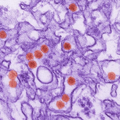 This is a transmission electron micrograph (TEM) of Zika virus, which is a member of the family Flaviviridae. Virus particles are 40 nm in diameter, with an outer envelope, and an inner dense core.[/caption]
MedicalResearch.com: What are the main findings?
This is a transmission electron micrograph (TEM) of Zika virus, which is a member of the family Flaviviridae. Virus particles are 40 nm in diameter, with an outer envelope, and an inner dense core.[/caption]
MedicalResearch.com: What are the main findings?
- Response: In our study of the 23 mothers, only one did not report rash during pregnancy (rash is a sign that can happen in Zika virus infection). However, Zika virus infection can be asymptomatic in three of every four infected patients. All of the 23 babies had the same clinical and epidemiological characteristics and other congenital infection diseases had been excluded. Of these 23 babies, six were tested for IgM antibodies, specific to Zika virus and all six proved positive. So, by deduction, the other 17 babies on whom it was not possible to make the IgM test, were considered as also having presumed congenital infection related to the Zika virus, after other congenital infections being excluded.
- All the babies showed malformations of cortical development and sulcation. The most frequent cortical malformation were: Microcephaly with a simplified cortical gyral pattern and areas of thick cortex of polymicrogyria or pachygyria which were located predominantly in the frontal lobes.
- Abnormalities of the corpus callósum (hypogenesis and hypoplasia) were common.
- Decreased brain volume was a common finding. Ventriculomegaly was present in all the babies, with a predominant enlargement of the posterior portions of the lateral ventricles,
- Delayed myelination were also common. The cisterna magna was enlarged in most of the cases, with or without cerebellar hypoplasia.
- Some of the babies showed a symmetrical enlargement of the anterior subarachnoid space of the supratentorial compartment, associated with severe ventriculomegaly.
13 Apr Ocular Herpes Zoster Can Be Chronic and Recurrent
MedicalResearch.com Interview with: Kimberly D Tran, MD Bascom Palmer Eye Institute MedicalResearch.com: What is the background and purpose for this study? Dr. Tran: Approximately 30% of the population will suffer from herpes zoster (also known as shingles) at some point in their lifetime, with an estimated 1 million cases in the U.S. each year (1). The most common long term complication of herpes zoster is postherpetic neuralgia (PHN), or persistent neuropathic pain lasting beyond three months after initial presentation of herpes zoster. PHN can negatively affect quality of life to a degree similar to congestive heart failure, depression, acute myocardial infarction,diabetes. Postherpetic neuralgia is a leading cause of suicide in patients over 70 with chronic pain.(3,4) Of all the cases of herpes zoster, an estimated 10-20% will have herpes zoster ophthalmicus (HZO), which is defined as shingles in the area of the face near the eye, and sometimes the eye itself becomes involved. Approximately 50% of individuals with HZO will develop ocular complications without antiviral treatment, while antiviral induction within the first 72 hours of rash onset reduces this number to 20-30% (2). Randomized control trial has demonstrated the efficacy antiviral therapy in the treatment of herpes zoster on first presentation.(6) What is less understood is the course of HZ after its initial presentation. Traditionally studied and treated in the acute phase,(5-7) recent data suggest that some patients experience a chronic or recurrent disease course. Based on this data, it is clear that more information is needed on the long term clinical course of herpes zoster ophthalmicus. The purpose of this study was to characterize the epidemiology of recurrent and chronic HZO in a unique South Florida population, with an ethnically and racially mixed, predominately male population.13 Apr 4-Dose Hepatitis B Vaccine Schedule For HIV Patients Induces Longer Protection
MedicalResearch.com Interview with: [caption id="attachment_23342" align="alignleft" width="160"] Dr. Odile Launay[/caption]
Odile Launay MD, PhD
Paris Descartes University
Assistance Publique Hôpitaux de Paris, Cochin Hospital
MedicalResearch.com: What is the background for this study? What are the main findings?
Dr. Launay: In patients with HIV infection, responses to standard HBV vaccination regimens remain suboptimal compared with responses in HIV seonegative individuals. We previously reported that alternative regimens (a 4 injection IMdouble dose regimen and a 4 injection intradermal low dose regimen) improve antibody response compared with the standard HBV vaccination regimen (ANRS HB03 VIHVAC-B study). Further precision on the duration of response achieved with alternative HBV vaccination regimes was needed.
We report in this paper the results from the follow-up of the study.
The results of this study show that the 4 dose IM regimen induces higher seroconversion rate but also higher long term seroprotection in HIV infected patients
Dr. Odile Launay[/caption]
Odile Launay MD, PhD
Paris Descartes University
Assistance Publique Hôpitaux de Paris, Cochin Hospital
MedicalResearch.com: What is the background for this study? What are the main findings?
Dr. Launay: In patients with HIV infection, responses to standard HBV vaccination regimens remain suboptimal compared with responses in HIV seonegative individuals. We previously reported that alternative regimens (a 4 injection IMdouble dose regimen and a 4 injection intradermal low dose regimen) improve antibody response compared with the standard HBV vaccination regimen (ANRS HB03 VIHVAC-B study). Further precision on the duration of response achieved with alternative HBV vaccination regimes was needed.
We report in this paper the results from the follow-up of the study.
The results of this study show that the 4 dose IM regimen induces higher seroconversion rate but also higher long term seroprotection in HIV infected patients
12 Apr 6-Step More Effective than 3-Step Hand Hygiene Technique
MedicalResearch.com Interview with: [caption id="attachment_23395" align="alignleft" width="180"] Dr. Jacqui Reilly[/caption]
Professor Jacqui Reilly PhD
Institute for Applied Health Research
Glasgow Caledonian University
Glasgow
MedicalResearch.com: What is the background for this study? What are the main findings?
Dr. Reilly: Hand hygiene is the single most important intervention to reduce avoidable illness and prevent infections. Two techniques have been reported for hand hygiene use with alcohol-based hand rub (ABHR) in international guidance: 6 step by the WHO and 3 step by the Center for Disease Control. Neither of these techniques have an evidence base to support their effectiveness.
The study provides the first evidence in a RCT that the 6 step technique is superior in reducing residual bacterial load on the hands. The reduction was not related to coverage, type of organism or staff group. The 6 step technique was microbiologically more effective at reducing the median log10 bacterial count (3.28 to 2.58)than the 3 step (3.08 to 2.88), (p=0.02), but did not increase the total hand coverage area (98.8% versus 99.0%, p=0.15) and required 25% (95% CI: 6%-24%) more time (42.50 seconds vs 35.0 seconds, p=0.002). Total hand coverage was not related to the reduction in bacterial count.
Dr. Jacqui Reilly[/caption]
Professor Jacqui Reilly PhD
Institute for Applied Health Research
Glasgow Caledonian University
Glasgow
MedicalResearch.com: What is the background for this study? What are the main findings?
Dr. Reilly: Hand hygiene is the single most important intervention to reduce avoidable illness and prevent infections. Two techniques have been reported for hand hygiene use with alcohol-based hand rub (ABHR) in international guidance: 6 step by the WHO and 3 step by the Center for Disease Control. Neither of these techniques have an evidence base to support their effectiveness.
The study provides the first evidence in a RCT that the 6 step technique is superior in reducing residual bacterial load on the hands. The reduction was not related to coverage, type of organism or staff group. The 6 step technique was microbiologically more effective at reducing the median log10 bacterial count (3.28 to 2.58)than the 3 step (3.08 to 2.88), (p=0.02), but did not increase the total hand coverage area (98.8% versus 99.0%, p=0.15) and required 25% (95% CI: 6%-24%) more time (42.50 seconds vs 35.0 seconds, p=0.002). Total hand coverage was not related to the reduction in bacterial count.
12 Apr Surgical Site Infection Reduction Program Penalizes Major Teaching and Advanced Care Hospitals
Posted at 11:23h
in Author Interviews, Hospital Acquired, JAMA, Outcomes & Safety, Surgical Research
MedicalResearch.com Interview with:
[caption id="attachment_23379" align="alignleft" width="128"] Dr. Christina Minami[/caption]
Christina A. Minami, MD
Surgical Outcomes and Quality Improvement Center
Department of Surgery, Feinberg School of Medicine,
Center for Healthcare Studies, Feinberg School of Medicine
Northwestern University, Chicago, Illinois
MedicalResearch.com: What is the background for this study?
Dr. Minami: An earlier study by our group demonstrated a seemingly paradoxical relationship between hospital quality and hospital penalization in the Hospital-Acquired Condition, or HAC, Reduction Program. Basically, of those hospitals that were penalized more frequently were those that were major teaching hospitals, had more quality accreditations, and had better performance on process and outcome measures. When CMS released that surgical-site infections were going to be added to the HAC scoring, we decided to see if these additional measures might exhibit the same paradoxical association between quality and penalization.
MedicalResearch.com: What are the main findings?
Dr. Minami: The SSI measures follow the same trend as was previously illustrated. Basically, the hospitals who were in the bottom 25% (that is, those who were the worst performers) were more often those that were major teaching hospitals, with more quality accreditations, and offered more advanced services. It’s possible that this is due in part to surveillance bias, or “the more you look, the more you find” phenomenon. Also, what do we really call an infection? The National Healthcare Safety Network has specific definitions and guidelines, but there are still different data collections used by different hospitals.
Dr. Christina Minami[/caption]
Christina A. Minami, MD
Surgical Outcomes and Quality Improvement Center
Department of Surgery, Feinberg School of Medicine,
Center for Healthcare Studies, Feinberg School of Medicine
Northwestern University, Chicago, Illinois
MedicalResearch.com: What is the background for this study?
Dr. Minami: An earlier study by our group demonstrated a seemingly paradoxical relationship between hospital quality and hospital penalization in the Hospital-Acquired Condition, or HAC, Reduction Program. Basically, of those hospitals that were penalized more frequently were those that were major teaching hospitals, had more quality accreditations, and had better performance on process and outcome measures. When CMS released that surgical-site infections were going to be added to the HAC scoring, we decided to see if these additional measures might exhibit the same paradoxical association between quality and penalization.
MedicalResearch.com: What are the main findings?
Dr. Minami: The SSI measures follow the same trend as was previously illustrated. Basically, the hospitals who were in the bottom 25% (that is, those who were the worst performers) were more often those that were major teaching hospitals, with more quality accreditations, and offered more advanced services. It’s possible that this is due in part to surveillance bias, or “the more you look, the more you find” phenomenon. Also, what do we really call an infection? The National Healthcare Safety Network has specific definitions and guidelines, but there are still different data collections used by different hospitals.
09 Apr New Antibiotic Class Under Development For Acne Treatment
MedicalResearch.com Interview with: [caption id="attachment_23322" align="alignleft" width="150"] Dr. Ramiz Boulos[/caption]
Dr Ramiz Boulos PhD
School of Chemical and Physical Sciences
Flinders University, Bedford Park
Chief Executive Officer
Boulos & Cooper Pharmaceuticals Pty Ltd
Port Adelaide, SA, Australia
MedicalResearch.com: What is the background for this study? What are the main findings?
Dr. Boulos: Zolav® is a first generation antibiotic belonging to a novel class of small molecule synthetic antibiotics that was developed using in-silico modelling. It targets the mechanosensitive ion channel of large conductance, a highly conserved ion channel in bacteria not found in the human genome, making it a well sought after target. The channels have evolved to rescue bacteria from a high osmotic environment by acting as an emergency valve, opening up, and preventing bacteria from lysis. Our antibiotics reduce the threshold at which the channels open and elongate their opening times, in essence causing bacteria to "vomit".
Acne affects about 650 million people worldwide making it one of the top ten most common diseases. Isotretinoin, a vitamin A derivative, is currently the standard of care for treatment. However, it has a number of side effects among which a well-established teratogenic activity is the most serious, a reason for the development of novel and low-risk treatment options for acne. Zolav®,like other antibiotics in this new class, have low cytotoxicity, antioxidant properties and high chemical stability. The very low concentrations needed to yield a therapeutic effect and reduce inflammation in the mouse intradermal acne infection model, and the low risk nature of a topical administration of the drug, makes Zolav® a potentially very attractive option for the treatment of acne.
Dr. Ramiz Boulos[/caption]
Dr Ramiz Boulos PhD
School of Chemical and Physical Sciences
Flinders University, Bedford Park
Chief Executive Officer
Boulos & Cooper Pharmaceuticals Pty Ltd
Port Adelaide, SA, Australia
MedicalResearch.com: What is the background for this study? What are the main findings?
Dr. Boulos: Zolav® is a first generation antibiotic belonging to a novel class of small molecule synthetic antibiotics that was developed using in-silico modelling. It targets the mechanosensitive ion channel of large conductance, a highly conserved ion channel in bacteria not found in the human genome, making it a well sought after target. The channels have evolved to rescue bacteria from a high osmotic environment by acting as an emergency valve, opening up, and preventing bacteria from lysis. Our antibiotics reduce the threshold at which the channels open and elongate their opening times, in essence causing bacteria to "vomit".
Acne affects about 650 million people worldwide making it one of the top ten most common diseases. Isotretinoin, a vitamin A derivative, is currently the standard of care for treatment. However, it has a number of side effects among which a well-established teratogenic activity is the most serious, a reason for the development of novel and low-risk treatment options for acne. Zolav®,like other antibiotics in this new class, have low cytotoxicity, antioxidant properties and high chemical stability. The very low concentrations needed to yield a therapeutic effect and reduce inflammation in the mouse intradermal acne infection model, and the low risk nature of a topical administration of the drug, makes Zolav® a potentially very attractive option for the treatment of acne.
07 Apr No Causal Association Found Between Vaccines and Deaths in Young People
MedicalResearch.com Interview with: Natalie L. McCarthy, MPH Centers for Disease Control and Prevention Atlanta, Georgia MedicalResearch.com: What is the background for this study? What are the main findings? Response: Recently, deaths immediately following 4vHPV vaccination have garnered intense media attention. Often, these media stories do not take into account the background rates of death in older children and young adults or disclose the potential for non-vaccine related causes of death. The publicity surrounding deaths temporally associated with HPV and the paucity of studies examining deaths in adolescents following vaccination, was the basis for our evaluation of deaths following vaccines administered to individuals 9 through 26 years of age in the Vaccine Safety Datalink (VSD). The VSD is a collaborative project between the Centers for Disease Control and Prevention and several integrated healthcare systems, which monitors the safety of vaccines in the U.S. This study assessed the risk of death in the first 30 days following vaccination, described the causes of death, and included an evaluation of the potential association of vaccination and death among older children and young adults. The risk of death was not increased during the 30 days following vaccination, and no deaths were found to be causally associated with vaccination. The causes of death were consistent with what would be expected for this age group.05 Apr Unusual Microorganisms and Antimicrobial Resistances in a Group of Syrian Migrants
MedicalResearch.com Interview with: Prof. Ciccozzi Massimo
Clinical Pathology and Microbiology Laboratory
University Hospital Campus Bio-Medico of Rome, Italy; Department of Infectious, Parasitic, and Immune-Mediated Diseases, Epidemiology Unit, Reference Centre on Phylogeny, Molecular Epidemiology, and Microbial Evolution (FEMEM), National Institute of Health, Rome, Italy.
MedicalResearch.com: What is the background for this study? What are the main findings?
Prof. Massimo: In the spring 2011 civil war becoming in Syria providing condition for diseases outbreaks In the Syrian Arab Republic before the crisis, the access to health services increased since the 1980s, with better equity between the rural populations and the middle class. the capacity of the health system, so as the quality of care, were not sufficient to improve the decrease the inequity. As normally happens the onset of civil war can led to the complete deterioration of the health infrastructure through the destruction of facilities.
We describe a group of 48 Syrian migrants arrived in the second week of October 2015 in the asylum seekers centre (ASC) in Rome (Italy) where they receive social, legal and health assistance. An internal healthcare facility (IHF) is operative where specialized personnel (e.g. infectivologist, nurses and psychologist) was prompt to receive the Syrian people making them all the tests for microbial agents presence (bacterial and virus agents).
This group is of importance not only because refugee from the tremendous civil war but also because stopped in this Centre for only twenty days. Our aim was the knowledge of their health status, this is important for people that have to travel in north Europe facing many kilometers again.
Rectal, nasal and pharyngeal swabs were collected from all refugees, whereas serum samples were available from 30/48 subjects. Eighteen refugees refused phlebotomy for blood collection for religious reasons.
All refugees resulted negative for HBV, HBC and HIV infections. Bacterial microorganism and fungi isolated from surveillance swabs were found with Gram-negative bacteria representing by a larger number of species than Gram-positive and fungi microorganisms.
These reports enforce the hypothesis that circulation of new emerging pathogens found, can be source of infection in susceptible patients or nosocomial settings.
Interestingly, in some subjects, polymicrobial colonization was found and in some cases until to six different microorganisms, potentially pathogens, were isolated in the same individual. The microbiological surveillance performed in this group of Syrian migrants upon their arrival in Italy evidenced the carriage of unusual microorganism, potentially pathogens and carriers of antimicrobial resistance in some cases, that could be introduced in the country giving asylum. These migrants moving from a country to another could promote the diffusion of these microorganisms within different settings during their traveling around the world.
Prof. Ciccozzi Massimo
Clinical Pathology and Microbiology Laboratory
University Hospital Campus Bio-Medico of Rome, Italy; Department of Infectious, Parasitic, and Immune-Mediated Diseases, Epidemiology Unit, Reference Centre on Phylogeny, Molecular Epidemiology, and Microbial Evolution (FEMEM), National Institute of Health, Rome, Italy.
MedicalResearch.com: What is the background for this study? What are the main findings?
Prof. Massimo: In the spring 2011 civil war becoming in Syria providing condition for diseases outbreaks In the Syrian Arab Republic before the crisis, the access to health services increased since the 1980s, with better equity between the rural populations and the middle class. the capacity of the health system, so as the quality of care, were not sufficient to improve the decrease the inequity. As normally happens the onset of civil war can led to the complete deterioration of the health infrastructure through the destruction of facilities.
We describe a group of 48 Syrian migrants arrived in the second week of October 2015 in the asylum seekers centre (ASC) in Rome (Italy) where they receive social, legal and health assistance. An internal healthcare facility (IHF) is operative where specialized personnel (e.g. infectivologist, nurses and psychologist) was prompt to receive the Syrian people making them all the tests for microbial agents presence (bacterial and virus agents).
This group is of importance not only because refugee from the tremendous civil war but also because stopped in this Centre for only twenty days. Our aim was the knowledge of their health status, this is important for people that have to travel in north Europe facing many kilometers again.
Rectal, nasal and pharyngeal swabs were collected from all refugees, whereas serum samples were available from 30/48 subjects. Eighteen refugees refused phlebotomy for blood collection for religious reasons.
All refugees resulted negative for HBV, HBC and HIV infections. Bacterial microorganism and fungi isolated from surveillance swabs were found with Gram-negative bacteria representing by a larger number of species than Gram-positive and fungi microorganisms.
These reports enforce the hypothesis that circulation of new emerging pathogens found, can be source of infection in susceptible patients or nosocomial settings.
Interestingly, in some subjects, polymicrobial colonization was found and in some cases until to six different microorganisms, potentially pathogens, were isolated in the same individual. The microbiological surveillance performed in this group of Syrian migrants upon their arrival in Italy evidenced the carriage of unusual microorganism, potentially pathogens and carriers of antimicrobial resistance in some cases, that could be introduced in the country giving asylum. These migrants moving from a country to another could promote the diffusion of these microorganisms within different settings during their traveling around the world.
01 Apr Patients Take Only Little More Than Half Prescribed Antibiotic After leaving Hospital For Skin Infections
MedicalResearch.com Interview with: [caption id="attachment_23027" align="alignleft" width="132"] Dr. Loren Miller[/caption]
Loren G. Miller, M.D., M.P.H.
Professor of Medicine,
David Geffen School of Medicine at UCLA
Division of Infectious Diseases
Los Angeles BioMedical Research Institute at Harbor-UCLA
Torrance CA 90502
MedicalResearch.com: What is the background for this study? What are the main findings?
Dr. Miller: We know that medication adherence (compliance) by patients to all sort of treatments for a variety of diseases is suboptimal. Adherence to medication varies a lot by disease state (e.g. it is typically high in cancer and low in hypertension), but adherence to antibiotics for skin infection is unstudied. We wanted to find out what adherence is to antibiotics for patients with skin infections is and whether it was associated with important clinical outcomes.
We measured patients adherence to antibiotic dosing by using medication containers fitted with electronic caps that reported when the patient opened the antibiotic container.
We followed 87 patients who had been hospitalized and suffered S. aureus associated skin and soft tissue infections
We found that patients with S. aureus skin and soft tissue infections, on average, took just 57% of their prescribed antibiotic doses after leaving the hospital. Lower antibiotic adherence was associated with a higher chance of skin infection relapse or recurrence.
Interestingly, we also found a large discrepancy in patient reports and the electronic measurement. Patients reported taking, on average, 96% of their medication, or nearly twice the 57% reported by the electronic caps. This suggests that asking patients how well they took their medication is highly problematic as non-adherent patients will typically vastly overstate their medication adherence.
We also found higher rates of non-adherence to antibiotic regimens among patients who were prescribed more than one antibiotic after leaving the hospital, didn’t see the same healthcare provider for follow-up visits or felt they didn’t have a regular healthcare provider
Dr. Loren Miller[/caption]
Loren G. Miller, M.D., M.P.H.
Professor of Medicine,
David Geffen School of Medicine at UCLA
Division of Infectious Diseases
Los Angeles BioMedical Research Institute at Harbor-UCLA
Torrance CA 90502
MedicalResearch.com: What is the background for this study? What are the main findings?
Dr. Miller: We know that medication adherence (compliance) by patients to all sort of treatments for a variety of diseases is suboptimal. Adherence to medication varies a lot by disease state (e.g. it is typically high in cancer and low in hypertension), but adherence to antibiotics for skin infection is unstudied. We wanted to find out what adherence is to antibiotics for patients with skin infections is and whether it was associated with important clinical outcomes.
We measured patients adherence to antibiotic dosing by using medication containers fitted with electronic caps that reported when the patient opened the antibiotic container.
We followed 87 patients who had been hospitalized and suffered S. aureus associated skin and soft tissue infections
We found that patients with S. aureus skin and soft tissue infections, on average, took just 57% of their prescribed antibiotic doses after leaving the hospital. Lower antibiotic adherence was associated with a higher chance of skin infection relapse or recurrence.
Interestingly, we also found a large discrepancy in patient reports and the electronic measurement. Patients reported taking, on average, 96% of their medication, or nearly twice the 57% reported by the electronic caps. This suggests that asking patients how well they took their medication is highly problematic as non-adherent patients will typically vastly overstate their medication adherence.
We also found higher rates of non-adherence to antibiotic regimens among patients who were prescribed more than one antibiotic after leaving the hospital, didn’t see the same healthcare provider for follow-up visits or felt they didn’t have a regular healthcare provider
29 Mar Vaginal Ultrasound Probes Should Be Covered With Condom To Reduce HPV Infection Risk
MedicalResearch.com Interview with: Andrew Combs MD Alan Fishman MD Obstetrix Medical Group San Jose, California MedicalResearch.com: What is the background for this study? Response: Vaginal ultrasound is a common procedure in gynecology and obstetrics. To perform vaginal ultrasound, an ultrasound probe is placed in the vagina in order to get a close-up view of a woman’s pelvic organs. In non-pregnant women, this is the preferred method for ultrasound of the uterus and ovaries. In early pregnancy, vaginal ultrasound often yields better images of the developing embryo than abdominal ultrasound. In later pregnancy, vaginal ultrasound gives more accurate pictures of the cervix and placenta than abdominal ultrasound. In order to prevent transmission of disease from patient to patient, it is mandatory to clean and disinfect the probe after each vaginal exam. The FDA has a list of “high level” disinfectants that neutralize or kill a variety of bacteria and viruses. Several manufacturers make disinfectant systems that are approved for disinfection of ultrasound probes. It is also mandatory to cover the probe with a barrier during each exam. Various companies manufacture ultrasound probe covers intended to be barriers against infection. MedicalResearch.com: What are the main findings? Response: Recent studies found that two widely-used disinfectants (glutaraldehyde and ortho-ophthalaldehyde) do not neutralize human papilloma virus (HPV) even though they are on the FDA list of high level disinfectants. HPV is the most common sexually transmitted infection in the USA, affecting over 8 million women of reproductive age. HPV is responsible for 60% of cervical cancer worldwide. Clearly, it is critical to neutralize this virus on vaginal ultrasound probes. A different high-level disinfectant system, sonicated hydrogen peroxide, was found to be highly effective at neutralizing HPV. Other studies show that commercial ultrasound probe covers have a high rate of leakage, 8-81%. Condoms are safer probe covers, with leakage rates of 0.9 to 2%.28 Mar Ear Infections Decreasing in Babies Due to Breastfeeding and Vaccines
MedicalResearch.com Interview with: [caption id="attachment_22955" align="alignleft" width="100"] Dr. Tasnee Chonmaitree[/caption]
Tasnee Chonmaitree, M.D.
Professor, Pediatrics and Pathology
Division of Pediatric Infectious Diseases
Department of Pediatrics
University of Texas Medical Branch
Galveston, TX 77555-0371
MedicalResearch.com: What is the background for this study?
Dr. Chonmaitree: Respiratory infections are common in infants and young children; they are caused by viruses and/or bacteria. Viral upper respiratory tract infection or the common cold is exceedingly common and leads to bacterial complications such as ear infection, which the leading cause of antibiotic prescription in the US and the most common reason children undergo surgery (ear tube placement). In the past few decades, some bacterial and viral vaccines have become available aiming to reduce respiratory infections in children.
MedicalResearch.com: What are the main findings?
Dr. Chonmaitree: Our study looked to update information on how often infants in the first year of life acquired the common cold, and ear infection in the new vaccine era. The study was performed between 2009 and 2014 and included 367 infants followed closely from near birth up to one year of age. We found that on average, an infant had about 3 colds in the first year of life, and almost half of infants had ear infection by age 1 year. This was less than what happened in the past few decades. The reduction of ear infection may have been the result of many factors from bacterial and viral vaccine use, to increased breastfeeding rate and reduction in household smoking. Risk factors for ear infection included carriage of bacteria in the nose, frequencies of common cold and lack of breastfeeding.
Dr. Tasnee Chonmaitree[/caption]
Tasnee Chonmaitree, M.D.
Professor, Pediatrics and Pathology
Division of Pediatric Infectious Diseases
Department of Pediatrics
University of Texas Medical Branch
Galveston, TX 77555-0371
MedicalResearch.com: What is the background for this study?
Dr. Chonmaitree: Respiratory infections are common in infants and young children; they are caused by viruses and/or bacteria. Viral upper respiratory tract infection or the common cold is exceedingly common and leads to bacterial complications such as ear infection, which the leading cause of antibiotic prescription in the US and the most common reason children undergo surgery (ear tube placement). In the past few decades, some bacterial and viral vaccines have become available aiming to reduce respiratory infections in children.
MedicalResearch.com: What are the main findings?
Dr. Chonmaitree: Our study looked to update information on how often infants in the first year of life acquired the common cold, and ear infection in the new vaccine era. The study was performed between 2009 and 2014 and included 367 infants followed closely from near birth up to one year of age. We found that on average, an infant had about 3 colds in the first year of life, and almost half of infants had ear infection by age 1 year. This was less than what happened in the past few decades. The reduction of ear infection may have been the result of many factors from bacterial and viral vaccine use, to increased breastfeeding rate and reduction in household smoking. Risk factors for ear infection included carriage of bacteria in the nose, frequencies of common cold and lack of breastfeeding.
28 Mar First Therapeutic Vaccine For Genital Herpes Shows Promise
MedicalResearch.com Interview with: [caption id="attachment_22946" align="alignleft" width="200"] Dr. Zeena Nawas[/caption]
Zeena Y. Nawas, MD
Clinical Research Fellow
Center for Clinical Studies
Houston, TX, 77004
MedicalResearch.com: What is the background for this study? What are the main findings?
Dr. Nawas: T cell immunity is believed to be particularly critical to the control of genital herpes, an incurable, lifelong sexually transmitted disease that affects roughly 500 million people worldwide. Genital herpes is characterized by recurrent, painful genital lesions and can be transmitted to sexual partners, even when there is no visible sign of the infection. Current genital herpes therapies only partially control the infection in some patients. These individuals continue to experience clinical symptoms and viral shedding, which drives disease transmission. Incomplete control of genital lesions and transmission risk, and the inconvenience of taking a daily medication are hurdles for effective long-term disease management.
GEN-003, is a first-in-class immunotherapy developed by Genocea Biosciences and is intended to treat genital herpes by inducing both a T cell and B cell (antibody) immune response. GEN-003 has demonstrated promising results to date by showing statistically significant reductions in the clinical signs of genital herpes and viral shedding, as well as safety and tolerability in its Phase 1/2 and Phase 2 clinical studies.
Dr. Zeena Nawas[/caption]
Zeena Y. Nawas, MD
Clinical Research Fellow
Center for Clinical Studies
Houston, TX, 77004
MedicalResearch.com: What is the background for this study? What are the main findings?
Dr. Nawas: T cell immunity is believed to be particularly critical to the control of genital herpes, an incurable, lifelong sexually transmitted disease that affects roughly 500 million people worldwide. Genital herpes is characterized by recurrent, painful genital lesions and can be transmitted to sexual partners, even when there is no visible sign of the infection. Current genital herpes therapies only partially control the infection in some patients. These individuals continue to experience clinical symptoms and viral shedding, which drives disease transmission. Incomplete control of genital lesions and transmission risk, and the inconvenience of taking a daily medication are hurdles for effective long-term disease management.
GEN-003, is a first-in-class immunotherapy developed by Genocea Biosciences and is intended to treat genital herpes by inducing both a T cell and B cell (antibody) immune response. GEN-003 has demonstrated promising results to date by showing statistically significant reductions in the clinical signs of genital herpes and viral shedding, as well as safety and tolerability in its Phase 1/2 and Phase 2 clinical studies.
28 Mar Infectious Tuberculosis Can Arrive With Temporary Visa Holders in Tourism Industry
MedicalResearch.com Interview with: Meghan Weinberg PhD Epidemic Intelligence Service CDC Michigan Department of Health and Human Services MedicalResearch.com: What is the background for this study? What are the main findings? Dr. Weinberg: Tuberculosis (TB) is a deadly disease. Once a leading killer in the United States, national, state, and local TB program efforts have dramatically reduced cases. With fewer cases occurring each year in the United States, health care providers might not consider TB when a patient has symptoms of TB disease. Every year, temporary visa holders come to the United States to work in a variety of tourist locations including amusement parks, ski lodges, national parks, and cultural or historical sites. TB testing is not required for persons entering the United States on a temporary visa. This report documents three cases of infectious TB disease among temporary workers in the tourism industry. Increased Tuberculosis awareness is needed among employers, health care providers, and public health officials.28 Mar How Well Do Packaging Systems Preserve Sterility of Hospital Instruments?
MedicalResearch.com Interview with: Peggy Luebbert, MS, MT, CIC, CHSP, CBSPD; Infection Preventionist at Nebraska Orthopaedic Hospital; Owner and Consultant at Healthcare Interventions, Inc.; and Brian Heimbuch, MS, Associate Division Manager/Sr. Bioaerosol Scientist, Applied Research Associates MedicalResearch: What is the background for this study? Mr. Heimbuch: The purpose of the study was to examine the ability of sterilization packaging systems to maintain sterility of surgical instruments and devices from the time of sterilization until use. Ms. Luebbert: Maintaining a sterile environment in the operating room is essential for preventing the estimated 300,000 surgical site infections (SSIs) that occur annually in U.S. hospitals and result in approximately 9,000 deaths.[i]-iii Sterilization packaging systems are designed to maintain the sterility of surgical instruments and devices from the time of sterilization until use in the operating room. The two primary types of sterilization packaging systems include trays covered in sterilization wrap and rigid containers. Sterilization wrap is composed of polypropylene or cloth and is disposed of after use. Rigid containers are reusable and come in a variety of materials (including metals, aluminum and polymers) and sizes.27 Mar Exploiting Pathway in Host Cells May Combat Emerging Viral Infections
MedicalResearch.com Interview with: [caption id="attachment_22813" align="alignleft" width="200"] Dr. Cameron Stewart[/caption]
Dr. Cameron Stewart PhD
Team Leader within the Emerging Infectious Diseases Program
CSIRO Biosecurity Flagship
Commonwealth Scientific and Industrial Research Organisation
MedicalResearch.com: What is the background for this study? What are the main findings?
Dr. Stewart: Hendra and Nipah viruses (referred to jointly as henipaviruses) are deadly cousins of the more common mumps, measles, and respiratory syncytial viruses, all members of the paramyxovirus family. Henipavirus outbreaks are on the rise, but little is known about the viruses, partly because research has to be undertaken under extreme containment conditions. Our team performs research at the largest high containment facility in the Asia-Pacific region, the CSIRO Australian Animal Health Laboratory in Geelong, Australia.
To understand the henipavirus infection cycle and to identify targets for new antiviral therapies, we performed a genome-wide screen to identify the host molecules required by henipaviruses for infection. The host gene with the largest impact, called fibrillarin, codes for a protein present in the nucleolus. Inhibiting fibrillarin impaired henipavirus infection greater than 1,000-fold in human cells. We examined closely which step of the viral life cycle was blocked by interfering with fibrillarin function, and found it was required for the early synthesis of viral RNA. Results from our study suggest that fibrillarin could be targeted therapeutically to combat henipavirus infections. This research was undertaken by an international team of researchers from CSIRO, the Victorian Centre for Functional Genomics, Duke-NUS, the University of Georgia and the Centers for Disease Control and Prevention.
Dr. Cameron Stewart[/caption]
Dr. Cameron Stewart PhD
Team Leader within the Emerging Infectious Diseases Program
CSIRO Biosecurity Flagship
Commonwealth Scientific and Industrial Research Organisation
MedicalResearch.com: What is the background for this study? What are the main findings?
Dr. Stewart: Hendra and Nipah viruses (referred to jointly as henipaviruses) are deadly cousins of the more common mumps, measles, and respiratory syncytial viruses, all members of the paramyxovirus family. Henipavirus outbreaks are on the rise, but little is known about the viruses, partly because research has to be undertaken under extreme containment conditions. Our team performs research at the largest high containment facility in the Asia-Pacific region, the CSIRO Australian Animal Health Laboratory in Geelong, Australia.
To understand the henipavirus infection cycle and to identify targets for new antiviral therapies, we performed a genome-wide screen to identify the host molecules required by henipaviruses for infection. The host gene with the largest impact, called fibrillarin, codes for a protein present in the nucleolus. Inhibiting fibrillarin impaired henipavirus infection greater than 1,000-fold in human cells. We examined closely which step of the viral life cycle was blocked by interfering with fibrillarin function, and found it was required for the early synthesis of viral RNA. Results from our study suggest that fibrillarin could be targeted therapeutically to combat henipavirus infections. This research was undertaken by an international team of researchers from CSIRO, the Victorian Centre for Functional Genomics, Duke-NUS, the University of Georgia and the Centers for Disease Control and Prevention.
24 Mar Dual and Concordant Vaginal and Oral HPV Infections in Women
MedicalResearch.com Interview with: [caption id="attachment_22854" align="alignleft" width="200"] Dr. Ryan Orosco[/caption]
Ryan K. Orosco, MD
Division of Head and Neck Surgery
Department of Surgery
University of California, San Diego
MedicalResearch.com: What is the background for this study? What are the main findings?
Dr. Orosco: Our group at UC San Diego is interested in HPV as it relates to diseases of the head and neck. HPV is a well-publicized cause of cervical cancer, and awareness about its link to throat (oropharynx) cancer is rapidly increasing.
Less well-known, is the relationship between HPV and benign (non-cancerous) diseases such as genital warts and papilloma of the throat. As we strive to understand how to best care for patients with HPV-related disorders, it is important to understand the entire process of disease progression, which begins with HPV infection. Our group wanted to explore the relationship between HPV infection in the two most commonly infected body sites: oral and vaginal.
Dr. Ryan Orosco[/caption]
Ryan K. Orosco, MD
Division of Head and Neck Surgery
Department of Surgery
University of California, San Diego
MedicalResearch.com: What is the background for this study? What are the main findings?
Dr. Orosco: Our group at UC San Diego is interested in HPV as it relates to diseases of the head and neck. HPV is a well-publicized cause of cervical cancer, and awareness about its link to throat (oropharynx) cancer is rapidly increasing.
Less well-known, is the relationship between HPV and benign (non-cancerous) diseases such as genital warts and papilloma of the throat. As we strive to understand how to best care for patients with HPV-related disorders, it is important to understand the entire process of disease progression, which begins with HPV infection. Our group wanted to explore the relationship between HPV infection in the two most commonly infected body sites: oral and vaginal.
18 Mar High School Girls Using Long-Acting Reversible Contraception Less Likely To Use Condoms
MedicalResearch.com Interview with: [caption id="attachment_22743" align="alignleft" width="200"] Riley Steiner[/caption] Riley Steiner MPH Epidemiologist in the Division of Adolescent and School Health Centers for Disease Control and Prevention MedicalResearch.com: What is the background for this study? What are the main findings? Response: Using data from the 2013 national Youth Risk Behavior Survey (YRBS), our analysis looks at...
16 Mar Hospital Stays Provide Opportunity to Vaccinate High Risk Patients
Posted at 13:48h
in Annals Internal Medicine, Author Interviews, Flu - Influenza, Kaiser Permanente, Surgical Research, Vaccine Studies
MedicalResearch.com Interview with:
[caption id="attachment_22564" align="alignleft" width="142"] Dr. Sarah Tartoff[/caption]
Sara Y. Tartof, PhD, MPH
Kaiser Permanente Southern California Department of Research & Evaluation
MedicalResearch.com: What is the background for this study? What are the main findings?
Dr. Tartof: The flu is a highly contagious respiratory infection that can cause serious complications, hospitalizations and, in some cases, even death. Some people, such as older adults, young children and people with certain health conditions, are at high risk for serious complications. In addition to recommending annual flu vaccination for people 6 months of age and older, the Centers for Disease Control and Prevention recommends that hospitalized patients who are eligible receive the flu vaccine before discharge.
Historically, inpatient rates of vaccination have been low. There has been concern among surgeons that vaccinating patients while they are in the hospital can contribute to increased risk of vaccine-related fever or muscle pain, which might be incorrectly attributed to surgical complications. However, there have been no data to support that concern. The objective of this study was to provide clinical evidence that would either substantiate or refute concerns about the safety of perioperative vaccination.
Dr. Sarah Tartoff[/caption]
Sara Y. Tartof, PhD, MPH
Kaiser Permanente Southern California Department of Research & Evaluation
MedicalResearch.com: What is the background for this study? What are the main findings?
Dr. Tartof: The flu is a highly contagious respiratory infection that can cause serious complications, hospitalizations and, in some cases, even death. Some people, such as older adults, young children and people with certain health conditions, are at high risk for serious complications. In addition to recommending annual flu vaccination for people 6 months of age and older, the Centers for Disease Control and Prevention recommends that hospitalized patients who are eligible receive the flu vaccine before discharge.
Historically, inpatient rates of vaccination have been low. There has been concern among surgeons that vaccinating patients while they are in the hospital can contribute to increased risk of vaccine-related fever or muscle pain, which might be incorrectly attributed to surgical complications. However, there have been no data to support that concern. The objective of this study was to provide clinical evidence that would either substantiate or refute concerns about the safety of perioperative vaccination.
16 Mar Dogs Strong Sense of Smell Can Detect Urinary Tract Infections
MedicalResearch.com Interview with: Maureen Maurer, MS American Institute for Research Makawao, HI MedicalResearch.com: What is the background for this study? Response: Complications from UTIs are a serious medical problem for many people with neurological impairment such as spinal cord injuries. Detection is often difficult in these patients, resulting in delayed diagnosis and more serious infections such as pyelonephritis and sepsis. UTIs are also the most common hospital acquired infection for all patients. Given the prevalence of UTIs, their complications, and increasing drug therapy resistance, improved early detection methods are needed. The olfactory acuity of dogs is over 100,000 times stronger than humans. Dogs’ superior olfactory capabilities have been employed to assist humans by detecting bombs, drugs, and more recently, cancer. Trained dogs may present a novel method for early UTI detection. Our objective was to determine whether canines could be trained to discriminate culture-positive from culture-negative urine samples.15 Mar Many Seniors Carry Superbugs Home From the Hospital
Posted at 10:40h
in Author Interviews, Hand Washing, Hospital Acquired, JAMA, University of Michigan
MedicalResearch.com Interview with:
Lona Mody, MD, MS
Veterans Affairs Healthcare System, Geriatric Research, Education, and Clinical Center
Division of Geriatric and Palliative Medicine, University of Michigan Medical School,
School of Public Health
University of Michigan, Ann Arbor
MedicalResearch.com: What is the background for this study?
Dr. Mody: Hand hygiene is considered to be the most important strategy to prevent infections and spread of drug resistant organisms. Surprisingly, all strategies and efforts have predominantly involved healthcare workers and that too mainly in acute care hospitals. We are now facing a tsunami of an aging population in our hospitals, post-acute care facilities and long-term care facilities. Hand hygiene falls off when patients are hospitalized compared to when they are at home. So, we were very interested, first, in hand colonization in older patients who have recently been transferred from the acute care hospital to a post-acute care (PAC) facility for rehabilitation or other medical care before fully returning home. We were also interested in evaluating whether these organisms persisted.
MedicalResearch.com: What are the main findings?
Dr. Mody: We recruited and followed 357 patients (54.9 percent female with an average age of 76 years). The dominant hands of patients were swabbed at baseline when they were first enrolled in a post-acute care facility, at day 14 and then monthly for up to 180 days or until discharge.
The study found:
- To our surprise, nearly one-quarter (86 of 357) of patients had at least one multi-drug resistant organism on their hands when they were transferred from the hospital to the post-acute care facility
- During follow-up, 34.2 percent of patients’ hands (122 of 357) were colonized with a resistant organism and 10.1 percent of patients (36 of 357) newly acquired one or more resistant organisms.
- Overall, 67.2 percent of colonized patients (82 of 122) remained colonized at discharge from PAC.
14 Mar Bacterial Infections in Pregnancy May Lead To Brain Changes in Baby
MedicalResearch.com Interview with: [caption id="attachment_22601" align="alignleft" width="200"] Dr. Elaine Tuomanen[/caption]
Elaine Tuomanen, MD
Chair and Full Member
Dept of Infectious Diseases
St Jude Children’s Research Hospital
262 Danny Thomas Place
Memphis, TN 38105
MedicalResearch.com: What is the background for this study? What are the main findings?
Dr. Tuomanen: While investigating mechanisms of brain repair during infection in a mouse model system, we found that components from the surfaces of bacteria could traffic from the mother to the fetus. The bacterial components moved across the placenta and into the fetal brain. To our surprise, the fetal brain did not respond with neuronal death like we see in children with meningitis. Rather, fetal neurons proliferated. This response involved the innate immune system (TLR2) inducing the neuronal transcription factor, FoxG1, which is known to drive proliferation. The newly born neurons migrated appropriately to the cortical plate, the area on the surface of the fetal brain that forms the cortex, a major part of the adult brain. Although the neurons moved to the right place in the brain, there were too many and they crowded together in the cortex, changing the architecture of the brain. At birth, affected mice seemed to have no abnormalities. However, when we tested if this change in architecture would affect brain function after birth, mice were shown to progressively develop defects in learning, memory and other cognitive functions. This indicates there is a window during pregnancy where components of bacteria from the mother can change fetal brain architecture and subsequent postnatal behavior
Dr. Elaine Tuomanen[/caption]
Elaine Tuomanen, MD
Chair and Full Member
Dept of Infectious Diseases
St Jude Children’s Research Hospital
262 Danny Thomas Place
Memphis, TN 38105
MedicalResearch.com: What is the background for this study? What are the main findings?
Dr. Tuomanen: While investigating mechanisms of brain repair during infection in a mouse model system, we found that components from the surfaces of bacteria could traffic from the mother to the fetus. The bacterial components moved across the placenta and into the fetal brain. To our surprise, the fetal brain did not respond with neuronal death like we see in children with meningitis. Rather, fetal neurons proliferated. This response involved the innate immune system (TLR2) inducing the neuronal transcription factor, FoxG1, which is known to drive proliferation. The newly born neurons migrated appropriately to the cortical plate, the area on the surface of the fetal brain that forms the cortex, a major part of the adult brain. Although the neurons moved to the right place in the brain, there were too many and they crowded together in the cortex, changing the architecture of the brain. At birth, affected mice seemed to have no abnormalities. However, when we tested if this change in architecture would affect brain function after birth, mice were shown to progressively develop defects in learning, memory and other cognitive functions. This indicates there is a window during pregnancy where components of bacteria from the mother can change fetal brain architecture and subsequent postnatal behavior