Author Interviews, Orthopedics, Transplantation / 25.02.2016
Larger Grafts Do Better For Biological Hip Joint Replacement
MedicalResearch.com Interview with:
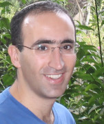
[caption id="attachment_22054" align="alignleft" width="87"] Dr. Brett Crist[/caption]
Dr. Brett Crist MD FACS
Associate Professor of Orthopaedic Surgery
Co-Chief, Orthopaedic Trauma Division
Associate Director, Joint Preservation Surgery
Director, Trauma Orthopaedic Fellowship
School of Medicine
University of Missouri Health
Medical Research: What is the background for this study? What are the main findings?
Dr. Crist: Some young patients have bone and/or cartilage problems on the femoral head due to disease or injury. Resurfacing the femoral head with donated bone and cartilage tissue is often a better option for these young patients with active lifestyles, who would otherwise require an artificial joint that would limit their activities and eventually wear out. However, there is no standard method for implantation. Our study provides initial clinical evidence that larger, size-matched grafts have the potential to improve outcomes when resurfacing cartilage defects of the femoral head in the hip joint.
Dr. Brett Crist[/caption]
Dr. Brett Crist MD FACS
Associate Professor of Orthopaedic Surgery
Co-Chief, Orthopaedic Trauma Division
Associate Director, Joint Preservation Surgery
Director, Trauma Orthopaedic Fellowship
School of Medicine
University of Missouri Health
Medical Research: What is the background for this study? What are the main findings?
Dr. Crist: Some young patients have bone and/or cartilage problems on the femoral head due to disease or injury. Resurfacing the femoral head with donated bone and cartilage tissue is often a better option for these young patients with active lifestyles, who would otherwise require an artificial joint that would limit their activities and eventually wear out. However, there is no standard method for implantation. Our study provides initial clinical evidence that larger, size-matched grafts have the potential to improve outcomes when resurfacing cartilage defects of the femoral head in the hip joint.


 Dr. Margaret Gourlay[/caption]
MedicalResearch.com Interview with:
Margaret L. Gourlay, MD, MPH
Assistant Professor
UNC Department of Family Medicine
Chapel Hill, NC 27599-7595
Medical Research: What is the background for this study? What are the main findings?
Dr. Gourlay: While clinical practice guidelines universally recommend bone density screening for fracture prevention in women aged 65 years and older, minimal data exist to guide bone density screening in older men. We studied how often bone density screening tests should be ordered in men, using data from the Osteoporotic Fractures in Men (MrOS) Study. MrOS is the largest and longest-running (since 2000) US study of bone density and fracture in men aged 65 and older.
After peak bone mass is reached in young adulthood, both men and women lose bone density as they get older. Based on our earlier findings in older women, we expected that men aged 65 and older with higher bone density T-score measurements (T-score >-1.50) on a first (baseline) bone density test would have a substantially longer estimated time to develop the lowest level of bone density (osteoporosis) than men with better baseline measurements. Clinicians want to know the time to osteoporosis because they prescribe osteoporosis treatments to prevent future fractures in elderly patients.
As expected, we found that the men with higher baseline bone density had a much slower transition to osteoporosis compared to men with lower bone density. In fact, only nine out of 4203 (0.2%) of men with higher baseline bone density developed osteoporosis after an average of 8.7 years of bone density follow-up. That was much lower than we expected and is good news for men who have favorable scores on their first bone density test. Men who had lower baseline bone density measurements developed osteoporosis faster.
Unfortunately, maintaining bone density above the osteoporosis range did not guarantee that men remained fracture-free. Most of the major osteoporotic fractures (broken hip, spine, wrist or upper arm/shoulder) occurred in men who did not have osteoporosis. This might be because they had accidents or injuries that broke their bones despite their bone density being above the thinnest range.
Dr. Margaret Gourlay[/caption]
MedicalResearch.com Interview with:
Margaret L. Gourlay, MD, MPH
Assistant Professor
UNC Department of Family Medicine
Chapel Hill, NC 27599-7595
Medical Research: What is the background for this study? What are the main findings?
Dr. Gourlay: While clinical practice guidelines universally recommend bone density screening for fracture prevention in women aged 65 years and older, minimal data exist to guide bone density screening in older men. We studied how often bone density screening tests should be ordered in men, using data from the Osteoporotic Fractures in Men (MrOS) Study. MrOS is the largest and longest-running (since 2000) US study of bone density and fracture in men aged 65 and older.
After peak bone mass is reached in young adulthood, both men and women lose bone density as they get older. Based on our earlier findings in older women, we expected that men aged 65 and older with higher bone density T-score measurements (T-score >-1.50) on a first (baseline) bone density test would have a substantially longer estimated time to develop the lowest level of bone density (osteoporosis) than men with better baseline measurements. Clinicians want to know the time to osteoporosis because they prescribe osteoporosis treatments to prevent future fractures in elderly patients.
As expected, we found that the men with higher baseline bone density had a much slower transition to osteoporosis compared to men with lower bone density. In fact, only nine out of 4203 (0.2%) of men with higher baseline bone density developed osteoporosis after an average of 8.7 years of bone density follow-up. That was much lower than we expected and is good news for men who have favorable scores on their first bone density test. Men who had lower baseline bone density measurements developed osteoporosis faster.
Unfortunately, maintaining bone density above the osteoporosis range did not guarantee that men remained fracture-free. Most of the major osteoporotic fractures (broken hip, spine, wrist or upper arm/shoulder) occurred in men who did not have osteoporosis. This might be because they had accidents or injuries that broke their bones despite their bone density being above the thinnest range.
 MedicalResearch.com Interview with:
Daniel Steffens, Ph.D.
The George Institute for Global Health
The University of Sydney
Medical Research: What is the background for this study?
Dr. Steffens: Back pain is a leading cause of disease burden globally. At present, a variety of interventions, such as getting a
MedicalResearch.com Interview with:
Daniel Steffens, Ph.D.
The George Institute for Global Health
The University of Sydney
Medical Research: What is the background for this study?
Dr. Steffens: Back pain is a leading cause of disease burden globally. At present, a variety of interventions, such as getting a  Dr. Zachary Kerr[/caption]
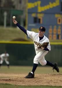
MedicalResearch.com Interview with:
Zachary Y. Kerr, PhD, MPH
Sports Injury Epidemiologist
Director, NCAA Injury Surveillance Program
Datalys Center for Sports Injury Research and Prevention
Indianapolis, IN 46202
Medical Research: What is the background for this study? What are the main findings?
Dr. Kerr: The NCAA Injury Surveillance Program has been ongoing since 1982, but the Datalys Center for Sports Injury Research and Prevention began management in 2009. We provide the NCAA sports and medical committees with evidence-based data they can use to make rule and policy decisions aimed at student-athlete health and safety. However, among the research community, there lacks current injury incidence data across the collegiate student-athlete population.
The main findings of this study is that the rate of injury was higher in competitions than in practices. However, the total number of
Dr. Zachary Kerr[/caption]
MedicalResearch.com Interview with:
Zachary Y. Kerr, PhD, MPH
Sports Injury Epidemiologist
Director, NCAA Injury Surveillance Program
Datalys Center for Sports Injury Research and Prevention
Indianapolis, IN 46202
Medical Research: What is the background for this study? What are the main findings?
Dr. Kerr: The NCAA Injury Surveillance Program has been ongoing since 1982, but the Datalys Center for Sports Injury Research and Prevention began management in 2009. We provide the NCAA sports and medical committees with evidence-based data they can use to make rule and policy decisions aimed at student-athlete health and safety. However, among the research community, there lacks current injury incidence data across the collegiate student-athlete population.
The main findings of this study is that the rate of injury was higher in competitions than in practices. However, the total number of 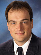 Dr. Schütz[/caption]
MedicalResearch.com Interview with:
Uwe Schütz, M.D.
Radiologist and specialist in orthopedics and trauma surgery
Department of Diagnostic and Interventional Radiology
University Hospital of Ulm
Germany
Medical Research: What is the background for this study? What are the main findings?
Dr. Schütz: In this study, which is a small part of the Trans Europe Foot Race (TEFR) TEFR-project, we investigated the question, what happens to the joints, in detail to the joint cartilage of the lower extremities, when running 4500 km without any day rest for nearly 10 weeks. Is there really a risk for developing an arthrosis when doing this, like some researches and many physicians postulate?
Well, what we find when accompanying 44 ultra-athletes with a modern 1.5Tesla MRI mounted on a custom made 38tonnes truck trailer day by day over 64 days on their way throughout whole Europe is, that the joint cartilage is initially altered by this running burden: It shows signals of cartilage matrix degradation beneath the first 1000 to 1500 km of running. But then the situation changes. When further running occurs, then the cartilage shows the ability to partially regenerate under ongoing running burden. This is a pretty new and astonishing finding, first time measured and observed in human joint in vivo. But knowledge of Scandinavian animal studies show the same behavior in dog cartilage.
Dr. Schütz[/caption]
MedicalResearch.com Interview with:
Uwe Schütz, M.D.
Radiologist and specialist in orthopedics and trauma surgery
Department of Diagnostic and Interventional Radiology
University Hospital of Ulm
Germany
Medical Research: What is the background for this study? What are the main findings?
Dr. Schütz: In this study, which is a small part of the Trans Europe Foot Race (TEFR) TEFR-project, we investigated the question, what happens to the joints, in detail to the joint cartilage of the lower extremities, when running 4500 km without any day rest for nearly 10 weeks. Is there really a risk for developing an arthrosis when doing this, like some researches and many physicians postulate?
Well, what we find when accompanying 44 ultra-athletes with a modern 1.5Tesla MRI mounted on a custom made 38tonnes truck trailer day by day over 64 days on their way throughout whole Europe is, that the joint cartilage is initially altered by this running burden: It shows signals of cartilage matrix degradation beneath the first 1000 to 1500 km of running. But then the situation changes. When further running occurs, then the cartilage shows the ability to partially regenerate under ongoing running burden. This is a pretty new and astonishing finding, first time measured and observed in human joint in vivo. But knowledge of Scandinavian animal studies show the same behavior in dog cartilage.