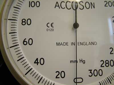
Alcohol, Author Interviews, Surgical Research, Weight Research / 20.11.2017
Weight Loss Procedures Can Double Blood Alcohol Absorption
MedicalResearch.com Interview with:
 Marta Yanina Pepino PhD
Department of Food Science and Human Nutrition
College of Agricultural, Consumer and Environmental Sciences
University of Illinois
Urbana, IL
MedicalResearch.com: What is the background for this study? What are the main findings?
Response: Our study is not the first to look at whether sleeve gastrectomy affects alcohol absorption and metabolism. Before our study, there were three published studies in the literature on this issue. However, findings from these studies were discrepant. Two of the studies found that sleeve gastrectomy did not affect blood alcohol levels and one of the studies did found that peak blood alcohol levels were higher when people drink after having a sleeve gastrectomy. All these three studies used a breathalyzer to estimate blood alcohol levels.
Our study tested the following two related hypothesis.
First, that similar to Roux-en-Y- gastric bypass (RYGB), sleeve gastrectomy accelerates alcohol absorption, which cause peak blood alcohol levels to be higher and much faster than before surgery. Because the breathalyzer requires a 15 min of waiting time between drinking the last sip of alcohol and the time that you can read a good estimate of blood alcohol levels from the breath, we hypothesized that the breathalyzer was not a good technique to estimate peak blood alcohol levels in people who may reach a peak blood alcohol level before those 15 min have passed, such as people who underwent sleeve gastrectomy or RYGB.
We found these two hypothesis to be truth:
1) Sleeve gastrectomy, similar to RYGB, can double blood alcohol levels; and
2) The breathalyzer technique is invalid to assess effects of gastric surgeries on pharmacokinetics of ingested alcohol (it underestimate blood alcohol levels by ~27% and it may miss peak blood alcohol levels).
Marta Yanina Pepino PhD
Department of Food Science and Human Nutrition
College of Agricultural, Consumer and Environmental Sciences
University of Illinois
Urbana, IL
MedicalResearch.com: What is the background for this study? What are the main findings?
Response: Our study is not the first to look at whether sleeve gastrectomy affects alcohol absorption and metabolism. Before our study, there were three published studies in the literature on this issue. However, findings from these studies were discrepant. Two of the studies found that sleeve gastrectomy did not affect blood alcohol levels and one of the studies did found that peak blood alcohol levels were higher when people drink after having a sleeve gastrectomy. All these three studies used a breathalyzer to estimate blood alcohol levels.
Our study tested the following two related hypothesis.
First, that similar to Roux-en-Y- gastric bypass (RYGB), sleeve gastrectomy accelerates alcohol absorption, which cause peak blood alcohol levels to be higher and much faster than before surgery. Because the breathalyzer requires a 15 min of waiting time between drinking the last sip of alcohol and the time that you can read a good estimate of blood alcohol levels from the breath, we hypothesized that the breathalyzer was not a good technique to estimate peak blood alcohol levels in people who may reach a peak blood alcohol level before those 15 min have passed, such as people who underwent sleeve gastrectomy or RYGB.
We found these two hypothesis to be truth:
1) Sleeve gastrectomy, similar to RYGB, can double blood alcohol levels; and
2) The breathalyzer technique is invalid to assess effects of gastric surgeries on pharmacokinetics of ingested alcohol (it underestimate blood alcohol levels by ~27% and it may miss peak blood alcohol levels).


 Dr. Jacobs[/caption]
Dr. Lisa K. Jacobs MD
Johns Hopkins School of Medicine
Baltimore, Maryland
MedicalResearch.com: What is the background for this study? What are the main findings?
Response: Breast preservation is the preferred treatment for many women diagnosed with breast cancer. The most common question that a patient will ask after the surgery is, “Did you get it all?” In the ideal case, this is accomplished in a single outpatient surgery with very good cosmetic results. In our study, Beyond the Margins-Economic Costs and Complications Associated with Repeated Breast-Conserving Surgeries we evaluated the detrimental effects of an unsuccessful initial surgery due to positive surgical margins. Using private insurance claims data, we found that 16% of patients planning breast preservation required a second breast-conserving surgery and an additional 7% converted to mastectomy. Of those patients that required additional surgery there was a 56% ($16,072) increase in cost and a 48% increase in complications. Those complications include infection, hematoma, seroma, and fat necrosis. This study demonstrates that repeated surgery has not only cosmetic consequences, but also has financial implications and increased risk.
Dr. Jacobs[/caption]
Dr. Lisa K. Jacobs MD
Johns Hopkins School of Medicine
Baltimore, Maryland
MedicalResearch.com: What is the background for this study? What are the main findings?
Response: Breast preservation is the preferred treatment for many women diagnosed with breast cancer. The most common question that a patient will ask after the surgery is, “Did you get it all?” In the ideal case, this is accomplished in a single outpatient surgery with very good cosmetic results. In our study, Beyond the Margins-Economic Costs and Complications Associated with Repeated Breast-Conserving Surgeries we evaluated the detrimental effects of an unsuccessful initial surgery due to positive surgical margins. Using private insurance claims data, we found that 16% of patients planning breast preservation required a second breast-conserving surgery and an additional 7% converted to mastectomy. Of those patients that required additional surgery there was a 56% ($16,072) increase in cost and a 48% increase in complications. Those complications include infection, hematoma, seroma, and fat necrosis. This study demonstrates that repeated surgery has not only cosmetic consequences, but also has financial implications and increased risk.