Author Interviews, MRI, Pain Research, Rheumatology / 11.10.2018

MedicalResearch.com Interview with: Daniel S. Albrecht, Ph.D. Research Fellow, Department of Radiology Harvard Medical School MedicalResearch.com: What is the background for this study? Can you briefly describe what is meant by fibromyalgia? Response: Fibromyalgia (FM) is a poorly understood chronic condition characterized by widespread musculoskeletal pain, fatigue, unrefreshing sleep, memory deficits and attention difficulties, among other symptoms. FM affects an estimated 4 million adults in the U.S., but despite this prevalence, effective therapies for treating FM are lacking. 
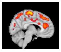
[caption id="attachment_45185" align="aligncenter" width="400"]This combined MR/PET image highlights areas of the brain in which patients with fibromyalgia were found to have increased glial activation, compared with unaffected control volunteers. Credit: Marco Loggia, PhD, Martinos Center for Biomedical Imaging, Massachusetts General Hospital This combined MR/PET image highlights areas of the brain in which patients with fibromyalgia were found to have increased glial activation, compared with unaffected control volunteers.
Credit: Marco Loggia, PhD, Martinos Center for Biomedical Imaging, Massachusetts General Hospital[/caption] Part of the reason for the paucity of effective therapeutics is insufficient knowledge of the underlying mechanisms contributing to FM. Previous work from co-senior author of the current manuscript, Eva Kosek, MD, PhD, and collaborators at the Karolinska Institute in Sweden found elevated inflammatory molecules in the cerebrospinal fluid of FM patients, which could be reflective of brain neuroinflammation in these patients. However, no study had directly assessed the presence of neuroinflammation in the brain of FM patients. Co-senior author of the study, Marco Loggia, PhD, and collaborators showed in a 2015 Brain publication that individuals with chronic low back pain (cLBP) exhibit evidence of brain neuroinflammation, specifically activation of glial cells. Our team utilized simultaneous MR/PET imaging to image brain levels of the 18 kDa translocator protein (TSPO), which is widely used as a marker of glial activation due to vast upregulation of TSPO in glial cells, e.g. microglia and astrocytes, in preclinical models of inflammation and neurological disease. Dr. Loggia sought to extend these finding in cLBP to FM, hypothesizing that activation of glial cells may also be associated with FM pathology. To this end, we used the same TSPO PET tracer to image 20 FM patients and 16 healthy controls. During a conference where I was presenting preliminary results of the fibromyalgia study, Dr. Loggia met with Dr. Kosek and discovered that, across the Atlantic, her group was performing a very similar study, imaging 11 FM patients and 11 controls with the same TSPO PET compound. They decided to form a collaboration, and logistic talks began to determine the best strategy to combine and analyze the separate datasets. In addition to PET imaging with the TSPO tracer, which is suggested to reflect activated microglia and astrocytes, Dr. Kosek’s group also collected PET scans using a tracer thought to bind specifically to astrocytes rather than microglia. This tracer was used in order to discern the relative contributions of microglia and astrocytes to any observed differences in TSPO PET signal.
Author Interviews, Brain Injury, CT Scanning, Emergency Care, JAMA, Pediatrics / 24.09.2018

MedicalResearch.com Interview with: [caption id="attachment_44691" align="alignleft" width="200"]Erik P. Hess MD MSc Professor and Vice Chair for Research Department of Emergency Medicine UAB Medicine | The University of Alabama at Birmingham Birmingham Alabama 35249 Dr. Hess[/caption] Erik P. Hess MD MSc Professor and Vice Chair for Research Department of Emergency Medicine UAB Medicine he University of Alabama at Birmingham Birmingham Alabama 35249 MedicalResearch.com: What is the background for this study? What are the main findings?  Response: 450,000 children present to U.S emergency departments each year for evaluation of head trauma.  Physicians obtain head computed tomography (CT) scans in 37%-50% of these patients, with less than 10% showing evidence of traumatic brain injury and only 0.2% that require neurosurgical treatment. In order to avoid unnecessary CT scans and to limit radiation exposure, the Pediatric Emergency Care Applied Research Network (PECARN) developed clinical prediction rules that consist of 6 readily available factors that can be assessed from the history and physical examination.  If none of these risk factors are present, a CT scan is not indicated. If either of 2 high risk factors such as signs of a skull fracture are present, CT scanning is indicated. If 1 or 2 non-high risk factors are present, then either CT scanning or observation are recommended, depending on considerations such as parental preference, clinician experience and/or symptom progression. In this study we designed a parent decision aid, “Head CT Choice” to educate the parent about the difference between a concussion – which does not show up on a CT scan – and a more serious brain injury causing bleeding in or around the brain.  The decision aid also shows parents their child’s risk for a serious brain injury – less than 1% risk in the majority of patients in our trial – what to observe their child at home for should they opt not to obtain a CT scan, and the advantages and disadvantages of CT scanning versus home observation. In our trial, we did not observe a difference in the rate of head CT scans obtained in the ED but did find that parents who were engaged in shared decision-making using Head CT Choice were more knowledgeable about their child’s risk for serious brain injury, has less difficulty making the decision because they were clearer about the advantages and disadvantages of the diagnostic options, and were more involved in decision-making by their physician.  Parents also less frequently sought additional testing for their child within 1 week of the emergency department visit.
Alzheimer's - Dementia, Author Interviews, CT Scanning, JAMA, Medical Imaging / 20.09.2018

MedicalResearch.com Interview with: PET scanner Wikipedia imageRik Ossenkoppele -PhD Lund University & VU University Medical Center Oskar Hansson - Lund University MedicalResearch.com: What is the background for this study? What are the main findings? Response: [18F]flortaucipir is a relatively novel PET tracer that can be used to detect tau pathology in the living human brain. Previous studies have shown a robust signal in patients with Alzheimer’s disease, but in patients with other types of dementia the signal was more variable. We aimed to assess the ability of [18F]flortaucipir PET to distinguish Alzheimer’s disease from other neurodegenerative disease in more than 700 study participants. T he main finding was that [18F]flortaucipir discriminated Alzheimer’s disease patients from patients with other neurodegenerative diseases with high accuracy. Furthermore, [18F]flortaucipir PET outperformed established MRI markers and showed higher specificity than amyloid-β PET. 
Author Interviews, CT Scanning, Heart Disease, NEJM / 25.08.2018

MedicalResearch.com Interview with: [caption id="attachment_44122" align="alignleft" width="150"]Prof David Newby FRSE FMedSci Personal Chair - BHF John Wheatley Chair of Cardiology University of Edinburgh Prof. Newby[/caption] Prof David Newby FRSE FMedSci Personal Chair - BHF John Wheatley Chair of Cardiology University of Edinburgh MedicalResearch.com: What is the background for this study? What are the main findings? Response: There are many tests that can try and determine whether a patient has heart disease. All are imperfect and do not directly see if the heart arteries are diseased. This study used a CT heart scan to see if there was any heart disease in patients who presented to the outpatient clinic with chest pains that could be due to coronary heart disease. The doctor use the scan result to decide whether they had heart disease and how to manage the patient. The study has found that if you use a CT heart scan then you are less likely to have a heart attack in the future. In the first year, you may require treatment with an angiogram and heart surgery (stent or heart bypass) but after the first year, you are less likely to need these treatments because the disease has already been treated promptly.
Author Interviews, Breast Cancer, Cancer Research, JAMA, Mammograms / 25.08.2018

MedicalResearch.com Interview with: [caption id="attachment_44044" align="alignleft" width="142"]Lisa A Newman, MD Director of the Breast Oncology Program for the multi-hospital  Henry Ford  Health System Dr. Newman[/caption] Lisa A Newman, MD Director of the Breast Oncology Program for the multi-hospital Henry Ford  Health System MedicalResearch.com: What is the background for this study? What are the main findings?  Response: In 2009 the United States Preventive Services Task Force published a guideline recommending that American women at average risk for breast cancer defer undergoing screening mammography until they reach the age of 50 years. Prior to this publication, women were widely-encouraged to initiate annual mammography at age 40 years. Women that have a history of breast cancer are automatically considered to be at increased risk for developing a new breast cancer, and so routine screening mammography guidelines do not apply to them. These women require annual mammography regardless of age, unless they have undergone a bilateral mastectomy. We utilized data from Michigan Blue Cross/Blue Shield to evaluate patterns of mammography utilization among women age 40-49 years, comparing rates before versus after 2009, when the USPSTF guideline was published. We analyzed women that had a prior history of breast cancer separately from those that had no history of breast cancer, and we excluded women that underwent bilateral mastectomy. Disturbingly, we found that mammography utilization rates declined among women with a history of breast cancer as well as among those with no history of breast cancer in the post-2009 timeline. This suggested to us that changes in screening recommendations may have had the unintended consequence of generating confusion and misunderstandings regarding the value of mammography among women that undeniably benefit from this imaging, such as those with a history of breast cancer. 
Author Interviews, Cancer Research, CT Scanning, JAMA, Lung Cancer / 16.08.2018

MedicalResearch.com Interview with: “CT Scan” by frankieleon is licensed under CC BY 2.0Dr. Bruno Heleno MD PhD Assistant Professor | Professor Auxiliar NOVA Medical School | Faculdade de Ciências Médicas Universidade Nova da Lisboa  MedicalResearch.com: What is the background for this study? What are the main findings? Response: The Danish Lung Cancer Screening Trial (DLCST) is a randomized controlled trial which enrolled 4104 participants (aged 50-70 years; current or former smokers; ≥20 pack years; former smokers must have quit <10 years before enrollment) to either 5 rounds of screening for lung cancer with low-dose CT-scans or to no screening. After 10 years of follow-up, there was a 2.10 percentage points lung cancer absolute risk increase with low-dose CT-screening. Overdiagnosis, i.e. the detection of cancer that would not progress to symptoms or death, was estimated at 67.2% of the screen-detected cancers.
Author Interviews, Breast Cancer, Cost of Health Care, Mammograms, Medical Imaging / 29.07.2018

MedicalResearch.com Interview with: [caption id="attachment_43571" align="alignleft" width="133"]Michal Horný PhD Assistant Professor Emory University School of Medicine, Department of Radiology and Imaging Sciences Emory University Rollins School of Public Health Department of Health Policy and Management Atlanta, GA 30322 Dr. Horný[/caption] Michal Horný PhD Assistant Professor Emory University School of Medicine, Department of Radiology and Imaging Sciences Emory University Rollins School of Public Health Department of Health Policy and Management Atlanta, GA 30322 MedicalResearch.com: What is the background for this study? Response: Increased breast tissue density is a common finding at screening mammography. Approximately 30-50% of women have so-called “dense breasts” but many of them are not aware of it. The problem is that the increased tissue density can potentially mask early cancers. In other words, if there is cancer hiding in dense breast tissue, it could be difficult to spot it. To improve the awareness of breast tissue density, a patient group called Are You Dense Advocacy, Inc., started lobbying state and federal policymakers to pass laws mandating health care providers to notify women about their breast density assessments. As a result, 31 states have already enacted some form of legislation regarding dense breast tissue.
Author Interviews, Emergency Care, Heart Disease, JACC, Medical Imaging / 21.06.2018

MedicalResearch.com Interview with: [caption id="attachment_42363" align="alignleft" width="125"]Jeffrey M. Levsky, M.D., Ph.D. Associate Professor, Department of Radiology Associate Professor, Department of Medicine (Cardiology) Albert Einstein College of Medicine Montefiore Medical Center  Dr. Levsky[/caption] Jeffrey M. Levsky, M.D., Ph.D. Associate Professor, Department of Radiology Associate Professor, Department of Medicine (Cardiology) Albert Einstein College of Medicine Montefiore Medical Center MedicalResearch.com: What is the background for this study? What are the main findings? Response: Millions of Americans are evaluated each year for acute chest pain in the Emergency Department.  There are multiple modalities that can be used to triage these patients and there have only been a few studies comparing different imaging methods. We chose to study Stress Echocardiography and Coronary CT Angiography, two exams that have not been compared directly in this population.  We found that Stress Echocardiography was able to discharge a higher proportion of patients in a shorter amount of time as compared to Coronary CTA. 
Alzheimer's - Dementia, Author Interviews, JAMA, Medical Imaging, Memory / 12.06.2018

MedicalResearch.com Interview with: Arno de Wilde, MD / PhD candidate Department of Neurology & Alzheimer Center Amsterdam Neuroscience VU University Medical Center Amsterdam, the Netherlands MedicalResearch.com: What is the background for this study? What are the main findings? Response: Previous studies assessing the clinical utility of amyloid imaging used very selected research populations, limiting the translatability to clinical practice. In contrast, we used an unselected memory clinic cohort, offering amyloid PET to ALL patients visiting our memory clinic, and for the purpose of this study, we implemented amyloid PET in our routine diagnostic work-up. Our results demonstrate that amyloid PET has important consequences, in terms of diagnosis and treatment changes, for a significant number of patients within a situation that closely resembles clinical practice. I think that these results are an important step in 'bridging the gap' between using amyloid PET in a research setting versus daily clinical practice.
Author Interviews, JAMA, MRI, Prostate Cancer / 11.06.2018

MedicalResearch.com Interview with: [caption id="attachment_32943" align="alignleft" width="180"]Lars Boesen MD PhD Department of Urology Herlev Gentofte University Hospital Herlev Dr. Boessen[/caption] Dr. Lars Boesen MD PhD Department of Urology Herlev Hospital MedicalResearch.com: What is the background for this study? What are the main findings?  Response: The current standard of care in prostate cancer diagnosis includes untargeted transrectal ultrasound-guided biopsies for all biopsy-naïve men with clinically suspicion of prostate cancer. However, this strategy that practically has remained unchanged for decades has limited diagnostic accuracy as significant cancers are missed or under-graded and insignificant cancers are unintendedly detected by the random sampling leading to possible overtreatment. Multiparametric MRI in the diagnosis of prostate cancer has been studied extensively in recent years and has improved detection, localization, staging and risk stratification. It has been suggested that if multiparametric MRIs were used as a triage test prior to biopsies, a significant proportion of men might safely avoid prostate biopsies and the diagnostic ratio of significant vs. insignificant cancer could be improved compared to performing standard biopsies in all men. However, multiparametric MRIs are generally time-consuming (~40 min scan time), expensive and include intravenous contrast media. This reduces its feasibility for widespread clinical implementation in larger patient populations in the western community with its high PCa prevalence. The development of a simpler and faster (~15 min) biparametric MRI protocol using less scan sequences and circumvents intravenous contrast-media seems to preserve adequate diagnostic accuracy in a detection setting and could facilitate dissemination of prostate MRI as a triage test before any biopsy. Here we present a large prospective study that assesses the diagnostic accuracy of a novel biparametric MRI to rule out significant prostate cancer in N=1020 biopsy-naive men with clinically suspicion of prostate cancer. We found that a low suspicion biparametric MRI had a very high negative predictive value (97%) for ruling out significant cancer on confirmatory biopsies. Furthermore, bpMRI suspicion scores were strongly associated with prostate cancer detection rates and restricting biopsies (targeted plus standard) to men with suspicious biparametric MRIs meant 30% could avoid prostate biopsies, improved significant prostate cancer diagnosis by 11%, and reduced insignificant prostate cancer diagnosis by 40% compared to our current diagnostic approach – standard biopsies for all men with clinically suspicion of prostate cancer. 
Author Interviews, JAMA, Medical Imaging, Prostate Cancer, Technology / 17.05.2018

MedicalResearch.com Interview with: [caption id="attachment_41807" align="alignleft" width="156"]Andrew J. Armstrong, MD ScM FACP Associate Professor of Medicine, Surgery, Pharmacology and Cancer Biology Associate Director for Clinical Research in Genitourinary Oncology Duke Cancer Institute Divisions of Medical Oncology and Urology Duke University Dr. Armstrong[/caption] Andrew J. Armstrong, MD ScM FACP Associate Professor of Medicine, Surgery, Pharmacology and Cancer Biology Associate Director for Clinical Research in Genitourinary Oncology Duke Cancer Institute Divisions of Medical Oncology and Urology Duke University MedicalResearch.com: What is the background for this study? What are the main findings?  Response: Men with prostate cancer commonly develop bone metastases and undergo nuclear medicine bone scans. However, these scans are non-quantitative, and disease burden has been challenging to assess over time and to relate to clinical outcomes. We developed a software program and measurement called the automated bone scan index that essentially reads a standard of care nuclear bone scan, provides a quantitative metric, and demonstrate in a phase 3 trial that this aBSI is highly associated with clinical outcomes including survival, time to symptomatic progression, and prostate cancer specific survival. We accomplished this within a prospective phase 3 international trial of men with metastatic hormone resistant prostate cancer who were followed over a long period of time.  All bone scans were read and measured using the aBSI at baseline, and we found that the aBSI was highly prognostic.  This work validates prior smaller phase 2 BSI studies, and demonstrates both the feasibility and clinical utility for incorporating the aBSI into clinical practice to provide this important prognostic information to patients and providers.
Author Interviews, Breast Cancer, Brigham & Women's - Harvard, JAMA, Mammograms / 03.05.2018

MedicalResearch.com Interview with: [caption id="attachment_41460" align="alignleft" width="200"]Anne Marie McCarthy, PhD Department of Medicine Massachusetts General Hospital and Harvard Medical School Boston Dr. McCarthy[/caption] Anne Marie McCarthy, PhD Department of Medicine Massachusetts General Hospital and Harvard Medical School Boston MedicalResearch.com: What is the background for this study? What are the main findings? Response: Mammography is effective in reducing breast cancer mortality. However, it is not perfect, and approximately 15% of breast cancers are diagnosed despite a negative mammogram before the next recommended screening. MedicalResearch.com: What should clinicians and patients take away from your report? Response: Using data from the NCI funded PROSPR (Population-Based Research Optimizing Screening through Personalized Regimens) Consortium, we determined the rates of cancer diagnosis within one year following a negative or positive screening mammogram. The rate of cancer diagnosis within one year of a negative mammogram was small (5.9 per 10,000 screenings), but those cancers were more likely to have poor prognosis than cancers diagnosed after a positive mammogram (43.8% vs. 26.9%). As expected, women with dense breasts were more likely to have cancer diagnosed within 1 year of a negative mammogram. However, breast density was not a good predictor of poor prognosis among women diagnosed with cancer after a negative mammogram. Younger women were more likely to be diagnosed with poor prognosis breast cancer after a negative screening mammogram.
Author Interviews, Cancer Research, MRI, Prostate Cancer / 21.03.2018

MedicalResearch.com Interview with: [caption id="attachment_40681" align="alignleft" width="200"]Veeru Kasivisvanathan MBBS BSc MRCS MSc PGCert Lead for CPD, Division of Surgery and Interventional Science, UCL Academic Section Committee, British Association of Urological Surgeons Twitter: @veerukasi PRECISION Study Coordinator https://clinicaltrials.gov/ct2/show/NCT02380027   Dr. Kasivisvanathan[/caption] Veeru Kasivisvanathan MBBS BSc MRCS MSc PGCert Lead for CPD, Division of Surgery and Interventional Science, UCL Academic Section Committee, British Association of Urological Surgeons Twitter: @veerukasi PRECISION Study Coordinator https://clinicaltrials.gov/ct2/show/NCT02380027   MedicalResearch.com: What is the background for this study? What are the main findings? 
  • We knew that there were limitations in the standard of care pathway for the diagnosis of prostate cancer, TRUS biopsy which missed harmful cancers and over diagnosed harmless cancers.
  • Emerging reports in the literature showed that using an alternative diagnostic pathway, MRI and MRI-targeted biopsy, showed promising prostate cancer detection rates
  • In 2012 we set out in an international working group to design a study that could change clinical practice and replace the standard of care with a pathway involving MRI 
Abuse and Neglect, Hematology, Lancet, Medical Imaging, Transplantation / 07.03.2018

MedicalResearch.com Interview with: [caption id="attachment_40425" align="alignleft" width="133"]Kirsten Williams, M.D. Blood and marrow transplant specialist Children’s National Health System Dr. Williams[/caption] Kirsten Williams, M.D. Blood and marrow transplant specialist Children’s National Health System  MedicalResearch.com: What is the background for this study? What are the main findings?  Response: This study addressed a life-threatening complication of bone marrow transplantation called bone marrow failure. Bone marrow transplantation has provided a cure for patients with aggressive leukemias or acquired or genetic marrow dysfunction. The process of bone marrow transplantation involves giving chemotherapy and/or radiation, which removes the diseased blood cells from the bone marrow. After this, new bone marrow stem cells are infused from a healthy individual. They travel to the bone marrow and start the slow process of remaking the blood system. Because these new cells start from infancy, it takes upwards of four to five weeks for new mature healthy cells to emerge into the blood, where they can be identified. Historically, there has been no timely way to determine if the new cells have successfully repopulated unless they can be seen in the blood compartment. This condition of bone marrow failure is life-threatening, because patients don't have white blood cells to protect them from infection. Once bone marrow failure is diagnosed, a second new set of stem cells are infused, often after more chemotherapy is given. However, for many individuals this re-transplantation is too late, because severe infections can be fatal while waiting cells to recover. We were the first group to use a new imaging test to understand how the newly infused bone marrow cells develop inside the patient. We have recently published a way to detect the new bone marrow cell growth as early as five days after the cells are given. We used an investigational nuclear medicine test to reveal this early cell growth, which could be detected weeks before the cells appear in the blood. This radiology test is safe, does not cause any problems and is not invasive. It is called FLT (18F-fluorothymidine) and the contrast is taken up by dividing hematopoietic stem cells. The patients could even see the growth of their new cells inside the bone marrow (which they very much enjoyed while waiting to see recovery of the cells in their blood). We could use the brightness of the image (called SUV) to determine approximately how many weeks remained before the cells were visible in the blood. Finally, we actually could see where the new cells went after they were infused, tracking their settling in various organs and bones. Through this, we could see that cells did not travel directly to all of the bones right away as was previously thought, but rather first went to the liver and spleen, then to the mid-spine (thorax), then to the remainder of the spine and breastplate, and finally to the arms and legs. This pattern of bone marrow development is seen in healthy developing fetuses. In this case, it occurs in a similar pattern in adults undergoing bone marrow transplant.
Author Interviews, Breast Cancer, Mammograms / 26.02.2018

MedicalResearch.com Interview with: [caption id="attachment_40252" align="alignleft" width="150"]Prof-Stephen-Duffy.jpg Prof. Duffy[/caption]

Stephen W. Duffy Professor of Cancer Screening Wolfson Institute of Preventive Medicine, Barts and The London School of Medicine and Dentistry Queen Mary University of London

MedicalResearch.com: What is the background for this study? What are the main findings? Response: The phenomenon of length bias, whereby screening has more chance of detecting slow growing tumours, has been known about for some years. This has led some colleagues to speculate that breast cancer screening only benefits those with slow-growing, less aggressive cancers, and does not reduce deaths from more aggressive, rapidly progressing cancers. In this study, we addressed this question directly using data from a randomised trial of mammographic screening. We calculated the reduction in mortality from grade 1 (less aggressive), grade 2 (intermediate) and grade 3 (most aggressive) cancers, as a result of screening. We found that the greatest reduction in breast cancer mortality was from the aggressive, fast-growing grade 3 cancers, contrary to what had been suspected. 
Author Interviews, Biomarkers, CT Scanning, MRI, Prostate Cancer / 07.02.2018

MedicalResearch.com Interview with: Jeremie Calais PhD Ahmanson Translational Imaging Division UCLA Nuclear Medicine Department Los Angeles, CA 90095Jeremie Calais MD Ahmanson Translational Imaging Division UCLA Nuclear Medicine Department Los Angeles, CA 90095  MedicalResearch.com: What is the background for this study? What are the main findings? Response: The only curative treatment for recurrent prostate cancer after radical prostatectomy is salvage radiotherapy. Unfortunately, current standard imaging modalities are too insensitive to visualize the location of the recurrence until it is too late. As a result, salvage radiotherapy is directed to areas only suspected to harbor the recurrence based upon a "best guess" approach according to standard guidelines that define radiotherapy treatment volumes. PSMA PET/CT is a new imaging technique with sensitivity sufficient to detect and localize the recurrent prostate cancer early enough to potentially guide salvage radiotherapy. The first sign of prostate cancer recurrence is a rising PSA. For salvage radiotherapy to be successful, it should be initiated before the PSA rises above 1 ng/mL, and ideally, closer to 0.2 ng/mL or lower. PSMA PET/CT localizes sites of prostate cancer recurrence in up to 70% of patients with low PSA, below < 1.0. In the US it is not yet FDA approved and currently only used for research purposes. In our current study we included 270 patients with early recurrence of prostate cancer after surgery from Germany and UCLA,  we found that 20 % of the patients had at least one lesion detected by  PSMA PET/CT which was NOT covered by the standard radiation fields. Obviously, salvage radiotherapy is only curative if recurrent disease is completely encompassed by the radiotherapy fields and would have failed in these patients.
Author Interviews, Environmental Risks, JAMA, MRI, Toxin Research / 31.01.2018

MedicalResearch.com Interview with: “Danger Carbon Monoxide” by SmartSign is licensed under CC BY 2.0Won Young Kim, MD PhD Department of Emergency Medicine Asan Medical Center University of Ulsan College of Medicine Seoul, Korea MedicalResearch.com: What is the background for this study? Response: Neurological symptoms of carbon monoxide (CO) poisoning can manifest not only immediately but also as late as 2 to 6 weeks after successful initial resuscitation as delayed neurological sequelae (DNS). To date, no reliable methods of assessing the probability of DNS after acute CO poisoning have been developed, which make it difficult to research the pathophysiology of DNS and targeting prevention.
Author Interviews, Cognitive Issues, Genetic Research, JAMA, Medical Imaging, Mental Health Research / 25.01.2018

MedicalResearch.com Interview with: “The Fourth Sex: Adolescent Extremes” by Victor Soto is licensed under CC BY 2.0Dag Alnaes, PhD Norwegian Centre for Mental Disorders Research KG Jebsen Centre for Psychosis Research Division of Mental Health and Addiction, Oslo University Hospital Oslo, Norway  MedicalResearch.com: What is the background for this study? What are the main findings? Response: The transition from childhood to adulthood is characterized by swift and dramatic changes, both in our environment and in our brains. This period of life also coincides with the onset of many mental disorders. To gain a better understanding of why, the clinical neurosciences must attempt to disentangle the complex and dynamic interactions between genes and the environment and how they shape our brains. The ultimate goal is to be able to predict which individuals are at risk before clinical symptoms appear. Advanced brain imaging has been proposed to represent one promising approach for such early detection, but there is currently no robust imaging marker that allows us to identify individuals at risk with any clinically relevant degree of certainty. Our study shows that self-reported early signs of mental illness are associated with specific patterns of brain fiber pathways in young people, even if they may not fulfill criteria for a formal diagnosis or are currently in need of treatment. 
Author Interviews, CT Scanning, Heart Disease, Technology / 23.01.2018

MedicalResearch.com Interview with: [caption id="attachment_39498" align="alignleft" width="165"]MedicalResearch.com Interview with: Cardiologist Mark Rabbat, MD, FSCCT Dr. Rabbat[/caption] Cardiologist Mark Rabbat, MD, FSCCT Who pioneered the use of FFRct at Loyola Medicine and was first author of an international expert panel of leading cardiologists and radiologists from centers in the United States, Canada, Denmark, Italy, Belgium and the Netherlands on how to interpret and report the tests published in the Journal of Cardiovascular Computed Tomography  MedicalResearch.com: What is the scope of the problem? Response: Coronary artery disease is a very large healthcare burden. Over sixteen million individuals in the United States have coronary artery disease.  Coronary artery disease may result in your heart not getting enough blood and increases your risk of a heart attack. Historically, we have been faced with either using tests we knew were not always accurate or putting a patient through an invasive angiogram just to determine whether they would need another invasive procedure to restore blood flow.  The CT-derived fractional flow reserve (FFRct) analysis is the first technology that bridges the gap between the non-invasive and invasive tests within one platform.  Any patient with symptoms such as chest pain, chest tightness, fatigue, or shortness of breath without known coronary artery disease may be a candidate for the FFRct study. 
Alzheimer's - Dementia, Author Interviews, Biomarkers, JAMA, MRI / 12.01.2018

MedicalResearch.com Interview with: [caption id="attachment_39274" align="alignleft" width="143"]Miguel A. Santos-Santos, MD Department of Neurology, Memory and Aging Center University of California San Francisco Autonomous University of Barcelona, Cerdanyola del Valles, Spain Dr. Miguel A. Santos-Santos[/caption] Miguel ASantos-SantosMD Department of Neurology, Memory and Aging Center University of California San Francisco Autonomous University of Barcelona, Cerdanyola del Valles, Spain MedicalResearch.com: What is the background for this study? Response: Primary progressive aphasia (PPA) is a clinically and pathologically heterogeneous (generally Frontotemporal lobar degeneration [FTLD, generally tau or tdp proteinopathies] or Alzheimer’s disease [AD] pathology) condition in which language impairment is the predominant cause of functional impairment during the initial phases of disease. Classification of PPA cases into clinical-anatomical phenotypes is of great importance because they are linked to different prevalence of underlying pathology and prediction of this pathology during life is of critical importance due to the proximity of molecule-specific therapies. The 2011 international consensus diagnostic criteria established a classification scheme for the three most common variants (the semantic [svPPA], non-fluent/agrammatic [nfvPPA], and logopenic [lvPPA]) of PPA and represent a collective effort to increase comparability between studies and improve the reliability of clinicopathologic correlations compared to the previous semantic dementia and progressive non-fluent aphasia criteria included in the 1998 consensus FTLD clinical diagnostic criteria. Since their publication, a few studies have reported amyloid imaging and pathological results in PPA, however most of these studies are retrospective in nature and the prevalence of FTLD and Alzheimer’s disease pathological findings or biomarkers in each variant has been inconsistent across the literature, therefore prospective validation with biomarker and autopsy data remains scarce and highly necessary.
Author Interviews, Brigham & Women's - Harvard, Gender Differences, Medical Imaging, Weight Research / 22.11.2017

MedicalResearch.com Interview with: [caption id="attachment_38089" align="alignleft" width="161"]Miriam Bredella, MD Associate Professor of Radiology, Harvard Medical School Department of Radiology Massachusetts General Hospital Boston, MA 02114 Dr. Bredella[/caption] Miriam Bredella, MD Associate Professor of Radiology Harvard Medical School Department of Radiology Massachusetts General Hospital Boston, MA 02114 MedicalResearch.com: What is the background for this study? What are the main findings? Response: It is well known that body composition differs between men and women, with women having proportionally more fat and men more muscle mass. But not the amount of fat but its distribution is an important determinant of cardiometabolic risk, with certain ectopic fat depots, such as visceral adipose tissue, fat within muscle cells - intramyocellular (IMCL), and liver fat, being more detrimental than others, such as femorogluteal subcutaneous adipose tissue. We therefore wanted to study sex differences in body composition and cardiometabolic risk in men and women with obesity. We found that at the same BMI, men had relatively higher visceral adipose tissue, IMCL, liver fat, muscle and lean mass, while women higher percent fat mass and higher subcutaneous adipose tissue. This female anthropometric phenotype was associated with a better cardiometabolic risk profile at similar BMI compared to men. However, ectopic fat depots were more strongly associated with adverse cardiometabolic risk factors in women compared to men
Author Interviews, CT Scanning, Emergency Care, Pain Research / 14.11.2017

MedicalResearch.com Interview with: [caption id="attachment_38261" align="alignleft" width="133"]Jeff Perry, MD, MSc, CCFP-EM Professor, Department of Emergency Medicine Senior Scientist, Ottawa Hospital Research Institute Research Chair in Emergency Neurological Research, University of Ottawa Emergency Physician and Epidemiology Program The Ottawa Hospital Ottawa, Ontario Dr. Perry[/caption] Jeff Perry, MD, MSc, CCFP-EM Professor, Department of Emergency Medicine Senior Scientist, Ottawa Hospital Research Institute Research Chair in Emergency Neurological Research, University of Ottawa Emergency Physician and Epidemiology Program The Ottawa Hospital Ottawa, Ontario MedicalResearch.com: What is the background for this study? What are the main findings? Response: Headache accounts for about 1-2% of all emergency department visits.  One of the most feared diagnosis within these patients is subarachnoid hemorrhage. While investigations are clearly warranted for patients with a diminished level of consciousness or new focal neurological deficits, approximately 50% of patients with subarachnoid hemorrhage (SAH) have no focal or global neurological findings. Deciding whether to image headache patients with no deficits is difficult, especially since timely diagnosis and treatment results in substantially better outcomes. The desire to never miss a subarachnoid hemorrhage, however, contributes to escalating neuroimaging rates and a dogmatic adherence to lumbar puncture, even if the scan is negative, despite the very high sensitivity of computed tomography. However, a recent population-based study suggested that over 5% of confirmed subarachnoid hemorrhages were missed at initial presentation, especially in smaller hospitals. Therefore, identifying which headache patients require investigations to rule-out SAH is of great importance. We have previously derived (N=1,999) and refined (N=2,131) the Ottawa SAH Rule. In this study, we conducted an multicenter prospective cohort study at six tertiary care hospitals, and found that the Ottawa SAH Rule performed well, with an 100% sensitivity, and specificity of 13.6%.
Author Interviews, JAMA, Lung Cancer, Medical Imaging, Surgical Research, Vanderbilt / 10.11.2017

MedicalResearch.com Interview with: [caption id="attachment_38163" align="alignleft" width="300"]PET Scan Vanderbilt Health PET Scan Vanderbilt Health[/caption] Amelia W. Maiga, MD MPH Vanderbilt General Surgery Resident VA Quality Scholar, TVHS MedicalResearch.com: What is the background for this study? What are the main findings? Response: Positron emission tomography (PET) combined with fludeoxyglucose F18 (FDG) is currently recommended for the noninvasive diagnosis of lung nodules suspicious for lung cancer. Our investigation adds to growing evidence that FDG-PET scans should be interpreted with caution in the diagnosis of lung cancer. Misdiagnosis of lung lesions driven by FDG-PET avidity can lead to unnecessary tests and surgeries for patients, along with potentially additional complications and mortality. To estimate FDG-PET diagnostic accuracy, we conducted a multi-center retrospective cohort study. The seven cohorts originating from Tennessee, Arizona, Massachusetts and Virginia together comprised 1188 nodules, 81 percent of which were malignant. Smaller nodules were missed by FDG-PET imaging. Surprisingly, negative PET scans were also not reliable indicators of the absence of disease, especially in patients with smaller nodules or who are known to have a high probability of lung cancer prior to the FDG-PET test. Our study supports a previous meta-analyses that found FDG-PET to be less reliable in regions of the country where fungal lung diseases are endemic. The most common fungal lung diseases in the United States are histoplasmosis, coccidioidomycosis and blastomycosis. All three fungi reside in soils. Histoplasmosis and blastomycosis are common across much of the Mississippi, Ohio and Missouri river valleys and coccidioidomycosis is prevalent in the southwestern U.S. These infections generate inflamed nodules in the lungs (granulomas), which can be mistaken for cancerous lesions by imaging.
Author Interviews, Breast Cancer, Brigham & Women's - Harvard, Cancer Research, Mammograms, Technology / 20.10.2017

MedicalResearch.com Interview with: [caption id="attachment_37640" align="alignleft" width="135"]Manisha Bahl, MD, MPH Director, Breast Imaging Fellowship Program, Massachusetts General Hospital Assistant Professor of Radiology, Harvard Medical School Dr. Bahl[/caption] Manisha Bahl, MD, MPH Director, Breast Imaging Fellowship Program, Massachusetts General Hospital Assistant Professor of Radiology, Harvard Medical School MedicalResearch.com: What is the background for this study? What are the main findings? Response: Image-guided biopsies that we perform based on suspicious findings on mammography can yield one of three pathology results: cancer, high-risk, or benign. Most high-risk breast lesions are noncancerous, but surgical excision is typically recommended because some high-risk lesions can be upgraded to cancer at surgery. Currently, there are no imaging or other features that reliably allow us to distinguish between high-risk lesions that warrant surgery from those that can be safely followed, which has led to unnecessary surgery of high-risk lesions that are not associated with cancer. We decided to apply machine learning algorithms to help us with this challenging clinical scenario: to distinguish between high-risk lesions that warrant surgery from those that can be safely followed. Machine learning allows us to incorporate the full spectrum of diverse and complex data that we have available, such as patient risk factors and imaging features, in order to predict which high-risk lesions are likely to be upgraded to cancer and, ultimately, to help our patients make more informed decisions about surgery versus surveillance. We developed the machine learning model with almost 700 high-risk lesions, then tested it with more than 300 high-risk lesions. Instead of surgical excision of all high-risk lesions, if those categorized with the model to be at low risk for upgrade were surveilled and the remainder were excised, then 97.4% malignancies would have been diagnosed at surgery, and 30.6% of surgeries of benign lesions could have been avoided.
Alzheimer's - Dementia, Author Interviews, Medical Imaging, MRI / 06.10.2017

MedicalResearch.com Interview with: Dr. Sanja Josef Golubic, dr. sc Department of Physics, Faculty of Science University of Zagreb, Croatia MedicalResearch.com: What is the background for this study? Response: Our study was aimed to search the topological biomarker of Alzheimer’s disease. A recent evidences suggest that the decades long progression of brain degeneration that is irreversible by the stage of symptomatic Alzheimer’s disease, may account for failures to develop successful disease-modifying therapies. Currently, there is a pressing worldwide search for a marker of very early, possibly reversible, pathological changes related to Alzheimer’s disease in still cognitively intact individuals, that could provide a critical opportunity for evolving of efficient therapeutic interventions. Three years ago we reported the discovery of the novel, fast brain pathway specialized for rapid processing of the simple tones. We named it gating loop. Gating loop directly links auditory brain areas to prefrontal brain area. We have also noticed the high sensitivity of the gating loop processing on AD pathology. It was inspiration to focus our Alzheimer’s disease biomarker search in the direction of prefrontal brain activation during listening of simple tones.
Author Interviews, MRI, Schizophrenia / 31.08.2017

MedicalResearch.com Interview with: [caption id="attachment_36710" align="alignleft" width="179"]Irina Rish PhD IBM T.J. Watson Research Center Yorktown Heights, NY 10598 Dr. Rish[/caption] Irina Rish PhD IBM T.J. Watson Research Center Yorktown Heights, NY 10598  MedicalResearch.com: What is the background for this study? What are the main findings? Response: Schizophrenia is a chronic and severe psychiatric disorder that affects roughly about 1% of population. Although it is not as common as other mental disorders, such as depression, anxiety, and attention deficit disorder (ADD), and so on, schizophrenia  is perhaps one of  the most debilitating psychiatric disorders,  preventing people from normal  functioning in daily life. It is characterized primarily by a range of psychotic symptoms, including hallucinations (false auditory, visual or tactile perceptions detached from reality), as well as delusions, disorganized thoughts, speech and behavior, and multiple other symptoms including difficulty showing (and recognizing) emotions, poor executive functioning, inattentiveness, problems with working memory,  and so one. Overall, schizophrenia has a devastating impact not only on patients and their families, but on the economy, as it was estimated to cost the US about 2% off  gross national product in treatment costs, missed work, etc. Thus, taking steps towards better understanding of the disease can potentially lead to more accurate early diagnosis and better treatments. In this work, the objective was to identify "statistical biomarkers' of schizophrenia from brain imaging data (specifically, functional MRI), i.e. brain activity patterns that would be capable of accurately discriminating between schizophrenic patients and controls, and reproducible (stable) across multiple datasets. The focus on both predictive accuracy (generalization to previously unseen subjects) as well as on stability (reproducibility) across multiple datsets differentiates our work from majority of similar studies in neuroimaging field that tend to focus only on statistically significant differences between such patterns on a fixed dataset, and may not reliably generalize to new data. Our prior work on neuroimaging-based analysis of schizoprenia http://journals.plos.org/plosone/article/related?id=10.1371/journal.pone.0050625, as well as other research in the field, suggest that disrupted functional connectivity can be a much more informative source of discriminative patterns than local changes in brain activations, since schizophrenia is well known to be a "network disease", rather than a localized one.
Author Interviews, Brain Injury, Cleveland Clinic, Cognitive Issues, MRI, Occupational Health / 29.07.2017

MedicalResearch.com Interview with: [caption id="attachment_36217" align="alignleft" width="160"]Virendra Mishra, Ph.D. Department of Imaging Research Cleveland Clinic Lou Ruvo Center for Brain Health in Las Vegas Dr. Virendra Mishra[/caption] Virendra Mishra, Ph.D. Department of Imaging Research Cleveland Clinic Lou Ruvo Center for Brain Health in Las Vegas MedicalResearch.com: What is the background for this study? What are the main findings? Response: Repetitive head trauma has been shown to be a risk factor for various neurodegenerative disorders, mood swings, depression and chronic traumatic encephalopathy. There has been a significant amount of research into identifying an imaging biomarker of mild traumatic brain injury (mTBI) due to repetitive head trauma. Unfortunately, most of the biomarkers have not been able to find a successful translation to clinics. Additionally, the quest for the mTBI imaging biomarker especially using Magnetic Resonance Imaging (MRI) techniques has been done by looking at either the gray matter (T1-weighted) or the white matter (Diffusion Tensor Imaging) independently; and both have shown changes that are associated with repetitive head trauma. Hence in this study, we wanted to investigate if combining gray matter and white matter information enables us to better predict the fighters who are more vulnerable to cognitive decline due to repetitive head trauma. Our method found seven imaging biomarkers that when combined together in a multivariate sense were able to predict with greater than 73% accuracy those fighters who are vulnerable to cognitive decline both at baseline and follow-up. The imaging biomarkers were indeed a combination of gray and white matter measures of regions reported previously in the literature. A key point in our study was we found the regions predicting cognitive decline without enforcing any assumptions on the regions previously reported.
Author Interviews, Medical Imaging, Prostate Cancer / 19.06.2017

MedicalResearch.com Interview with: Dr. Susanne Lütje Ärztlicher Dienst Universitätsklinikum Essen (AöR) Klinik für Nuklearmedizin Essen Germany MedicalResearch.com: What is the background for this study? What are the main findings? Response: Prostate cancer (PCa) is the most common cancer in men and accounts for a significant amount of morbidity and mortality. At present, the curative treatment option of choice for localized stages of PCa is radical prostatectomy, which may include extended lymph node dissection. Unfortunately, surgical procedures can be accompanied by complications such as urinary incontinence. Most importantly, small tumor deposits may not be seen by the surgeon during surgery and could ultimately lead to disease recurrence. To overcome these issues, new and innovative treatments are needed. The prostate-specific membrane antigen (PSMA) is a surface protein that is overexpressed in prostate cancer and can be used as a target to guide new therapies. Photodynamic therapy (PDT) is an ablative procedure in which tumor cells can be destroyed effectively by irradiation of light of a specific wavelength, which activates previously administered photosensitizers. The photosensitizers can respond by emitting fluorescence or emitting oxygen radicals which can cause cellular damage. Coupling the photosensitizer to an agent that targets PSMA on the tumor surface offers the possibility to selectively and effectively destroy prostate tumor remnants and micrometastases, while surrounding healthy tissues remain unaffected. In our study, the PSMA targeting antibody D2B was coupled to the photosensitizer IRDye700DX and radiolabeled with 111In. In a mouse model, this multi-modality agent was used to preoperatively visualize tumor lesions with SPECT/CT to allow rough localization of the tumors. During surgery, the fluorescent signal originating from the photosensitizer facilitates visualization of tumors and residual tumor tissue, so the surgeon can be guided towards accurate resection of the entire tumors and metastases. In addition, the PSMA-targeted PDT can be applied to destroy small tumor deposits in cases where close proximity of the tumors.
Author Interviews, Autism, MRI / 09.06.2017

MedicalResearch.com Interview with: [caption id="attachment_35164" align="alignleft" width="150"]Joseph Piven, MD The Thomas E. Castelloe Distinguished Professor of Psychiatry UNC School of Medicine Director of the Carolina Institute for Developmental Disabilities Co-senior author of the study Dr. Piven[/caption] Joseph Piven, MD The Thomas E. Castelloe Distinguished Professor of Psychiatry UNC School of Medicine Director of the Carolina Institute for Developmental Disabilities Co-senior author of the study MedicalResearch.com: What is the background for this study? What are the main findings? Response: Babies with older siblings with autism are at an increased risk (20%) of getting autism over the general population (1%).  Infants who later are diagnosed with autism don’t have any of the stigmata of autism in the first year of life. The symptoms of autism unfold in the first and particularly in the second year of life and beyond. We have evidence to support the idea that behavioral symptoms of autism arise from changes in the brain that occur very early in life. So we have employed MRI and computer analyses to study those early brain changes and abnormalities in infancy to see if early brain changes at 6 months of age can predict whether babies at high-risk of developing autism will indeed develop the condition at age two. For this particular study, we used data from MRIs of six-month olds to show the pattern of synchronization or connection across brain regions throughout the brain and then predict which babies at high familial risk of developing autism would be most likely to be diagnosed with the condition at age two.