MedicalResearch.com Interview with:
[caption id="attachment_52550" align="alignleft" width="200"]

Dr. Clarke[/caption]
Robert Clarke MD, FRCP, FFPH, FFPHI, MSc, DCH
Professor of Epidemiology and Public Health Medicine
Clinical Trial Service Unit (CTSU)
Nuffield Department of Population Health
University of Oxford
MedicalResearch.com: What is the background for this study?
Response: Approximately 1 in 2 women and 1 in 5 men aged 50 years or older will suffer from an osteoporotic fracture in their remaining lifetime. Hip fracture is the most serious type of osteoporotic fracture with an approximately 30% risk of death in the year following a hip fracture
. Vitamin D is essential for optimal musculoskeletal health by promotion of calcium absorption, and mineralisation of osteoid tissue formation in bone and maintenance of muscle function. Low vitamin D status causes secondary hyperparathyroidism, bone loss and muscle weakness. Observational studies have reported that lower blood concentrations of vitamin D are associated with higher risks of falls and fractures.
Combined supplementation with 800 IU/day vitamin D and 1200 mg/day calcium has been recommended for prevention of fractures in older adults living in institutions and in those with low vitamin D status. However, previous trials and meta-analyses of vitamin D alone, or in combination with calcium for prevention of fracture in either community-dwelling or general population settings reported conflicting results, with some reporting protective effects against fractures, but others demonstrated no beneficial effects. However, most of the previous trials had only limited power to detect differences in risk of fracture predicted by the observational studies, largely because of a combination of small sample size, relatively low equivalent daily doses of vitamin D, intermittent dosing regimens (>1 month), and short duration of follow-up. In addition, interpretation of the results of previous meta-analyses of such trials is complicated by use of variable inclusion criteria, inappropriate statistical methods, inclusion of multiple small trials with very few fracture events, in addition to failure to report achieved differences in blood 25(OH)D concentrations.
We summarised the available evidence to guide clinical practice and future research, by conducting parallel meta-analyses of:
- (i) observational studies of risks of fracture associated with prolonged differences in blood concentrations of 25(OH)D;
- (ii) randomised trials of vitamin D alone versus placebo or no treatment for prevention of fracture; and
- (iii) randomised trials of calcium and vitamin D versus placebo or no treatment for prevention of fracture.In addition, we reviewed the design of the ongoing randomised trials assessing the effects of higher doses of vitamin D alone or in combination with calcium for prevention of fracture.


 Dr. Anderson[/caption]
MedicalResearch.com Interview with:
Matilda Anderson MBBS MBS
General Surgery Trainee/Public Health/Researcher
Footscray, Victoria, Australia
MedicalResearch.com: What is the background for this study?
Response: Female representation in the surgical workforce is increasing. Previous studies have shown higher infertility rates and adverse pregnancy outcomes in this population. We aimed to accurately define the available research in this area and provide some basic recommendations about how workplaces can support their female surgical residents and surgeons.
On a more personal note- Dr Anderson is a female surgical resident and have seen countless pregnant colleagues remove themselves from operating rooms with the concern about how the conditions may affect their pregnancies. Dr. Anderson met Associate Professor Goldman at Harvard University (a leading expert on occupational reproductive hazards) and collaborated to explore this area further.
Dr. Anderson[/caption]
MedicalResearch.com Interview with:
Matilda Anderson MBBS MBS
General Surgery Trainee/Public Health/Researcher
Footscray, Victoria, Australia
MedicalResearch.com: What is the background for this study?
Response: Female representation in the surgical workforce is increasing. Previous studies have shown higher infertility rates and adverse pregnancy outcomes in this population. We aimed to accurately define the available research in this area and provide some basic recommendations about how workplaces can support their female surgical residents and surgeons.
On a more personal note- Dr Anderson is a female surgical resident and have seen countless pregnant colleagues remove themselves from operating rooms with the concern about how the conditions may affect their pregnancies. Dr. Anderson met Associate Professor Goldman at Harvard University (a leading expert on occupational reproductive hazards) and collaborated to explore this area further.


 Dr. Kistler[/caption]
Professor Peter M Kistler MBBS, PhD, FRACP
Head of Clinical Electrophysiology Research
Baker Heart and Diabetes Institute
Head of Electrophysiology at The Alfred hospital
Professor of Medicine
University of Melbourne.
MedicalResearch.com: What is the background for this study?
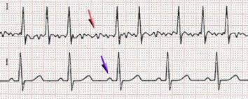
Response: There is a well known association between alcohol intake and atrial fibrillation form population based studies which demonstrate that for every 1 standard drink the incidence of AFib increases by 8%.
This is the first randomised study to determine of alcohol reduction/abstinence leads to a reduction in AFib episodes and time to recurrence.
Dr. Kistler[/caption]
Professor Peter M Kistler MBBS, PhD, FRACP
Head of Clinical Electrophysiology Research
Baker Heart and Diabetes Institute
Head of Electrophysiology at The Alfred hospital
Professor of Medicine
University of Melbourne.
MedicalResearch.com: What is the background for this study?
Response: There is a well known association between alcohol intake and atrial fibrillation form population based studies which demonstrate that for every 1 standard drink the incidence of AFib increases by 8%.
This is the first randomised study to determine of alcohol reduction/abstinence leads to a reduction in AFib episodes and time to recurrence.